
Canterbury > Private Hospitals & Specialists >
Family Eye Centre
Private Service, Ophthalmology, Optometry
Today
9:00 AM to 4:30 PM.
Description
Services provided include:
- Cataract surgery (adults and children)
- Strabismus (squint) correction
- Paediatric Eye Surgery
- Neurological vision problems
- Glaucoma care (medical, laser, surgery and MIGS)
- Retinal disease (intravitreal injection, laser)
- Comprehensive eye care (all common eye issues)
- Strabismus surgery (Squint repair)
- Vision correction (glasses, lazy eye, amblyopia)
- School vision issues
- Tracking difficulties
- Progressive myopia
- Contact lenses
- Cataract and glaucoma diagnosis, treatment and surgery
- Nasolacrimal duct obstruction (watering & discharge)
- Headache, papilledema, cerebral visual impairment (CVI)
- Autism spectrum and developmental delay (eye examination)
- Retinal problems in children and teenagers
- Familial (inherited) eye problems
- Inflammatory eye problems
- Retinal disease
What is Ophthalmology?
Staff
- Marie Yeo: Optometrist (not registered). Runs the myopia clinic.
- Lora: Orthoptist. Cares for poor vision in young children.
- Kaye: Technician. Manages diabetic retinal screening & Eyescreen.
- Emma: Tests children & fits glasses.
Consultants
-
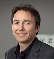
Dr Antony Bedggood
Ophthalmologist
Optometrists
-

Miss Catherine Small
Optometrist
Ages
Child / Tamariki, Youth / Rangatahi, Adult / Pakeke, Older adult / Kaumātua
How do I access this service?
Website / App
Our screening eye tests can be booked online, please click here for:
- children’s eye test ages 6 months to 6 years
- myopia progression test
- Adult Glaucoma, Cataract, or Diabetic retinal photograph
Eyescreen, gives access to affordable screening tests. Performed by an experienced technician, Eyescreen tests cost little when compared with a Specialist visit, but they provide the same quality, using the same complex medical equipment.
Referral
Adults: for a full assessment we need either a new referral, or a past letter or report.
Children: aged 0 - 16 years can be seen without a referral.
Anyone can access
Make an appointment
Contact us here to book an appointment or phone (03) 595 1360
Eyescreen, gives access to affordable screening tests. Performed by an experienced technician, Eyescreen tests cost little when compared with a Specialist visit, but they provide the same quality, using the same complex medical equipment.
Referral Expectations
Visit length can vary from 10 minutes to 45 minutes, depending on the type of appointment. Click here for more details
Fees and Charges Categorisation
Fees apply, Free
Fees and Charges Description
Find infomation about fees here
We are a Southern Cross Affiliated Provider and ACC providers.
Hours
9:00 AM to 4:30 PM.
| Mon – Fri | 9:00 AM – 4:30 PM |
|---|
Catherine Small's clinics run on Wednesday every week and Thursday morning every fortnight.
Procedures / Treatments
These conditions cause distance blur. In myopia, the eye has a resting focus at a near distance so that people will be able to see objects clearly at some point close to them, whilst the distance is blurry. Hyperopia also causes distance blur but often does not become noticeable until the eye loses its ability to change focus, frequently in middle age. The loss of focus for near distance (presbyopia or “aged sight”) is also related to a decreased ability to change focus but only affects reading. Astigmatism causes an image to be blurry at all distances, but does not affect clarity of images unless it is severe. An optometrist or ophthalmologist can test for these conditions. Treatment is usually glasses or contact lenses which are only obtainable through an optometrist or dispensing optician. Laser surgery and other corrective surgical techniques can also be used to change the focus of the eye to give clarity of sight in suitable patients.
These conditions cause distance blur. In myopia, the eye has a resting focus at a near distance so that people will be able to see objects clearly at some point close to them, whilst the distance is blurry. Hyperopia also causes distance blur but often does not become noticeable until the eye loses its ability to change focus, frequently in middle age. The loss of focus for near distance (presbyopia or “aged sight”) is also related to a decreased ability to change focus but only affects reading. Astigmatism causes an image to be blurry at all distances, but does not affect clarity of images unless it is severe. An optometrist or ophthalmologist can test for these conditions. Treatment is usually glasses or contact lenses which are only obtainable through an optometrist or dispensing optician. Laser surgery and other corrective surgical techniques can also be used to change the focus of the eye to give clarity of sight in suitable patients.
These conditions cause distance blur. In myopia, the eye has a resting focus at a near distance so that people will be able to see objects clearly at some point close to them, whilst the distance is blurry. Hyperopia also causes distance blur but often does not become noticeable until the eye loses its ability to change focus, frequently in middle age. The loss of focus for near distance (presbyopia or “aged sight”) is also related to a decreased ability to change focus but only affects reading. Astigmatism causes an image to be blurry at all distances, but does not affect clarity of images unless it is severe. An optometrist or ophthalmologist can test for these conditions. Treatment is usually glasses or contact lenses which are only obtainable through an optometrist or dispensing optician. Laser surgery and other corrective surgical techniques can also be used to change the focus of the eye to give clarity of sight in suitable patients.
Cataracts are the most common age-related occurrence in eyes. The lens becomes thicker and stiffer and appears yellow and cloudy. Eventually it may turn white, changing the colour of the pupil. A cataract may cause your vision to become fuzzy in a progressive fashion and may also be the cause of disabling glare. Once a cataract affects vision too much, a cataract removal operation is generally advised. This decision is usually made in consultation with an eye specialist. The operation is almost always done under local anaesthetic. Once the cataract has been removed an artificial lens is put in to replace it. It is relatively short in duration and an overnight stay in hospital is not required. Post-operative care consists of eye drops and a check at 1-2 days then after 2-4 weeks.
Cataracts are the most common age-related occurrence in eyes. The lens becomes thicker and stiffer and appears yellow and cloudy. Eventually it may turn white, changing the colour of the pupil. A cataract may cause your vision to become fuzzy in a progressive fashion and may also be the cause of disabling glare. Once a cataract affects vision too much, a cataract removal operation is generally advised. This decision is usually made in consultation with an eye specialist. The operation is almost always done under local anaesthetic. Once the cataract has been removed an artificial lens is put in to replace it. It is relatively short in duration and an overnight stay in hospital is not required. Post-operative care consists of eye drops and a check at 1-2 days then after 2-4 weeks.
A weakness in one or more of the muscles of the eye will cause the eye to turn or move away from the normal focusing position. This is commonly known as a squint. A squint can be corrected by surgery, or by using glasses. Rarely, children may grow out of a squint. Surgical correction of squint usually involves a general anaesthetic. In the procedure, the muscles involved are repositioned to correct the alignment. It is important to recognise and treat a squint as, if left uncorrected, it can result in permanent impairment of vision.
A weakness in one or more of the muscles of the eye will cause the eye to turn or move away from the normal focusing position. This is commonly known as a squint. A squint can be corrected by surgery, or by using glasses. Rarely, children may grow out of a squint. Surgical correction of squint usually involves a general anaesthetic. In the procedure, the muscles involved are repositioned to correct the alignment. It is important to recognise and treat a squint as, if left uncorrected, it can result in permanent impairment of vision.
Glaucoma is a group of diseases that can damage the eye’s optic nerve and may result in vision loss and blindness. Multiple factors are often important in causing glaucoma, but it is most commonly related to in an increase in pressure in the eye. Symptoms are generally absent until the condition has progressed to an advanced stage. Very occasionally, a rarer form of glaucoma can develop suddenly and symptoms may then include: headaches and aches around the affected eye, seeing halos around lights, sensitivity to light, blurred vision, nausea and vomiting. You may be more likely to develop glaucoma if you: have someone else in your family with glaucoma already have high pressure in your eye have experienced injury to your eye have or have had certain other eye problems have migraine or circulation problems. Glaucoma is more common in people over 50 years of age and more common in women than men. Diagnosis usually comes after consultation with an eye doctor. Signs of glaucoma may also be picked up at an optometrist’s eye examination. The following tests are used to diagnose and monitor glaucoma: Tonometry – measures eye pressure. It is often the first screening test for glaucoma. The eyes are numbed with eye drops and then examined. Dilated eye exam - this is done with an ophthalmoscope (which is a medical instrument that allows the doctor to look through the pupil to the back of the eye).The retina and optic nerve are then examined for any sign of damage. Visual acuity test – test to check distance vision using an eye chart. Visual field test – test to measure side (peripheral) vision. Pachymetry – test to measure the thickness of the cornea. Many other new techniques are emerging to help identify the likelihood of glaucoma and help determine its rate of worsening. Although glaucoma cannot be cured, early treatment can prevent further worsening of the condition and vision loss. Regular eye examinations will need to be continued life-long. Eye drops to decrease eye pressure are the most common early treatment. Surgery may be required, especially if medications are not taking adequate effect. Laser trabeculoplasty, in which a surgeon uses a laser to help the fluid drain from the eye, may be considered in some cases, but has limited effectiveness. More commonly, a trabeculectomy may be performed when other methods have failed to adequately control pressure. This is a medium length operation that makes a new opening for fluid to drain from the eye.
Glaucoma is a group of diseases that can damage the eye’s optic nerve and may result in vision loss and blindness. Multiple factors are often important in causing glaucoma, but it is most commonly related to in an increase in pressure in the eye. Symptoms are generally absent until the condition has progressed to an advanced stage. Very occasionally, a rarer form of glaucoma can develop suddenly and symptoms may then include: headaches and aches around the affected eye, seeing halos around lights, sensitivity to light, blurred vision, nausea and vomiting. You may be more likely to develop glaucoma if you: have someone else in your family with glaucoma already have high pressure in your eye have experienced injury to your eye have or have had certain other eye problems have migraine or circulation problems. Glaucoma is more common in people over 50 years of age and more common in women than men. Diagnosis usually comes after consultation with an eye doctor. Signs of glaucoma may also be picked up at an optometrist’s eye examination. The following tests are used to diagnose and monitor glaucoma: Tonometry – measures eye pressure. It is often the first screening test for glaucoma. The eyes are numbed with eye drops and then examined. Dilated eye exam - this is done with an ophthalmoscope (which is a medical instrument that allows the doctor to look through the pupil to the back of the eye).The retina and optic nerve are then examined for any sign of damage. Visual acuity test – test to check distance vision using an eye chart. Visual field test – test to measure side (peripheral) vision. Pachymetry – test to measure the thickness of the cornea. Many other new techniques are emerging to help identify the likelihood of glaucoma and help determine its rate of worsening. Although glaucoma cannot be cured, early treatment can prevent further worsening of the condition and vision loss. Regular eye examinations will need to be continued life-long. Eye drops to decrease eye pressure are the most common early treatment. Surgery may be required, especially if medications are not taking adequate effect. Laser trabeculoplasty, in which a surgeon uses a laser to help the fluid drain from the eye, may be considered in some cases, but has limited effectiveness. More commonly, a trabeculectomy may be performed when other methods have failed to adequately control pressure. This is a medium length operation that makes a new opening for fluid to drain from the eye.
- have someone else in your family with glaucoma
- already have high pressure in your eye
- have experienced injury to your eye
- have or have had certain other eye problems
- have migraine or circulation problems.
- Tonometry – measures eye pressure. It is often the first screening test for glaucoma. The eyes are numbed with eye drops and then examined.
- Dilated eye exam - this is done with an ophthalmoscope (which is a medical instrument that allows the doctor to look through the pupil to the back of the eye).The retina and optic nerve are then examined for any sign of damage.
- Visual acuity test – test to check distance vision using an eye chart.
- Visual field test – test to measure side (peripheral) vision.
- Pachymetry – test to measure the thickness of the cornea.
This is a complication of diabetes and is caused by small blood vessel damage within the retina of the eye. It commonly affects both eyes and may cause permanent loss of vision. Macular oedema is sometimes also present with diabetic retinopathy. Macular oedema is when fluid leaks into the retina and causes swelling and blurred vision. This may occur at any stage of diabetic retinopathy, but is more common as the disease progresses. There are often no symptoms in the early stages but as the condition progresses vision may begin to become impaired. Often visual loss may be sudden and without warning. This is why it is imperative that at-risk diabetics have frequent eye checks. Poorly controlled diabetes and pregnancy in diabetes are risk factors for developing this condition. Often, first-stage diabetic retinopathy requires no active treatment on the eye but requires stabilisation of diabetes and regular eye examinations. With progressive retinopathy, a laser treatment called the PRP laser can be used. This works by shrinking enlarged blood vessels to prevent further bleeding into the retina. Severe bleeding may require a surgical procedure called a vitrectomy, where blood is surgically removed from the eye. Treatment of macular oedema, if present, is by focal laser treatment. Vision is stabilised by reducing the degree of fluid leakage into the retina. Often more than one treatment is required.
This is a complication of diabetes and is caused by small blood vessel damage within the retina of the eye. It commonly affects both eyes and may cause permanent loss of vision. Macular oedema is sometimes also present with diabetic retinopathy. Macular oedema is when fluid leaks into the retina and causes swelling and blurred vision. This may occur at any stage of diabetic retinopathy, but is more common as the disease progresses. There are often no symptoms in the early stages but as the condition progresses vision may begin to become impaired. Often visual loss may be sudden and without warning. This is why it is imperative that at-risk diabetics have frequent eye checks. Poorly controlled diabetes and pregnancy in diabetes are risk factors for developing this condition. Often, first-stage diabetic retinopathy requires no active treatment on the eye but requires stabilisation of diabetes and regular eye examinations. With progressive retinopathy, a laser treatment called the PRP laser can be used. This works by shrinking enlarged blood vessels to prevent further bleeding into the retina. Severe bleeding may require a surgical procedure called a vitrectomy, where blood is surgically removed from the eye. Treatment of macular oedema, if present, is by focal laser treatment. Vision is stabilised by reducing the degree of fluid leakage into the retina. Often more than one treatment is required.
This is when the retina detaches, meaning it is lifted or separated from its normal position within the eye. An acute retinal detachment requires urgent assessment and appropriate treatment. Unless prompt and effective treatment is given, some forms of retinal detachment may lead to irreversible blindness. Signs and symptoms include: a sudden or gradual increase in floaters, deterioration in vision, cobwebs or specks with the visual field, light flashes in the eye or the appearance of curtains over the visual field. You are more likely to have a retinal detachment if you are very short-sighted or have had an injury or previous surgery to the eye. For minor detachments, a laser or freeze treatment (cryopexy) are used. Both therapies re-attach the retina. For major detachment, surgery will be necessary. A band is often put around the back of the eye to prevent further detachment. Surgical treatment is usually a vitrectomy, where the jelly (vitreous) is removed from the eye. This often involves a hospital stay. It can take several months post-surgery to see the final visual result.
This is when the retina detaches, meaning it is lifted or separated from its normal position within the eye. An acute retinal detachment requires urgent assessment and appropriate treatment. Unless prompt and effective treatment is given, some forms of retinal detachment may lead to irreversible blindness. Signs and symptoms include: a sudden or gradual increase in floaters, deterioration in vision, cobwebs or specks with the visual field, light flashes in the eye or the appearance of curtains over the visual field. You are more likely to have a retinal detachment if you are very short-sighted or have had an injury or previous surgery to the eye. For minor detachments, a laser or freeze treatment (cryopexy) are used. Both therapies re-attach the retina. For major detachment, surgery will be necessary. A band is often put around the back of the eye to prevent further detachment. Surgical treatment is usually a vitrectomy, where the jelly (vitreous) is removed from the eye. This often involves a hospital stay. It can take several months post-surgery to see the final visual result.
Services Provided
During an eye test you are assessed for different things. A Snellen chart, which contains rows of letters starting with one huge letter then groups of letters getting progressively smaller, is used for visual acuity. An optometrist will establish how well you see without spectacles or contact lenses by getting you to read letters until they are too small or blurred to be recognised. They test each eye on its own and both together. Then the test is repeated while wearing your existing eye wear, this is known as your best corrected vision. Then the optometrist will ask you to remove your eye wear again and will see if your ability to read letters can be improved using lenses of different strengths for each eye until your eye sight is as good as it can be. If these new lenses are a different strength from your current spectacles you are given a new prescription and new lenses will be made for you. People usually wear spectacles to correct a refraction error. In these cases the person’s eye lens is incorrectly focused, making that person either long or short sighted. This fault is corrected by placing an external lens, spectacle lens or contact lens, in front of the eye so that what is seen reaches the correct area on the retina in the back of the eye. There are other tests to look for other conditions, so an optometrist can find a corrective lens that gets a person’s sight near to, or in excess of, 20/20 vision. We use central vision to read and look at thing in front of us. We also need side or peripheral vision to see things that are above and below and to the sides. This is also assessed during an eye test as you need a good field of vision to drive a car and it is a requirement for certain professions like the police.
During an eye test you are assessed for different things. A Snellen chart, which contains rows of letters starting with one huge letter then groups of letters getting progressively smaller, is used for visual acuity. An optometrist will establish how well you see without spectacles or contact lenses by getting you to read letters until they are too small or blurred to be recognised. They test each eye on its own and both together. Then the test is repeated while wearing your existing eye wear, this is known as your best corrected vision. Then the optometrist will ask you to remove your eye wear again and will see if your ability to read letters can be improved using lenses of different strengths for each eye until your eye sight is as good as it can be. If these new lenses are a different strength from your current spectacles you are given a new prescription and new lenses will be made for you. People usually wear spectacles to correct a refraction error. In these cases the person’s eye lens is incorrectly focused, making that person either long or short sighted. This fault is corrected by placing an external lens, spectacle lens or contact lens, in front of the eye so that what is seen reaches the correct area on the retina in the back of the eye. There are other tests to look for other conditions, so an optometrist can find a corrective lens that gets a person’s sight near to, or in excess of, 20/20 vision. We use central vision to read and look at thing in front of us. We also need side or peripheral vision to see things that are above and below and to the sides. This is also assessed during an eye test as you need a good field of vision to drive a car and it is a requirement for certain professions like the police.
During an eye test you are assessed for different things. A Snellen chart, which contains rows of letters starting with one huge letter then groups of letters getting progressively smaller, is used for visual acuity. An optometrist will establish how well you see without spectacles or contact lenses by getting you to read letters until they are too small or blurred to be recognised. They test each eye on its own and both together. Then the test is repeated while wearing your existing eye wear, this is known as your best corrected vision. Then the optometrist will ask you to remove your eye wear again and will see if your ability to read letters can be improved using lenses of different strengths for each eye until your eye sight is as good as it can be. If these new lenses are a different strength from your current spectacles you are given a new prescription and new lenses will be made for you.
People usually wear spectacles to correct a refraction error. In these cases the person’s eye lens is incorrectly focused, making that person either long or short sighted. This fault is corrected by placing an external lens, spectacle lens or contact lens, in front of the eye so that what is seen reaches the correct area on the retina in the back of the eye. There are other tests to look for other conditions, so an optometrist can find a corrective lens that gets a person’s sight near to, or in excess of, 20/20 vision.
We use central vision to read and look at thing in front of us. We also need side or peripheral vision to see things that are above and below and to the sides. This is also assessed during an eye test as you need a good field of vision to drive a car and it is a requirement for certain professions like the police.
During an eye test optometrists routinely examine the internal and external structures of your eye. The optometrist will look into your eye by shining a light source through your pupil to illuminate the retina. This may take some time as the optometrist will want to ensure the fundus is viewed as completely as possible and all signs and symptoms of eye disease are noted. It is important that the causes of poor vision are determined before any treatment, including spectacles, is prescribed.
During an eye test optometrists routinely examine the internal and external structures of your eye. The optometrist will look into your eye by shining a light source through your pupil to illuminate the retina. This may take some time as the optometrist will want to ensure the fundus is viewed as completely as possible and all signs and symptoms of eye disease are noted. It is important that the causes of poor vision are determined before any treatment, including spectacles, is prescribed.
During an eye test optometrists routinely examine the internal and external structures of your eye. The optometrist will look into your eye by shining a light source through your pupil to illuminate the retina. This may take some time as the optometrist will want to ensure the fundus is viewed as completely as possible and all signs and symptoms of eye disease are noted. It is important that the causes of poor vision are determined before any treatment, including spectacles, is prescribed.
Optometrists are able to detect early signs of glaucoma even before you are aware of any symptoms. They are specifically looking for changes when doing the health check part of an eye test. Since the optic nerve can be affected by this condition, they will inspect the nerve head in each eye to see if the disc shape has changed and if there are any variations in colour. If picked up early, damage from this condition can be prevented from becoming serious and sight threatening.
Optometrists are able to detect early signs of glaucoma even before you are aware of any symptoms. They are specifically looking for changes when doing the health check part of an eye test. Since the optic nerve can be affected by this condition, they will inspect the nerve head in each eye to see if the disc shape has changed and if there are any variations in colour. If picked up early, damage from this condition can be prevented from becoming serious and sight threatening.
Optometrists are able to detect early signs of glaucoma even before you are aware of any symptoms. They are specifically looking for changes when doing the health check part of an eye test. Since the optic nerve can be affected by this condition, they will inspect the nerve head in each eye to see if the disc shape has changed and if there are any variations in colour. If picked up early, damage from this condition can be prevented from becoming serious and sight threatening.
It is vital children have their eyes tested early to screen for abnormalities. If there are problems like strabismus, where eyes are misaligned, or amblyopia, commonly called a lazy eye, the earlier treatment starts the better the outcome. Poor eyesight will also reduce a child’s ability to learn at home as well as at school. As well as testing for these conditions optometrists test for near and far vision; if needed, corrective glasses will make a great difference to a child’s ability to see and interact with the world. Children are also tested for colour blindness.
It is vital children have their eyes tested early to screen for abnormalities. If there are problems like strabismus, where eyes are misaligned, or amblyopia, commonly called a lazy eye, the earlier treatment starts the better the outcome. Poor eyesight will also reduce a child’s ability to learn at home as well as at school. As well as testing for these conditions optometrists test for near and far vision; if needed, corrective glasses will make a great difference to a child’s ability to see and interact with the world. Children are also tested for colour blindness.
It is vital children have their eyes tested early to screen for abnormalities. If there are problems like strabismus, where eyes are misaligned, or amblyopia, commonly called a lazy eye, the earlier treatment starts the better the outcome. Poor eyesight will also reduce a child’s ability to learn at home as well as at school. As well as testing for these conditions optometrists test for near and far vision; if needed, corrective glasses will make a great difference to a child’s ability to see and interact with the world. Children are also tested for colour blindness.
Frames: there is a baffling range of spectacle frames available in most optical outlets. They are made in many shapes, sizes, colours and materials. They are also made for different uses, reflecting the lifestyle and work of individual wearers. Dispensing opticians and optometrists are able to help you find a frame that complements the shape of your face, colouring and suits your needs. They will also take a set of measurements to ensure that the frame built for you fits correctly and that you get maximum use from the lenses. Lenses: at the end of an eye test, if your vision can be improved by wearing glasses, the optometrist will write a prescription for a set of lenses. In some cases you will have a different prescription for each eye. Lenses are designed to bring your eyesight up to 20/20 vision and middle aged and older patients may be advised to have varifocals so that they do not require a second pair of glasses for reading or close work. Lens technology is improving all the time with new lightweight material becoming available that can self adjust to reduce light. Contact lenses: contact lenses have been around for more than half a century. The material has changed over this period: hard or soft lenses being replaced by gas permeable and now disposable contact lenses. These lenses are able to compensate for long or short sight and are worn for a day then thrown away to minimise the risk of infecting the eye. Because a foreign object is placed on the eye, contact lens users are advised to have an annual eye check so that an optometrist can check the cornea to ensure there are no abrasions or other long term complications. While most wearers use contact lenses to improve their eyesight there are products that can change the colour of your eyes or make them a darker or lighter shade of the same colour. Sunglasses: your dispensing optician and optometrist will be able to advise you on the most suitable sunglasses to lessen damage caused by ultraviolet light. Choosing: dispensing opticians and optometrists are trained to help you select spectacle frames and lenses that suit your look, lifestyle and pocket. They will ask about your work, interests and preferences and then find you a selection of models to consider. They are aware of a set of principles that help them find frames they think will suit the shape of your face and colouring. The final choice is yours. Care of glasses/lenses: as part of the service offered by dispensing opticians and optometrists, patients are given advice and tips about how to look after their spectacles. Things that will extend the working lives of eyewear like using both hands to take off or put them on, correct storage, best methods of cleaning and so forth. In the past they will also have advised contact lens wearers on the best way to sterilise these objects but most wearers today use disposable contact lenses so this is no longer necessary. Adjusting: wear and tear during everyday use means that arm hinges become loose and the frame is bent out of shape. Your dispensing optician or optometrist will usually readjust the glasses he has sold you free of charge. Warranty: generally your spectacle frames will have a manufacturer’s one or two year warranty which will cover things like faulty soldering or arm hinges. It does not cover damage done if your specs are chewed by a dog or they are run over by a car. Lenses also have a one or two year warranty. Repair: dispensing opticians and optometrists not only help you find the most suitable pair of glasses but are also able to repair them. Most commonly, arms get loose when they are pushed to the top of the head and need tightening up, nose pads break off and spectacles become bent when they are sat on. Many of these repairs can be done in the workshop most dispensing opticians have behind the shop or, when parts are broken, new components can be ordered in to replace the damaged part.
Frames: there is a baffling range of spectacle frames available in most optical outlets. They are made in many shapes, sizes, colours and materials. They are also made for different uses, reflecting the lifestyle and work of individual wearers. Dispensing opticians and optometrists are able to help you find a frame that complements the shape of your face, colouring and suits your needs. They will also take a set of measurements to ensure that the frame built for you fits correctly and that you get maximum use from the lenses. Lenses: at the end of an eye test, if your vision can be improved by wearing glasses, the optometrist will write a prescription for a set of lenses. In some cases you will have a different prescription for each eye. Lenses are designed to bring your eyesight up to 20/20 vision and middle aged and older patients may be advised to have varifocals so that they do not require a second pair of glasses for reading or close work. Lens technology is improving all the time with new lightweight material becoming available that can self adjust to reduce light. Contact lenses: contact lenses have been around for more than half a century. The material has changed over this period: hard or soft lenses being replaced by gas permeable and now disposable contact lenses. These lenses are able to compensate for long or short sight and are worn for a day then thrown away to minimise the risk of infecting the eye. Because a foreign object is placed on the eye, contact lens users are advised to have an annual eye check so that an optometrist can check the cornea to ensure there are no abrasions or other long term complications. While most wearers use contact lenses to improve their eyesight there are products that can change the colour of your eyes or make them a darker or lighter shade of the same colour. Sunglasses: your dispensing optician and optometrist will be able to advise you on the most suitable sunglasses to lessen damage caused by ultraviolet light. Choosing: dispensing opticians and optometrists are trained to help you select spectacle frames and lenses that suit your look, lifestyle and pocket. They will ask about your work, interests and preferences and then find you a selection of models to consider. They are aware of a set of principles that help them find frames they think will suit the shape of your face and colouring. The final choice is yours. Care of glasses/lenses: as part of the service offered by dispensing opticians and optometrists, patients are given advice and tips about how to look after their spectacles. Things that will extend the working lives of eyewear like using both hands to take off or put them on, correct storage, best methods of cleaning and so forth. In the past they will also have advised contact lens wearers on the best way to sterilise these objects but most wearers today use disposable contact lenses so this is no longer necessary. Adjusting: wear and tear during everyday use means that arm hinges become loose and the frame is bent out of shape. Your dispensing optician or optometrist will usually readjust the glasses he has sold you free of charge. Warranty: generally your spectacle frames will have a manufacturer’s one or two year warranty which will cover things like faulty soldering or arm hinges. It does not cover damage done if your specs are chewed by a dog or they are run over by a car. Lenses also have a one or two year warranty. Repair: dispensing opticians and optometrists not only help you find the most suitable pair of glasses but are also able to repair them. Most commonly, arms get loose when they are pushed to the top of the head and need tightening up, nose pads break off and spectacles become bent when they are sat on. Many of these repairs can be done in the workshop most dispensing opticians have behind the shop or, when parts are broken, new components can be ordered in to replace the damaged part.
Frames: there is a baffling range of spectacle frames available in most optical outlets. They are made in many shapes, sizes, colours and materials. They are also made for different uses, reflecting the lifestyle and work of individual wearers. Dispensing opticians and optometrists are able to help you find a frame that complements the shape of your face, colouring and suits your needs. They will also take a set of measurements to ensure that the frame built for you fits correctly and that you get maximum use from the lenses.
Lenses: at the end of an eye test, if your vision can be improved by wearing glasses, the optometrist will write a prescription for a set of lenses. In some cases you will have a different prescription for each eye. Lenses are designed to bring your eyesight up to 20/20 vision and middle aged and older patients may be advised to have varifocals so that they do not require a second pair of glasses for reading or close work. Lens technology is improving all the time with new lightweight material becoming available that can self adjust to reduce light.
Contact lenses: contact lenses have been around for more than half a century. The material has changed over this period: hard or soft lenses being replaced by gas permeable and now disposable contact lenses. These lenses are able to compensate for long or short sight and are worn for a day then thrown away to minimise the risk of infecting the eye. Because a foreign object is placed on the eye, contact lens users are advised to have an annual eye check so that an optometrist can check the cornea to ensure there are no abrasions or other long term complications. While most wearers use contact lenses to improve their eyesight there are products that can change the colour of your eyes or make them a darker or lighter shade of the same colour.
Sunglasses: your dispensing optician and optometrist will be able to advise you on the most suitable sunglasses to lessen damage caused by ultraviolet light.
Choosing: dispensing opticians and optometrists are trained to help you select spectacle frames and lenses that suit your look, lifestyle and pocket. They will ask about your work, interests and preferences and then find you a selection of models to consider. They are aware of a set of principles that help them find frames they think will suit the shape of your face and colouring. The final choice is yours.
Care of glasses/lenses: as part of the service offered by dispensing opticians and optometrists, patients are given advice and tips about how to look after their spectacles. Things that will extend the working lives of eyewear like using both hands to take off or put them on, correct storage, best methods of cleaning and so forth. In the past they will also have advised contact lens wearers on the best way to sterilise these objects but most wearers today use disposable contact lenses so this is no longer necessary.
Adjusting: wear and tear during everyday use means that arm hinges become loose and the frame is bent out of shape. Your dispensing optician or optometrist will usually readjust the glasses he has sold you free of charge.
Warranty: generally your spectacle frames will have a manufacturer’s one or two year warranty which will cover things like faulty soldering or arm hinges. It does not cover damage done if your specs are chewed by a dog or they are run over by a car. Lenses also have a one or two year warranty.
Repair: dispensing opticians and optometrists not only help you find the most suitable pair of glasses but are also able to repair them. Most commonly, arms get loose when they are pushed to the top of the head and need tightening up, nose pads break off and spectacles become bent when they are sat on. Many of these repairs can be done in the workshop most dispensing opticians have behind the shop or, when parts are broken, new components can be ordered in to replace the damaged part.
Diabetic retinopathy (DR) can be detected by systematic retinal screening. Diabetes retinal screening is funded by the DHB and some optometrists are contracted to provide the service in the community. Currently this community service is only available in the Greater Wellington Regional Area, Lakes District Health Board (DHB), Tairawhiti DHB, and Counties Manukau DHB. The aim of the service is to ensure each individual diagnosed with diabetes has received a retinal screen during the preceding 24 months and that their data is transferred to the primary care provider they are enrolled with, and DHB. Entry to this free service is by referral from your general practitioner (GP), who will know which optometrists are able to provide systematic retinal screening. If diabetic eye disease is detected by your regular optometrist, they will refer you to your GP for diabetes management. This will include regular systematic retinal screening. The majority of patients who have had diabetes for more than a decade will show signs of damage. Early detection helps as patients can be advised on changes to their diet and lifestyle to control blood sugar levels and minimise damage caused to this vital organ.
Diabetic retinopathy (DR) can be detected by systematic retinal screening. Diabetes retinal screening is funded by the DHB and some optometrists are contracted to provide the service in the community. Currently this community service is only available in the Greater Wellington Regional Area, Lakes District Health Board (DHB), Tairawhiti DHB, and Counties Manukau DHB. The aim of the service is to ensure each individual diagnosed with diabetes has received a retinal screen during the preceding 24 months and that their data is transferred to the primary care provider they are enrolled with, and DHB. Entry to this free service is by referral from your general practitioner (GP), who will know which optometrists are able to provide systematic retinal screening. If diabetic eye disease is detected by your regular optometrist, they will refer you to your GP for diabetes management. This will include regular systematic retinal screening. The majority of patients who have had diabetes for more than a decade will show signs of damage. Early detection helps as patients can be advised on changes to their diet and lifestyle to control blood sugar levels and minimise damage caused to this vital organ.
Diabetic retinopathy (DR) can be detected by systematic retinal screening. Diabetes retinal screening is funded by the DHB and some optometrists are contracted to provide the service in the community. Currently this community service is only available in the Greater Wellington Regional Area, Lakes District Health Board (DHB), Tairāwhiti DHB, and Counties Manukau DHB. The aim of the service is to ensure each individual diagnosed with diabetes has received a retinal screen during the preceding 24 months and that their data is transferred to the primary care provider they are enrolled with, and DHB. Entry to this free service is by referral from your general practitioner (GP), who will know which optometrists are able to provide systematic retinal screening.
If diabetic eye disease is detected by your regular optometrist, they will refer you to your GP for diabetes management. This will include regular systematic retinal screening. The majority of patients who have had diabetes for more than a decade will show signs of damage. Early detection helps as patients can be advised on changes to their diet and lifestyle to control blood sugar levels and minimise damage caused to this vital organ.
A visual field establishes how good your peripheral vision (PV) is. This is what you see above and below and to the side when you are looking straight ahead. Generally your PV will not be as good as what is known as your central vision but it is important for driving and a good visual field is required to become a New Zealand police officer. There are many conditions that can affect your PV including high blood pressure, glaucoma, strokes, an overactive thyroid, and diabetes. In glaucoma, nerve cells in the retina are damaged and die so messages are unable to be transmitted to the brain. Remarkably up to 40% of these cells can be lost before a person becomes aware of reduced vision.
A visual field establishes how good your peripheral vision (PV) is. This is what you see above and below and to the side when you are looking straight ahead. Generally your PV will not be as good as what is known as your central vision but it is important for driving and a good visual field is required to become a New Zealand police officer. There are many conditions that can affect your PV including high blood pressure, glaucoma, strokes, an overactive thyroid, and diabetes. In glaucoma, nerve cells in the retina are damaged and die so messages are unable to be transmitted to the brain. Remarkably up to 40% of these cells can be lost before a person becomes aware of reduced vision.
A visual field establishes how good your peripheral vision (PV) is. This is what you see above and below and to the side when you are looking straight ahead. Generally your PV will not be as good as what is known as your central vision but it is important for driving and a good visual field is required to become a New Zealand police officer. There are many conditions that can affect your PV including high blood pressure, glaucoma, strokes, an overactive thyroid, and diabetes. In glaucoma, nerve cells in the retina are damaged and die so messages are unable to be transmitted to the brain. Remarkably up to 40% of these cells can be lost before a person becomes aware of reduced vision.
Disability Assistance
Mobility parking space
Public Transport
We are on a major bus and cycle route.
Parking
Free parking available. Find details here
Pharmacy
Find your nearest pharmacy here
Website

Contact Details
211 Ferry Road, Waltham, Christchurch
Canterbury
9:00 AM to 4:30 PM.
-
Phone
0800 439383 or (03) 595 1360
Email
Website
Unit 15/211 Ferry Road
Waltham
Christchurch
Street Address
Unit 15/211 Ferry Road
Waltham
Christchurch
Was this page helpful?
This page was last updated at 11:14AM on December 6, 2023. This information is reviewed and edited by Family Eye Centre.