Waikato > Private Hospitals & Specialists >
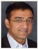
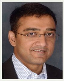
Dr Madhav Menon - Hamilton Cardiologist
Private Service, Cardiology
Today
9:00 AM to 5:00 PM.
Description
Dr Madhav Menon completed his cardiology specialist training at Waikato Hospital. followed by a two year interventional cardiology fellowship at University of Minnesota, Minneapolis, USA . He worked as a consultant cardiologist at Waikato Hospital from 2008 to 2025 November as Head of Coronary Interventions. Dr Menon now directs Cardiac Services, a cardiology group where he currently works full time. He consults and performs various investigative tests along with an established group of colleagues. His procedures are done at Midland Cardiovascular, Braemar Hospital in Hamilton. He is a board director at Midland Cardiovascular Ltd and is a member of the advisory board in Pharmac for coronary interventions.
Dr Menon's referrals have a high cohort of complex coronary angioplasty. He is a competent user of the various atherectomy devices, coronary imaging and does in depth coronary physiology studies. He is one of the lead operators for chronic total occlusion angioplasty in the country. He has conducted teaching programmes in these areas and has performed procedures with the best in the world. He regularly performs most of the high risk angioplasties in the region and has developed his expertise around this internationally.
He directs the REACT course, a new course in the world which is a simulated emergency management training for the cath lab.
Dr Menon performs Transcatheter Aortic Valve Implantation (TAVI). This is a minimally invasive procedure to replace diseased aortic valves. He is now offering this service from Braemar Hospital and is accepting referrals for aortic valve pathology.
Cardiac Services Ltd's team of specialists, doctors, nurses, cardiac technicians, sonographers and receptionists offers the complete cardiology assessment and delivers world class heart care to all their patients. They are located in Dawson Street within walking distance of the city and Hamilton East Village. Southern Cross Hospital is just around the corner.
Services offered include:
- cardiorespiratory testing
- exercise treadmill tests
- respiratory testing
- ECG with interpretation
- event recording
- 24/48 hour digital Holter and blood pressure monitoring
- oxygen saturation levels.
What is Cardiology?
Cardiology is the specialty within medicine that looks at the heart and blood vessels. Your heart consists of four chambers, which are responsible for pumping blood to your lungs and then the rest of your body. The study of the heart includes the heart muscle (the myocardium), the valves within the heart between the chambers, the blood vessels that supply blood (and hence oxygen and nutrients) to the heart muscle, and the electrical system of the heart which is what controls the heart rate.
Consultants
-

Dr Madhav Menon
Cardiologist
Referral Expectations
For patients:
Your GP will refer you to Dr Menon if they are concerned about your heart and want a specialist opinion.
For referrers:
Our commitment to support general practitioners in the community is reflected in our "FAX A QUESTION" service. We will answer complex cardiology questions within 24 hours and/or the option for direct discussion with a cardiologist (if available). Fax: (07) 839 7663.
URGENT appointments by referral or direct contact by GP.
Fees and Charges Description
Payment is expected on the day of appointment. EFTPOS and VISA facilities are available.
Hours
9:00 AM to 5:00 PM.
| Mon – Fri | 9:00 AM – 5:00 PM |
|---|
Procedures / Treatments
An ECG is a recording of your heart's electrical activity. Electrode patches are attached to your skin to measure the electrical impulses given off by your heart. The result is a trace that can be read by a doctor. It can give information of previous heart attacks or problems with the heart rhythm. Ambulatory ECG - this can be performed with a Holter monitor which monitors your heart for rhythm abnormalities during normal activity for an uninterrupted 24-hour period. During the test, electrodes attached to your chest are connected to a portable recorder - about the size of a paperback book - that's attached to your belt or hung from a shoulder strap. Another form of ambulatory ECG test is an Event recorder which covers 1-2 weeks. You wear a monitor (much smaller than a Holter monitor) and if you have any symptoms, such as dizziness, you press a button on a recording device which saves the recording of your heart rhythm made in the minutes leading up to and during your symptoms. Because you can wear this for a longer period of time it has a higher rate of catching your abnormal rhythm.
An ECG is a recording of your heart's electrical activity. Electrode patches are attached to your skin to measure the electrical impulses given off by your heart. The result is a trace that can be read by a doctor. It can give information of previous heart attacks or problems with the heart rhythm. Ambulatory ECG - this can be performed with a Holter monitor which monitors your heart for rhythm abnormalities during normal activity for an uninterrupted 24-hour period. During the test, electrodes attached to your chest are connected to a portable recorder - about the size of a paperback book - that's attached to your belt or hung from a shoulder strap. Another form of ambulatory ECG test is an Event recorder which covers 1-2 weeks. You wear a monitor (much smaller than a Holter monitor) and if you have any symptoms, such as dizziness, you press a button on a recording device which saves the recording of your heart rhythm made in the minutes leading up to and during your symptoms. Because you can wear this for a longer period of time it has a higher rate of catching your abnormal rhythm.
An ECG is a recording of your heart's electrical activity. Electrode patches are attached to your skin to measure the electrical impulses given off by your heart. The result is a trace that can be read by a doctor. It can give information of previous heart attacks or problems with the heart rhythm.
Ambulatory ECG - this can be performed with a Holter monitor which monitors your heart for rhythm abnormalities during normal activity for an uninterrupted 24-hour period. During the test, electrodes attached to your chest are connected to a portable recorder - about the size of a paperback book - that's attached to your belt or hung from a shoulder strap.
Another form of ambulatory ECG test is an Event recorder which covers 1-2 weeks. You wear a monitor (much smaller than a Holter monitor) and if you have any symptoms, such as dizziness, you press a button on a recording device which saves the recording of your heart rhythm made in the minutes leading up to and during your symptoms. Because you can wear this for a longer period of time it has a higher rate of catching your abnormal rhythm.
An ECG done when you are resting may be normal even when you have cardiovascular disease. During an exercise ECG the heart is made to work harder so that if there is any narrowing of the blood vessels resulting in poor blood supply it is more likely to be picked up on the tracing as your heart goes faster. For this test you have to work harder which involves walking on a treadmill while your heart is monitored. The treadmill gets faster with time but you can stop at anytime. This test is supervised and interpreted by a doctor as you go. This test is used to see if you have any evidence of cardiovascular disease and can give the doctor some idea as to how severe it might be so as to direct further tests and possible treatment.
An ECG done when you are resting may be normal even when you have cardiovascular disease. During an exercise ECG the heart is made to work harder so that if there is any narrowing of the blood vessels resulting in poor blood supply it is more likely to be picked up on the tracing as your heart goes faster. For this test you have to work harder which involves walking on a treadmill while your heart is monitored. The treadmill gets faster with time but you can stop at anytime. This test is supervised and interpreted by a doctor as you go. This test is used to see if you have any evidence of cardiovascular disease and can give the doctor some idea as to how severe it might be so as to direct further tests and possible treatment.
An ECG done when you are resting may be normal even when you have cardiovascular disease. During an exercise ECG the heart is made to work harder so that if there is any narrowing of the blood vessels resulting in poor blood supply it is more likely to be picked up on the tracing as your heart goes faster. For this test you have to work harder which involves walking on a treadmill while your heart is monitored. The treadmill gets faster with time but you can stop at anytime. This test is supervised and interpreted by a doctor as you go. This test is used to see if you have any evidence of cardiovascular disease and can give the doctor some idea as to how severe it might be so as to direct further tests and possible treatment.
Cardiology blood tests help doctors check for heart disease, monitor risk factors, and assess how well the heart is working. Some tests check for damage right now (like troponin), while others check your long-term risks (like cholesterol and blood sugar) or stress on the heart (like BNP).
Cardiology blood tests help doctors check for heart disease, monitor risk factors, and assess how well the heart is working. Some tests check for damage right now (like troponin), while others check your long-term risks (like cholesterol and blood sugar) or stress on the heart (like BNP).
Cardiology blood tests help doctors check for heart disease, monitor risk factors, and assess how well the heart is working. Some tests check for damage right now (like troponin), while others check your long-term risks (like cholesterol and blood sugar) or stress on the heart (like BNP).
Echocardiography (or cardiac ultrasound) is a test that uses high frequency sound waves to generate pictures of your heart. During the test, you generally lie on your back, gel is applied to your skin and a technician then moves the small, plastic transducer over your chest. The test is painless and can take from 10 minutes to an hour. The machine then develops images of your heart which are seen on a monitor. This is referred to as an echocardiogram. Echocardiography can help in the diagnosis of many heart problems including cardiovascular disease, previous heart attacks, valve disorders, weakened heart muscle, holes between heart chambers, fluid around the heart (pericardial effusion). If doctors are looking for evidence of coronary artery disease, they may perform variations of this test which include: Exercise echocardiography - compares how your heart works when stressed by exercise versus when it is at rest. The ultrasound is conducted before you exercise and immediately after you stop. Either a stationary bicycle or standard treadmill is used. Dobutamine stress echocardiography - if you’re unable to exercise for the above test, you might be given medication to simulate the effects of exercise. During this test, an echocardiogram initially is performed when you’re at rest. Then dobutamine is given to you via a needle into a vein in your arm. Its effect is to make your heart work harder and faster just like with exercise. After it has taken effect, the echocardiogram is repeated. The effect wears off very quickly.
Echocardiography (or cardiac ultrasound) is a test that uses high frequency sound waves to generate pictures of your heart. During the test, you generally lie on your back, gel is applied to your skin and a technician then moves the small, plastic transducer over your chest. The test is painless and can take from 10 minutes to an hour. The machine then develops images of your heart which are seen on a monitor. This is referred to as an echocardiogram. Echocardiography can help in the diagnosis of many heart problems including cardiovascular disease, previous heart attacks, valve disorders, weakened heart muscle, holes between heart chambers, fluid around the heart (pericardial effusion). If doctors are looking for evidence of coronary artery disease, they may perform variations of this test which include: Exercise echocardiography - compares how your heart works when stressed by exercise versus when it is at rest. The ultrasound is conducted before you exercise and immediately after you stop. Either a stationary bicycle or standard treadmill is used. Dobutamine stress echocardiography - if you’re unable to exercise for the above test, you might be given medication to simulate the effects of exercise. During this test, an echocardiogram initially is performed when you’re at rest. Then dobutamine is given to you via a needle into a vein in your arm. Its effect is to make your heart work harder and faster just like with exercise. After it has taken effect, the echocardiogram is repeated. The effect wears off very quickly.
Echocardiography (or cardiac ultrasound) is a test that uses high frequency sound waves to generate pictures of your heart. During the test, you generally lie on your back, gel is applied to your skin and a technician then moves the small, plastic transducer over your chest. The test is painless and can take from 10 minutes to an hour.
The machine then develops images of your heart which are seen on a monitor. This is referred to as an echocardiogram.
Echocardiography can help in the diagnosis of many heart problems including cardiovascular disease, previous heart attacks, valve disorders, weakened heart muscle, holes between heart chambers, fluid around the heart (pericardial effusion).
If doctors are looking for evidence of coronary artery disease, they may perform variations of this test which include:
- Exercise echocardiography - compares how your heart works when stressed by exercise versus when it is at rest. The ultrasound is conducted before you exercise and immediately after you stop. Either a stationary bicycle or standard treadmill is used.
- Dobutamine stress echocardiography - if you’re unable to exercise for the above test, you might be given medication to simulate the effects of exercise. During this test, an echocardiogram initially is performed when you’re at rest. Then dobutamine is given to you via a needle into a vein in your arm. Its effect is to make your heart work harder and faster just like with exercise. After it has taken effect, the echocardiogram is repeated. The effect wears off very quickly.
This test is performed by a cardiologist in a sterile operating theatre environment. Most people will need to have routine tests before the procedure. These tests may require separate appointments and are usually planned the day before or the day of the procedure. You will be asked not to eat or drink after midnight the evening before the procedure. You are not given a general anaesthetic but may have some medication to relax you if needed. Local anaesthetic is put into an area of skin to the side of your groin or in your arm. A needle and then tube are fed into an artery here and advanced through the blood vessels to the heart. Dye is then injected so that the heart and blood vessels can be seen on X-ray. X-rays and measurements are then taken giving the doctors information about the state of your heart and the exact nature of any narrowed blood vessels. This allows them to plan the best form of treatment to prevent heart attacks and control any symptoms you may have. After the procedure you will have to lay flat for several hours to prevent bleeding.
This test is performed by a cardiologist in a sterile operating theatre environment. Most people will need to have routine tests before the procedure. These tests may require separate appointments and are usually planned the day before or the day of the procedure. You will be asked not to eat or drink after midnight the evening before the procedure. You are not given a general anaesthetic but may have some medication to relax you if needed. Local anaesthetic is put into an area of skin to the side of your groin or in your arm. A needle and then tube are fed into an artery here and advanced through the blood vessels to the heart. Dye is then injected so that the heart and blood vessels can be seen on X-ray. X-rays and measurements are then taken giving the doctors information about the state of your heart and the exact nature of any narrowed blood vessels. This allows them to plan the best form of treatment to prevent heart attacks and control any symptoms you may have. After the procedure you will have to lay flat for several hours to prevent bleeding.
This test is performed by a cardiologist in a sterile operating theatre environment.
Most people will need to have routine tests before the procedure. These tests may require separate appointments and are usually planned the day before or the day of the procedure. You will be asked not to eat or drink after midnight the evening before the procedure.
You are not given a general anaesthetic but may have some medication to relax you if needed. Local anaesthetic is put into an area of skin to the side of your groin or in your arm. A needle and then tube are fed into an artery here and advanced through the blood vessels to the heart. Dye is then injected so that the heart and blood vessels can be seen on X-ray. X-rays and measurements are then taken giving the doctors information about the state of your heart and the exact nature of any narrowed blood vessels. This allows them to plan the best form of treatment to prevent heart attacks and control any symptoms you may have.
After the procedure you will have to lay flat for several hours to prevent bleeding.
Cardiovascular disease is a general term for any condition that affects the heart or blood vessels. Common types of cardiovascular disease include: Coronary artery disease – narrowing or blockage of the arteries that supply the heart, which can lead to chest pain or heart attack. Heart failure – when the heart can’t pump blood effectively. Arrhythmias – abnormal heart rhythms. Stroke – damage to the brain caused by blocked or burst blood vessels. Peripheral artery disease – narrowing of arteries in the legs or arms. Heart valve problems – issues with valves that control blood flow through the heart. Risk factors: High blood pressure High cholesterol Smoking Diabetes Obesity Family history of heart disease Are older (your risk increases as you get older)
Cardiovascular disease is a general term for any condition that affects the heart or blood vessels. Common types of cardiovascular disease include: Coronary artery disease – narrowing or blockage of the arteries that supply the heart, which can lead to chest pain or heart attack. Heart failure – when the heart can’t pump blood effectively. Arrhythmias – abnormal heart rhythms. Stroke – damage to the brain caused by blocked or burst blood vessels. Peripheral artery disease – narrowing of arteries in the legs or arms. Heart valve problems – issues with valves that control blood flow through the heart. Risk factors: High blood pressure High cholesterol Smoking Diabetes Obesity Family history of heart disease Are older (your risk increases as you get older)
Cardiovascular disease is a general term for any condition that affects the heart or blood vessels.
Common types of cardiovascular disease include:
- Coronary artery disease – narrowing or blockage of the arteries that supply the heart, which can lead to chest pain or heart attack.
- Heart failure – when the heart can’t pump blood effectively.
- Arrhythmias – abnormal heart rhythms.
- Stroke – damage to the brain caused by blocked or burst blood vessels.
- Peripheral artery disease – narrowing of arteries in the legs or arms.
- Heart valve problems – issues with valves that control blood flow through the heart.
Risk factors:
- High blood pressure
- High cholesterol
- Smoking
- Diabetes
- Obesity
- Family history of heart disease
- Are older (your risk increases as you get older)
Heart failure refers to the heart failing to pump efficiently. There are many diseases that cause this including cardiovascular disease, high blood pressure, viral infections, alcohol, and diseases affecting the valves of the heart. When the heart is inefficient a number of symptoms occur depending on the cause and severity of the condition. The main symptoms are tiredness, breathlessness on exertion or lying flat, and ankle swelling. Doctors often refer to oedema, which means fluid retention usually in your feet or lungs as a result of the heart not pumping efficiently. Tests looking for possible causes of heart failure include: Chest x-ray, Electrocardiogram (ECG), Echocardiogram (Cardiac ultrasound), Angiogram. You are likely to be given several medications over time, started and monitored by your cardiologist and GP. These include medication to control the amount of fluid that builds up (diuretics), medication to protect your heart and slow it down as well as to thin your blood. You will often be referred to a dietitian or given advice about restricting the amount of fluid and salt you take as this can contribute to symptoms.
Heart failure refers to the heart failing to pump efficiently. There are many diseases that cause this including cardiovascular disease, high blood pressure, viral infections, alcohol, and diseases affecting the valves of the heart. When the heart is inefficient a number of symptoms occur depending on the cause and severity of the condition. The main symptoms are tiredness, breathlessness on exertion or lying flat, and ankle swelling. Doctors often refer to oedema, which means fluid retention usually in your feet or lungs as a result of the heart not pumping efficiently. Tests looking for possible causes of heart failure include: Chest x-ray, Electrocardiogram (ECG), Echocardiogram (Cardiac ultrasound), Angiogram. You are likely to be given several medications over time, started and monitored by your cardiologist and GP. These include medication to control the amount of fluid that builds up (diuretics), medication to protect your heart and slow it down as well as to thin your blood. You will often be referred to a dietitian or given advice about restricting the amount of fluid and salt you take as this can contribute to symptoms.
Heart failure refers to the heart failing to pump efficiently. There are many diseases that cause this including cardiovascular disease, high blood pressure, viral infections, alcohol, and diseases affecting the valves of the heart. When the heart is inefficient a number of symptoms occur depending on the cause and severity of the condition. The main symptoms are tiredness, breathlessness on exertion or lying flat, and ankle swelling. Doctors often refer to oedema, which means fluid retention usually in your feet or lungs as a result of the heart not pumping efficiently.
Tests looking for possible causes of heart failure include: Chest x-ray, Electrocardiogram (ECG), Echocardiogram (Cardiac ultrasound), Angiogram.
You are likely to be given several medications over time, started and monitored by your cardiologist and GP. These include medication to control the amount of fluid that builds up (diuretics), medication to protect your heart and slow it down as well as to thin your blood. You will often be referred to a dietitian or given advice about restricting the amount of fluid and salt you take as this can contribute to symptoms.
Heart rhythm refers to the electrical source that is driving the heart rate and whether or not it is regular or irregular. Heart rhythm can be affected by a number of conditions. Some common terms Sinus rhythm is the normal rhythm Arrhythmia means abnormal rhythm Fibrillation means irregular rhythm or quivering of one part of the heart Bradycardia means slow heart rate Tachycardia means fast heart rate Paroxysmal means the arrhythmia comes and goes Tachycardia The most common form of this is atrial fibrillation. This is where the heart rhythm is irregular and often too fast. Symptoms include fatigue, palpitations (where you are aware of your heart racing or pounding), dizziness and breathlessness. Other tachycardias include supraventricular tachycardia (SVT) or ventricular tachycardia (VT). These have similar symptoms as atrial fibrillation but can also cause you to lose consciousness (faint). Bradycardia The most common form of this is called heart block. This is because messages from the electrical generator of the heart don't get through efficiently to the rest of the heart and hence it goes very slowly or can pause. Symptoms of the heart going too slowly include feeling tired, breathless or fainting. Tests Tests to diagnose what sort of arrhythmia you have include an electrocardiogram (ECG) and an ambulatory ECG (Holter monitor or Event recorder). Treatment Most treatments for tachycardias consist of medication to stop the abnormal rhythm or make it slower if and when it occurs. Atrial fibrillation, if you have other problems, can increase your risk of stroke so blood-thinning medication is often used as well. If you have bradycardia, you may be referred to the surgeons for a pacemaker. This is a small operation where a battery powered device is placed under the skin with wires that lead to your heart and provide it with electrical stimulation to prevent it from going too slowly. You can't feel it doing this but will be aware of a small flat lump under your skin just below your collar bone.
Heart rhythm refers to the electrical source that is driving the heart rate and whether or not it is regular or irregular. Heart rhythm can be affected by a number of conditions. Some common terms Sinus rhythm is the normal rhythm Arrhythmia means abnormal rhythm Fibrillation means irregular rhythm or quivering of one part of the heart Bradycardia means slow heart rate Tachycardia means fast heart rate Paroxysmal means the arrhythmia comes and goes Tachycardia The most common form of this is atrial fibrillation. This is where the heart rhythm is irregular and often too fast. Symptoms include fatigue, palpitations (where you are aware of your heart racing or pounding), dizziness and breathlessness. Other tachycardias include supraventricular tachycardia (SVT) or ventricular tachycardia (VT). These have similar symptoms as atrial fibrillation but can also cause you to lose consciousness (faint). Bradycardia The most common form of this is called heart block. This is because messages from the electrical generator of the heart don't get through efficiently to the rest of the heart and hence it goes very slowly or can pause. Symptoms of the heart going too slowly include feeling tired, breathless or fainting. Tests Tests to diagnose what sort of arrhythmia you have include an electrocardiogram (ECG) and an ambulatory ECG (Holter monitor or Event recorder). Treatment Most treatments for tachycardias consist of medication to stop the abnormal rhythm or make it slower if and when it occurs. Atrial fibrillation, if you have other problems, can increase your risk of stroke so blood-thinning medication is often used as well. If you have bradycardia, you may be referred to the surgeons for a pacemaker. This is a small operation where a battery powered device is placed under the skin with wires that lead to your heart and provide it with electrical stimulation to prevent it from going too slowly. You can't feel it doing this but will be aware of a small flat lump under your skin just below your collar bone.
Heart rhythm refers to the electrical source that is driving the heart rate and whether or not it is regular or irregular. Heart rhythm can be affected by a number of conditions.
Some common terms
- Sinus rhythm is the normal rhythm
- Arrhythmia means abnormal rhythm
- Fibrillation means irregular rhythm or quivering of one part of the heart
- Bradycardia means slow heart rate
- Tachycardia means fast heart rate
- Paroxysmal means the arrhythmia comes and goes
Tachycardia
The most common form of this is atrial fibrillation. This is where the heart rhythm is irregular and often too fast. Symptoms include fatigue, palpitations (where you are aware of your heart racing or
pounding), dizziness and breathlessness.
Other tachycardias include supraventricular tachycardia (SVT) or ventricular tachycardia (VT). These have similar symptoms as atrial fibrillation but can also cause you to lose consciousness (faint).
Bradycardia
The most common form of this is called heart block. This is because messages from the electrical generator of the heart don't get through efficiently to the rest of the heart and hence it goes very slowly or can pause. Symptoms of the heart going too slowly include feeling tired, breathless or fainting.
Tests
Tests to diagnose what sort of arrhythmia you have include an electrocardiogram (ECG) and an ambulatory ECG (Holter monitor or Event recorder).
Treatment
Most treatments for tachycardias consist of medication to stop the abnormal rhythm or make it slower if and when it occurs. Atrial fibrillation, if you have other problems, can increase your risk of stroke so blood-thinning medication is often used as well.
If you have bradycardia, you may be referred to the surgeons for a pacemaker. This is a small operation where a battery powered device is placed under the skin with wires that lead to your heart and provide it with electrical stimulation to prevent it from going too slowly. You can't feel it doing this but will be aware of a small flat lump under your skin just below your collar bone.
Your heart consists of 4 chambers that receive and send blood to the lungs and body. Disorders affecting valves can either cause stenosis (a narrowing) or regurgitation (leakage after the valve has closed). Depending on what valve is involved and how severe the damage is it may result in symptoms of heart failure (see above), as it makes the heart pump inefficiently. Suspicion of a heart valve problem is usually picked up by your doctor when they listen to your heart and hear a murmur. A murmur is heard with the stethoscope and is turbulence of blood flow that occurs through a narrowed or leaky valve. Not all heart murmurs mean serious problems but are best investigated further. The echocardiogram is the main test to diagnose what valve is involved and how severe it is. Treatment depends on the type and severity of the valve lesion. You may simply be monitored over years to see if anything changes. Some conditions require medication to thin the blood or treat any complicating heart problems. You may be referred to a heart surgeon for consideration of a valve replacement or dilatation of a narrowed valve.
Your heart consists of 4 chambers that receive and send blood to the lungs and body. Disorders affecting valves can either cause stenosis (a narrowing) or regurgitation (leakage after the valve has closed). Depending on what valve is involved and how severe the damage is it may result in symptoms of heart failure (see above), as it makes the heart pump inefficiently. Suspicion of a heart valve problem is usually picked up by your doctor when they listen to your heart and hear a murmur. A murmur is heard with the stethoscope and is turbulence of blood flow that occurs through a narrowed or leaky valve. Not all heart murmurs mean serious problems but are best investigated further. The echocardiogram is the main test to diagnose what valve is involved and how severe it is. Treatment depends on the type and severity of the valve lesion. You may simply be monitored over years to see if anything changes. Some conditions require medication to thin the blood or treat any complicating heart problems. You may be referred to a heart surgeon for consideration of a valve replacement or dilatation of a narrowed valve.
Your heart consists of 4 chambers that receive and send blood to the lungs and body.
Disorders affecting valves can either cause stenosis (a narrowing) or regurgitation (leakage after the valve has closed). Depending on what valve is involved and how severe the damage is it may result in symptoms of heart failure (see above), as it makes the heart pump inefficiently.
Suspicion of a heart valve problem is usually picked up by your doctor when they listen to your heart and hear a murmur. A murmur is heard with the stethoscope and is turbulence of blood flow that occurs through a narrowed or leaky valve. Not all heart murmurs mean serious problems but are best investigated further.
The echocardiogram is the main test to diagnose what valve is involved and how severe it is.
Treatment depends on the type and severity of the valve lesion. You may simply be monitored over years to see if anything changes. Some conditions require medication to thin the blood or treat any complicating heart problems. You may be referred to a heart surgeon for consideration of a valve replacement or dilatation of a narrowed valve.
Document Downloads
- Hybrid Coronary Revascularisation (PDF, 89.3 KB)
- Rapid Exchange MicroCatheter (PDF, 9.4 MB)
-
Chronic Total Occlusion Workshop
(DOCX, 374.8 KB)
Live case demonstration of managing chronic total occlusion. Live cases were performed by Dr Menon and Professor Ochiai who is a world leader in chronic total occlusion angioplasty procedures. Attended by high end operators from Australia and New Zealand.
Parking
Off-street parking is provided.
Website
Contact Details
1 Dawson Street, Hamilton
Waikato
9:00 AM to 5:00 PM.
-
Phone
(07) 839 7664
-
Fax
(07) 839 7663
Healthlink EDI
charlesn
Website
Or contact us online here
URGENT appointments by referral or direct contact by GP.
Cardiac Services Limited
1 Dawson Street
Hamilton
Street Address
Cardiac Services Limited
1 Dawson Street
Hamilton
Was this page helpful?
This page was last updated at 9:22AM on October 20, 2025. This information is reviewed and edited by Dr Madhav Menon - Hamilton Cardiologist.

