Wellington > Private Hospitals & Specialists >
Anwyl Specialist Medical Centre Dermatology Team
Private Service, Dermatology, Skin Cancer
Today
Description
Anwyl Specialist Medical Centre Dermatology Team's services include:
Staff
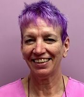
Rosa McLean: Phototherapist
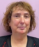
Liz Thach: Dermatology Nurse
Alam Judd: Health Care Assistant
Marcela Foos: Health Care Assistant
Consultants
-

Dr Lissa Judd
Dermatologist
Doctors
-

Dr Helen Fieldes
Specialist General Practitioner
Ages
Child / Tamariki, Youth / Rangatahi, Adult / Pakeke, Older adult / Kaumātua
How do I access this service?
Referral, Make an appointment, Contact us
Fees and Charges Categorisation
Fees apply
Fees and Charges Description
Find fees information here
Hours
| Mon – Fri | 8:00 AM – 7:00 PM |
|---|---|
| Sat | 8:00 AM – 4:00 PM |
Languages Spoken
English
Procedures / Treatments
Narrow Band UVB Phototherapy is a form of ultraviolet treatment used to treat a wide variety of skin diseases, including psoriasis, eczema, polymorphous light eruption, lichen planus, urticaria and vitiligo. Often we will do a ’test strip’ first to determine the best starting dose of treatment. This involves exposing several small squares of skin on the back to ultraviolet - each square getting a bigger dose than the one before. The next day these squares are checked to see if any of these doses have produced a mild sunburn - and one then commences treatment with the biggest dose which DID NOT produce a sunburn. The patient stands in front of a bank of lights (which look like fluorescent tubes but which emit a special wavelength of light from the ultraviolet B range) to receive their dose of light as determined by their test strip. The first dose usually only takes a minute. Protective goggles or a faceshield are worn (unless the face or eyelids require treatment). Treatments are done twice a week, and the dose is increased by 15-20% each treatment unless faint sunburn occurs. Most skin diseases show improvement after 3 weeks of treatment, and the treatment is continued until the skin problem has entirely resolved. Although the treatment is usually administered by a phototherapist or one of our clinical assistants, the progress is carefully monitored by the dermatologist.
Narrow Band UVB Phototherapy is a form of ultraviolet treatment used to treat a wide variety of skin diseases, including psoriasis, eczema, polymorphous light eruption, lichen planus, urticaria and vitiligo. Often we will do a ’test strip’ first to determine the best starting dose of treatment. This involves exposing several small squares of skin on the back to ultraviolet - each square getting a bigger dose than the one before. The next day these squares are checked to see if any of these doses have produced a mild sunburn - and one then commences treatment with the biggest dose which DID NOT produce a sunburn. The patient stands in front of a bank of lights (which look like fluorescent tubes but which emit a special wavelength of light from the ultraviolet B range) to receive their dose of light as determined by their test strip. The first dose usually only takes a minute. Protective goggles or a faceshield are worn (unless the face or eyelids require treatment). Treatments are done twice a week, and the dose is increased by 15-20% each treatment unless faint sunburn occurs. Most skin diseases show improvement after 3 weeks of treatment, and the treatment is continued until the skin problem has entirely resolved. Although the treatment is usually administered by a phototherapist or one of our clinical assistants, the progress is carefully monitored by the dermatologist.
Narrow Band UVB Phototherapy is a form of ultraviolet treatment used to treat a wide variety of skin diseases, including psoriasis, eczema, polymorphous light eruption, lichen planus, urticaria and vitiligo.
Often we will do a ’test strip’ first to determine the best starting dose of treatment. This involves exposing several small squares of skin on the back to ultraviolet - each square getting a bigger dose than the one before. The next day these squares are checked to see if any of these doses have produced a mild sunburn - and one then commences treatment with the biggest dose which DID NOT produce a sunburn.
The patient stands in front of a bank of lights (which look like fluorescent tubes but which emit a special wavelength of light from the ultraviolet B range) to receive their dose of light as determined by their test strip. The first dose usually only takes a minute. Protective goggles or a faceshield are worn (unless the face or eyelids require treatment).
Treatments are done twice a week, and the dose is increased by 15-20% each treatment unless faint sunburn occurs.
Most skin diseases show improvement after 3 weeks of treatment, and the treatment is continued until the skin problem has entirely resolved. Although the treatment is usually administered by a phototherapist or one of our clinical assistants, the progress is carefully monitored by the dermatologist.
When someone has noticed a suspicious lesion this can be checked, using a dermatoscope (a magnifying device for checking the features just below the surface of the skin). If necessary the lesion can be excised or sampled under local anaesthetic. All specimens are sent to the laboratory to be checked by a pathologist. Most skin cancers can be excised here at Anwyl Specialist Medical Centre. There is a small operating theatre for this purpose. Some people are at high risk of skin cancer because either they have 100's of moles they have multiple family members with melanoma they have a previous history of melanoma or multiple keratinocytic skin cancers they take immunosupressing medication eg after an organ transplant and in these circumstances we encourage regular skin checks. Such patients can be put on an annual recall list for skin checks. We can also do total body photography of moles - whereby the patient is re-photographed annually and their photos are compared side by side on the computer to determine whether or not a mole is changing over time, or a new lesion has developed. In addition, advice is given regarding self checks and sun protection.
When someone has noticed a suspicious lesion this can be checked, using a dermatoscope (a magnifying device for checking the features just below the surface of the skin). If necessary the lesion can be excised or sampled under local anaesthetic. All specimens are sent to the laboratory to be checked by a pathologist. Most skin cancers can be excised here at Anwyl Specialist Medical Centre. There is a small operating theatre for this purpose. Some people are at high risk of skin cancer because either they have 100's of moles they have multiple family members with melanoma they have a previous history of melanoma or multiple keratinocytic skin cancers they take immunosupressing medication eg after an organ transplant and in these circumstances we encourage regular skin checks. Such patients can be put on an annual recall list for skin checks. We can also do total body photography of moles - whereby the patient is re-photographed annually and their photos are compared side by side on the computer to determine whether or not a mole is changing over time, or a new lesion has developed. In addition, advice is given regarding self checks and sun protection.
When someone has noticed a suspicious lesion this can be checked, using a dermatoscope (a magnifying device for checking the features just below the surface of the skin). If necessary the lesion can be excised or sampled under local anaesthetic. All specimens are sent to the laboratory to be checked by a pathologist.
Most skin cancers can be excised here at Anwyl Specialist Medical Centre. There is a small operating theatre for this purpose.
Some people are at high risk of skin cancer because either
- they have 100's of moles
- they have multiple family members with melanoma
- they have a previous history of melanoma or multiple keratinocytic skin cancers
- they take immunosupressing medication eg after an organ transplant
and in these circumstances we encourage regular skin checks. Such patients can be put on an annual recall list for skin checks. We can also do total body photography of moles - whereby the patient is re-photographed annually and their photos are compared side by side on the computer to determine whether or not a mole is changing over time, or a new lesion has developed.
In addition, advice is given regarding self checks and sun protection.
There are literally thousands of skin diseases. A large part of a dermatologist's role is the diagnosis of rashes, lumps, spots, and skin blemishes of all descriptions (and also abnormalities of hair and nails). Once a diagnosis is made, then advice can be given about the nature and prognosis of the condition, and the kinds of treatment that are available. Information on conditions such as acne, psoriasis, eczema, keratosis pilaris, lichen planus, folliculitis, scabies, pemphigoid, alopecia areata, fungal infections, tinea versicolor (to name just a very few) are available on numerous internet sites. The New Zealand Dermatological Society website http://www.dermnet.org.nz is an excellent source of information, as is the emedicine site http://www.emedicine.com/specialties.htm.
There are literally thousands of skin diseases. A large part of a dermatologist's role is the diagnosis of rashes, lumps, spots, and skin blemishes of all descriptions (and also abnormalities of hair and nails). Once a diagnosis is made, then advice can be given about the nature and prognosis of the condition, and the kinds of treatment that are available. Information on conditions such as acne, psoriasis, eczema, keratosis pilaris, lichen planus, folliculitis, scabies, pemphigoid, alopecia areata, fungal infections, tinea versicolor (to name just a very few) are available on numerous internet sites. The New Zealand Dermatological Society website http://www.dermnet.org.nz is an excellent source of information, as is the emedicine site http://www.emedicine.com/specialties.htm.
There are literally thousands of skin diseases. A large part of a dermatologist's role is the diagnosis of rashes, lumps, spots, and skin blemishes of all descriptions (and also abnormalities of hair and nails). Once a diagnosis is made, then advice can be given about the nature and prognosis of the condition, and the kinds of treatment that are available.
Information on conditions such as acne, psoriasis, eczema, keratosis pilaris, lichen planus, folliculitis, scabies, pemphigoid, alopecia areata, fungal infections, tinea versicolor (to name just a very few) are available on numerous internet sites. The New Zealand Dermatological Society website http://www.dermnet.org.nz is an excellent source of information, as is the emedicine site http://www.emedicine.com/specialties.htm.
A patch test is used to diagnose contact allergy, where a person is allergic to a chemical which is in contact with the skin - for example a person might develop dermatitis on the hands due to allergy to their rubber gloves, or dermatitis on the face due to allergy to their hair dye. The majority of allergies occur after frequent exposure to the chemical. For example, hair dye allergy is most likely to occur in someone who has dyed their hair numerous times before, without previous problems (but once the person is allergic, then every subsequent time they dye their hair they will react with a dermatitis). Common allergens include hair colouring agents and bleaches, nickel in jewellery, chromate in cement, a large number of plants, the adhesive used with 'fake' nails, rubber, preservatives in creams and lotions and shampoos, epoxy products.....but there are hundreds of others too. When a contact allergy is suspected a thorough history is taken, to determine which chemicals (allergens) need to be tested. The test substances are then placed on special chambers on non allergenic tape, and these tapes (each containing 10 allergen samples) are placed on the person's back. Most people having allergy testing done will have 50 or more substances tested. The tapes are left in place for 2 days during which time the person must not get the back wet. Then the tapes are removed, having carefully marked their location. The sites are checked again in a further 2 days, and again in another 2 or 3 days. In total the test involves four visits. If a reaction occurs, an area of eczema about 10mm in size develops at the site of the allergen. The severity of the reaction is graded by the therapist or doctor. A copy of the test results and a report explaining the significance of these results is usually provided to both the patient and the referring doctor.
A patch test is used to diagnose contact allergy, where a person is allergic to a chemical which is in contact with the skin - for example a person might develop dermatitis on the hands due to allergy to their rubber gloves, or dermatitis on the face due to allergy to their hair dye. The majority of allergies occur after frequent exposure to the chemical. For example, hair dye allergy is most likely to occur in someone who has dyed their hair numerous times before, without previous problems (but once the person is allergic, then every subsequent time they dye their hair they will react with a dermatitis). Common allergens include hair colouring agents and bleaches, nickel in jewellery, chromate in cement, a large number of plants, the adhesive used with 'fake' nails, rubber, preservatives in creams and lotions and shampoos, epoxy products.....but there are hundreds of others too. When a contact allergy is suspected a thorough history is taken, to determine which chemicals (allergens) need to be tested. The test substances are then placed on special chambers on non allergenic tape, and these tapes (each containing 10 allergen samples) are placed on the person's back. Most people having allergy testing done will have 50 or more substances tested. The tapes are left in place for 2 days during which time the person must not get the back wet. Then the tapes are removed, having carefully marked their location. The sites are checked again in a further 2 days, and again in another 2 or 3 days. In total the test involves four visits. If a reaction occurs, an area of eczema about 10mm in size develops at the site of the allergen. The severity of the reaction is graded by the therapist or doctor. A copy of the test results and a report explaining the significance of these results is usually provided to both the patient and the referring doctor.
A patch test is used to diagnose contact allergy, where a person is allergic to a chemical which is in contact with the skin - for example a person might develop dermatitis on the hands due to allergy to their rubber gloves, or dermatitis on the face due to allergy to their hair dye.
The majority of allergies occur after frequent exposure to the chemical. For example, hair dye allergy is most likely to occur in someone who has dyed their hair numerous times before, without previous problems (but once the person is allergic, then every subsequent time they dye their hair they will react with a dermatitis).
Common allergens include hair colouring agents and bleaches, nickel in jewellery, chromate in cement, a large number of plants, the adhesive used with 'fake' nails, rubber, preservatives in creams and lotions and shampoos, epoxy products.....but there are hundreds of others too.
When a contact allergy is suspected a thorough history is taken, to determine which chemicals (allergens) need to be tested. The test substances are then placed on special chambers on non allergenic tape, and these tapes (each containing 10 allergen samples) are placed on the person's back. Most people having allergy testing done will have 50 or more substances tested.
The tapes are left in place for 2 days during which time the person must not get the back wet. Then the tapes are removed, having carefully marked their location. The sites are checked again in a further 2 days, and again in another 2 or 3 days. In total the test involves four visits.
If a reaction occurs, an area of eczema about 10mm in size develops at the site of the allergen. The severity of the reaction is graded by the therapist or doctor.
A copy of the test results and a report explaining the significance of these results is usually provided to both the patient and the referring doctor.
Botox (Botulinum toxin) injections are a very successful treatment for axillary hyperhidrosis (excessive perspiration from the armpits). The procedure involves first mapping the exact area in the armpit where the sweating occurs. To do this one paints the armpit with iodine and then applies a talc. When perspiration occurs the area is stained black, and this area can be marked out with a felt tip pen. It is commonplace for sweating to not occur uniformly throughout the entire armpit. Sometimes the excessive perspiration can occur from quite a localised area, which literally runs like a tap. It is important to identify the area where the sweat glands are overactive so that this area or areas can be treated (there is no point in injecting Botox into areas of armpit that are not going to perspire and which are not contributing to the problem). We strongly advise people having this procedure to not wear their best white blouse or shirt for the occasion, in case it is stained with the iodine or black dye despite our best attempts to clean it all off following the procedure! Once the area of perspiration is mapped out then we draw a grid pattern on the armpit and then inject a tiny amount of Botox into the skin at each point in the grid. It is common to have 20 injections into each armpit, but the needle that is used is so tiny that most people find that the procedure is quite tolerable. Sweating usually ceases after a couple of days, and the benefit may last several months or sometimes even a year or more. Dr Judd does not use Botox to treat frown or forehead lines (or other facial lines).
Botox (Botulinum toxin) injections are a very successful treatment for axillary hyperhidrosis (excessive perspiration from the armpits). The procedure involves first mapping the exact area in the armpit where the sweating occurs. To do this one paints the armpit with iodine and then applies a talc. When perspiration occurs the area is stained black, and this area can be marked out with a felt tip pen. It is commonplace for sweating to not occur uniformly throughout the entire armpit. Sometimes the excessive perspiration can occur from quite a localised area, which literally runs like a tap. It is important to identify the area where the sweat glands are overactive so that this area or areas can be treated (there is no point in injecting Botox into areas of armpit that are not going to perspire and which are not contributing to the problem). We strongly advise people having this procedure to not wear their best white blouse or shirt for the occasion, in case it is stained with the iodine or black dye despite our best attempts to clean it all off following the procedure! Once the area of perspiration is mapped out then we draw a grid pattern on the armpit and then inject a tiny amount of Botox into the skin at each point in the grid. It is common to have 20 injections into each armpit, but the needle that is used is so tiny that most people find that the procedure is quite tolerable. Sweating usually ceases after a couple of days, and the benefit may last several months or sometimes even a year or more. Dr Judd does not use Botox to treat frown or forehead lines (or other facial lines).
Botox (Botulinum toxin) injections are a very successful treatment for axillary hyperhidrosis (excessive perspiration from the armpits).
The procedure involves first mapping the exact area in the armpit where the sweating occurs. To do this one paints the armpit with iodine and then applies a talc. When perspiration occurs the area is stained black, and this area can be marked out with a felt tip pen. It is commonplace for sweating to not occur uniformly throughout the entire armpit. Sometimes the excessive perspiration can occur from quite a localised area, which literally runs like a tap. It is important to identify the area where the sweat glands are overactive so that this area or areas can be treated (there is no point in injecting Botox into areas of armpit that are not going to perspire and which are not contributing to the problem).
We strongly advise people having this procedure to not wear their best white blouse or shirt for the occasion, in case it is stained with the iodine or black dye despite our best attempts to clean it all off following the procedure!
Once the area of perspiration is mapped out then we draw a grid pattern on the armpit and then inject a tiny amount of Botox into the skin at each point in the grid. It is common to have 20 injections into each armpit, but the needle that is used is so tiny that most people find that the procedure is quite tolerable. Sweating usually ceases after a couple of days, and the benefit may last several months or sometimes even a year or more.
Dr Judd does not use Botox to treat frown or forehead lines (or other facial lines).
Targeted phototherapy includes technologies such as the excimer laser, and UV light sources delivered via fibre optic cables, such as Dualight. At Anwyl we use Dualight, which provides targeted broadband UVB (290-330nm with peak emission in the narrowband wavelengths) or broadband UVA (330-380nm with peak emission about 365nm ie in UVA1 range). There is a fibreoptic cable for light delivery with a 3.62 cm2 exit aperture. For UVA one can select doses up to 5 J/cm2, and for UVB up to 3120 mJ/cm2. There are several advantages to targeted phototherapy: uninvolved areas are spared, minimising side effects such as burns, and long term risks such as skin cancer higher doses can be used especially with UVB treatment of psoriasis, where 5 or 6 times the minimal erythema dose can be used, rather than starting with 50-70% of the minimal erythema dose - this results in a much faster treatment response the manoeuverable handpiece allows for difficult areas such as the ears, or armpits, to be treated. The disadvantage is that widespread skin disease cannot be treated in this fashion - one or two large plaques (roughly equal to an A4 page in their total combined size) or multiple small plaques (roughly equal to an A5 page in their total combined size) are about the maximal area that could be treated. Treatment is usually carried out twice a week, following phototesting to determine the starting dose.
Targeted phototherapy includes technologies such as the excimer laser, and UV light sources delivered via fibre optic cables, such as Dualight. At Anwyl we use Dualight, which provides targeted broadband UVB (290-330nm with peak emission in the narrowband wavelengths) or broadband UVA (330-380nm with peak emission about 365nm ie in UVA1 range). There is a fibreoptic cable for light delivery with a 3.62 cm2 exit aperture. For UVA one can select doses up to 5 J/cm2, and for UVB up to 3120 mJ/cm2. There are several advantages to targeted phototherapy: uninvolved areas are spared, minimising side effects such as burns, and long term risks such as skin cancer higher doses can be used especially with UVB treatment of psoriasis, where 5 or 6 times the minimal erythema dose can be used, rather than starting with 50-70% of the minimal erythema dose - this results in a much faster treatment response the manoeuverable handpiece allows for difficult areas such as the ears, or armpits, to be treated. The disadvantage is that widespread skin disease cannot be treated in this fashion - one or two large plaques (roughly equal to an A4 page in their total combined size) or multiple small plaques (roughly equal to an A5 page in their total combined size) are about the maximal area that could be treated. Treatment is usually carried out twice a week, following phototesting to determine the starting dose.
Targeted phototherapy includes technologies such as the excimer laser, and UV light sources delivered via fibre optic cables, such as Dualight.
At Anwyl we use Dualight, which provides targeted broadband UVB (290-330nm with peak emission in the narrowband wavelengths) or broadband UVA (330-380nm with peak emission about 365nm ie in UVA1 range). There is a fibreoptic cable for light delivery with a 3.62 cm2 exit aperture. For UVA one can select doses up to 5 J/cm2, and for UVB up to 3120 mJ/cm2.
There are several advantages to targeted phototherapy:
- uninvolved areas are spared, minimising side effects such as burns, and long term risks such as skin cancer
- higher doses can be used especially with UVB treatment of psoriasis, where 5 or 6 times the minimal erythema dose can be used, rather than starting with 50-70% of the minimal erythema dose - this results in a much faster treatment response
- the manoeuverable handpiece allows for difficult areas such as the ears, or armpits, to be treated.
The disadvantage is that widespread skin disease cannot be treated in this fashion - one or two large plaques (roughly equal to an A4 page in their total combined size) or multiple small plaques (roughly equal to an A5 page in their total combined size) are about the maximal area that could be treated.
Treatment is usually carried out twice a week, following phototesting to determine the starting dose.
Blue and Red Light Phototherapy is suitable for the treatment of mild to moderate acne, affecting the face, chest, or back. It is not suitable for treating nodular or cystic acne. Treatments are given twice a week for 4 weeks. Ideally there should be 2 or 3 days in between treatments eg Monday and Thursday or Tuesday and Saturday schedules. Blue (420nm) and Red (615nm) treatments are alternated, with the first treatment being a Blue light treatment. In other words there are 4 Blue treatments and 4 Red treatments. Each treatment lasts 20 minutes. There is no discomfort. Some people will notice slight warmth during the Red treatment. There is no swelling or peeling or redness afterwards. There is no ultraviolet in the lamps used, and so no sunburn or photoaging occurs. In fact Red Light can be used as a treatment (photo-rejuvenation) for sun-accelerated aging changes. You can use normal skin care following the treatment, however we usually advise against using acne washes and gels in conjunction with light treatment. Acne is primarily a disorder of keratinisation of the pilosebaceous (hair follicle/sebaceous gland) units. Bacteria called Propionibacterium acnes get trapped in the blocked pilosebaceous units and promote inflammation. P acnes produce porphyrins, which can be activated by light, which results in the bacteria being killed by their own products. Blue light works best to do this, but Red light penetrates deeper into the skin. It is likely that other mechanisms are at work also, since Red light can be beneficial in acne without altering bacterial populations. At the completion of 4 weeks of treatment 50% of patients have a marked improvement; 8 weeks later (without further treatment) about 75% of patients have a marked improvement. In other words, the greatest benefit is seen 2 months after the treatment is completed. This improvement is usually maintained for a further 2 or 3 months. Patients may refer themselves directly to our therapists for this treatment.
Blue and Red Light Phototherapy is suitable for the treatment of mild to moderate acne, affecting the face, chest, or back. It is not suitable for treating nodular or cystic acne. Treatments are given twice a week for 4 weeks. Ideally there should be 2 or 3 days in between treatments eg Monday and Thursday or Tuesday and Saturday schedules. Blue (420nm) and Red (615nm) treatments are alternated, with the first treatment being a Blue light treatment. In other words there are 4 Blue treatments and 4 Red treatments. Each treatment lasts 20 minutes. There is no discomfort. Some people will notice slight warmth during the Red treatment. There is no swelling or peeling or redness afterwards. There is no ultraviolet in the lamps used, and so no sunburn or photoaging occurs. In fact Red Light can be used as a treatment (photo-rejuvenation) for sun-accelerated aging changes. You can use normal skin care following the treatment, however we usually advise against using acne washes and gels in conjunction with light treatment. Acne is primarily a disorder of keratinisation of the pilosebaceous (hair follicle/sebaceous gland) units. Bacteria called Propionibacterium acnes get trapped in the blocked pilosebaceous units and promote inflammation. P acnes produce porphyrins, which can be activated by light, which results in the bacteria being killed by their own products. Blue light works best to do this, but Red light penetrates deeper into the skin. It is likely that other mechanisms are at work also, since Red light can be beneficial in acne without altering bacterial populations. At the completion of 4 weeks of treatment 50% of patients have a marked improvement; 8 weeks later (without further treatment) about 75% of patients have a marked improvement. In other words, the greatest benefit is seen 2 months after the treatment is completed. This improvement is usually maintained for a further 2 or 3 months. Patients may refer themselves directly to our therapists for this treatment.
Blue and Red Light Phototherapy is suitable for the treatment of mild to moderate acne, affecting the face, chest, or back. It is not suitable for treating nodular or cystic acne.
Treatments are given twice a week for 4 weeks. Ideally there should be 2 or 3 days in between treatments eg Monday and Thursday or Tuesday and Saturday schedules. Blue (420nm) and Red (615nm) treatments are alternated, with the first treatment being a Blue light treatment. In other words there are 4 Blue treatments and 4 Red treatments.
Each treatment lasts 20 minutes. There is no discomfort. Some people will notice slight warmth during the Red treatment. There is no swelling or peeling or redness afterwards. There is no ultraviolet in the lamps used, and so no sunburn or photoaging occurs. In fact Red Light can be used as a treatment (photo-rejuvenation) for sun-accelerated aging changes.
You can use normal skin care following the treatment, however we usually advise against using acne washes and gels in conjunction with light treatment.
Acne is primarily a disorder of keratinisation of the pilosebaceous (hair follicle/sebaceous gland) units. Bacteria called Propionibacterium acnes get trapped in the blocked pilosebaceous units and promote inflammation. P acnes produce porphyrins, which can be activated by light, which results in the bacteria being killed by their own products. Blue light works best to do this, but Red light penetrates deeper into the skin. It is likely that other mechanisms are at work also, since Red light can be beneficial in acne without altering bacterial populations.
At the completion of 4 weeks of treatment 50% of patients have a marked improvement; 8 weeks later (without further treatment) about 75% of patients have a marked improvement. In other words, the greatest benefit is seen 2 months after the treatment is completed. This improvement is usually maintained for a further 2 or 3 months.
Patients may refer themselves directly to our therapists for this treatment.
Services Provided
Skin cancer services provide management of common skin cancers, such as basal cell carcinoma, squamous cell carcinoma, and malignant melanoma. Skin cancer services can provide: Diagnosis: skin checks and biopsies can be used to identify suspicious moles, lesions, or spots and diagnose different types of skin cancer. Treatment: treatment options include surgical removal, cryotherapy, and topical treatments. Specialist Referral: for more complex cases providers can refer you to specialists for advanced care and treatment.
Skin cancer services provide management of common skin cancers, such as basal cell carcinoma, squamous cell carcinoma, and malignant melanoma. Skin cancer services can provide: Diagnosis: skin checks and biopsies can be used to identify suspicious moles, lesions, or spots and diagnose different types of skin cancer. Treatment: treatment options include surgical removal, cryotherapy, and topical treatments. Specialist Referral: for more complex cases providers can refer you to specialists for advanced care and treatment.
Skin cancer services provide management of common skin cancers, such as basal cell carcinoma, squamous cell carcinoma, and malignant melanoma.
Skin cancer services can provide:
- Diagnosis: skin checks and biopsies can be used to identify suspicious moles, lesions, or spots and diagnose different types of skin cancer.
- Treatment: treatment options include surgical removal, cryotherapy, and topical treatments.
- Specialist Referral: for more complex cases providers can refer you to specialists for advanced care and treatment.
Parking
We have plenty of free parks
Pharmacy
Website
Contact Details
13A Wall Place, Kenepuru, Porirua
Wellington
-
Phone
(04) 233 8584
-
Fax
(04) 233 1497
Healthlink EDI
anwylspm
Email
Website
Street Address
13A Wall Place
Kenepuru
Porirua
Wellington 5022
Was this page helpful?
This page was last updated at 8:48AM on December 23, 2024. This information is reviewed and edited by Anwyl Specialist Medical Centre Dermatology Team.


