
Central Auckland, East Auckland, North Auckland, South Auckland, West Auckland > Private Hospitals & Specialists >
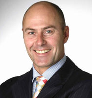
Mr John Chaplin - Thyroid and Head & Neck Surgeon, Otolaryngologist
Private Service, ENT/ Head & Neck Surgery
In emergency call 0272970971
if unavailable call ambulance and go to ED at Auckland City Hospital
Today
8:00 AM to 5:30 PM.
Description
Mr John M. Chaplin is an FRACS qualified Head and Neck, Endocrine and Facial Plastic and Reconstructive Surgeon, internationally fellowship trained in Head and Neck Surgery and Facial Plastic and Reconstructive surgery.
John works in private practice at Mauranui Clinic, Epsom and also at the Auckland Regional Head and Neck Service at Auckland City Hospital.
John's areas of interest are in thyroid, parathyroid and head & neck surgery, parotidectomy and salivary gland surgery and management of neck lumps. He is an expert in mouth and throat cancer and skin cancers and melanoma that have spread to the lymph nodes in the parotid gland and neck.
John has undergone training in Thyroid and Neck Ultrasound including Ultrasound Guided Fine Needle Aspiration Biopsy (USGFNA). John's practice at Gillies Hospital and Clinic has an ultrasound machine and John now performs in-office thyroid and neck lump ultrasound and also does USGFNA of thyroid lumps and nodules, neck lumps and salivary gland lumps. Along with in-office flexible endoscopy this makes a comprehensive one-stop referral point for assessment for neck masses and thyroid nodules and goitres.
He also has expertise in facial plastic and reconstructive surgery, including free tissue transfer, cosmetic and functional rhinoplasty and cosmetic facial surgery.
Video: Dr John Chaplin discusses his practice and talks about thyroid lumps and nodules and the types of surgery he performs.
For more information see www.nosesurgery.co.nz and www.thyroid.co.nz.
Staff
Mr John Chaplin
Head and Neck Surgeon
Ms Lisa Strachan
Practice Manager
Ms Sharon Moorhouse
Office Assistant
Consultants
-
Mr John Chaplin
Head & Neck Surgeon, Otolaryngologist
Ages
Youth / Rangatahi, Adult / Pakeke, Older adult / Kaumātua
How do I access this service?
Contact us
- Call: 09 6311948
Make an appointment
- Call: 09 6311948
Referral
- Call : 096311948
Website / App
https://www.thyroid.co.nz
Referral Expectations
- Thyroid
- Parathyroid
- One stop Neck Lump Clinic
- Parotid swellings, lumps and tumours
- Metastatic skin cancer and melanoma
- Tongue cancer
- Throat cancer
- Salivary gland surgery
- Facial plastic surgery
- Nose and nasal surgery
- Sinus surgery
- Snoring and OSA
Fees and Charges Description
Southern Cross Affiliated Provider
NIB first choice
Hours
8:00 AM to 5:30 PM.
| Mon – Fri | 8:00 AM – 5:30 PM |
|---|
Clinics:
Tues am/pm alt weeks
Wed am alt weeks
Thurs am every week
OR
Tues am Alt weeks
Wed Am/ all day alt weeks
Thurs pm every week
Public Holidays: Closed Waitangi Day (6 Feb), Good Friday (3 Apr), Easter Sunday (5 Apr), Easter Monday (6 Apr), ANZAC Day (observed) (27 Apr), King's Birthday (1 Jun), Matariki (10 Jul), Labour Day (26 Oct).
Languages Spoken
English
Procedures / Treatments
The thyroid gland is a small organ in the midline of the neck, just below the Adam's apple. It consists of a right and left lobe joined across the front of the trachea (windpipe) by a narrow bridge of thyroid tissue called the isthmus. The job of the thyroid gland is to make a hormone called thyroxine - the gland requires a small amount of iodine in the diet to produce thyroxine. Thyroidectomy is performed for thyroid nodules or thyroid lumps, cancers, Goitre with compressive symptoms, Graves Disease and thyroid cysts that involve the gland. Either the whole gland (total thyroidectomy) or a single lobe (hemithyroidectomy or lobectomy) is removed. Occasionally most of the gland is removed but a small portion left behind (subtotal thyroidectomy). Thyroid surgery is performed under general anaesthesia through a horizontal incision low down in the front of the neck. The incision can vary in length depending on the size of the thyroid gland. Sometimes if thyroid cancer is involved lymph nodes also need to be removed and this operation is called a neck dissection. There are several important structures near the thyroid gland that are at risk of injury during this type of surgery. Your surgeon will explain the risks of the operation to you. For more information please see www.thyroid.co.nz Watch the Youtube video below of a right hemithyroidectomy. Right thyroid lobectomy for a suspicious follicular thyroid nodule using a harmonic scalpel with audio commentary by Dr John M Chaplin, Head and Neck Surgeon, Auckland, New Zealand. Watch the Youtube video below of total thyroidectomy A total thyroidectomy for removal of a compressive, multinodular goitre with retrosternal extension particularly on the right side. Performed by Dr John M Chaplin, Head and Neck, Endocrine and Reconstructive Surgeon, Auckland New Zealand. The procedure demonstrates the use of the Focus Harmonic Scalpel. Audio commentary by Dr Chaplin. Watch the Youtube video below of thyroid lobectomy using the Focus Harmonic Scalpel handpiece Dr John Chaplin, Auckland, New Zealand, demonstrates the use of the Focus Harmonic Scalpel Handpiece to perform a right thyroid lobectomy. As well as haemostasis the device can also be used as a dissecting tool which reduces instrument exchange and increases efficiency.
The thyroid gland is a small organ in the midline of the neck, just below the Adam's apple. It consists of a right and left lobe joined across the front of the trachea (windpipe) by a narrow bridge of thyroid tissue called the isthmus. The job of the thyroid gland is to make a hormone called thyroxine - the gland requires a small amount of iodine in the diet to produce thyroxine. Thyroidectomy is performed for thyroid nodules or thyroid lumps, cancers, Goitre with compressive symptoms, Graves Disease and thyroid cysts that involve the gland. Either the whole gland (total thyroidectomy) or a single lobe (hemithyroidectomy or lobectomy) is removed. Occasionally most of the gland is removed but a small portion left behind (subtotal thyroidectomy). Thyroid surgery is performed under general anaesthesia through a horizontal incision low down in the front of the neck. The incision can vary in length depending on the size of the thyroid gland. Sometimes if thyroid cancer is involved lymph nodes also need to be removed and this operation is called a neck dissection. There are several important structures near the thyroid gland that are at risk of injury during this type of surgery. Your surgeon will explain the risks of the operation to you. For more information please see www.thyroid.co.nz Watch the Youtube video below of a right hemithyroidectomy. Right thyroid lobectomy for a suspicious follicular thyroid nodule using a harmonic scalpel with audio commentary by Dr John M Chaplin, Head and Neck Surgeon, Auckland, New Zealand. Watch the Youtube video below of total thyroidectomy A total thyroidectomy for removal of a compressive, multinodular goitre with retrosternal extension particularly on the right side. Performed by Dr John M Chaplin, Head and Neck, Endocrine and Reconstructive Surgeon, Auckland New Zealand. The procedure demonstrates the use of the Focus Harmonic Scalpel. Audio commentary by Dr Chaplin. Watch the Youtube video below of thyroid lobectomy using the Focus Harmonic Scalpel handpiece Dr John Chaplin, Auckland, New Zealand, demonstrates the use of the Focus Harmonic Scalpel Handpiece to perform a right thyroid lobectomy. As well as haemostasis the device can also be used as a dissecting tool which reduces instrument exchange and increases efficiency.
Thyroidectomy is performed for thyroid nodules or thyroid lumps, cancers, Goitre with compressive symptoms, Graves Disease and thyroid cysts that involve the gland. Either the whole gland (total thyroidectomy) or a single lobe (hemithyroidectomy or lobectomy) is removed. Occasionally most of the gland is removed but a small portion left behind (subtotal thyroidectomy).
Thyroid surgery is performed under general anaesthesia through a horizontal incision low down in the front of the neck. The incision can vary in length depending on the size of the thyroid gland. Sometimes if thyroid cancer is involved lymph nodes also need to be removed and this operation is called a neck dissection.
There are several important structures near the thyroid gland that are at risk of injury during this type of surgery. Your surgeon will explain the risks of the operation to you. For more information please see www.thyroid.co.nz
Right thyroid lobectomy for a suspicious follicular thyroid nodule using a harmonic scalpel with audio commentary by Dr John M Chaplin, Head and Neck Surgeon, Auckland, New Zealand.
Watch the Youtube video below of total thyroidectomy
A total thyroidectomy for removal of a compressive, multinodular goitre with retrosternal extension particularly on the right side. Performed by Dr John M Chaplin, Head and Neck, Endocrine and Reconstructive Surgeon, Auckland New Zealand. The procedure demonstrates the use of the Focus Harmonic Scalpel. Audio commentary by Dr Chaplin.
Watch the Youtube video below of thyroid lobectomy using the Focus Harmonic Scalpel handpiece
Dr John Chaplin, Auckland, New Zealand, demonstrates the use of the Focus Harmonic Scalpel Handpiece to perform a right thyroid lobectomy. As well as haemostasis the device can also be used as a dissecting tool which reduces instrument exchange and increases efficiency.
Hypercalcaemia is the medical word for a high level of calcium in the blood. This condition can occur in a number of different ways. The most common cause is primary hyperparathyroidism (over-active parathyroid glands). Secondary and tertiary hyperparathyroidism occur in renal dysfunction and results in enlargement of all four parathyroid glands. Parathyroidectomy The object of the operation is to locate and remove the overactive parathyroid tissue (usually one gland). The operation is performed through an incision in the front of the neck low down and usually in a skin crease. The thyroid gland is exposed and partially lifted out of the way to expose the parathyroid glands. The enlarged gland(s) are then removed completely. There are several important structures near the parathyroid glands that are at risk of injury during this type of surgery. Your surgeon will explain the risks of the operation to you. For more information please see www.thyroid.co.nz
Hypercalcaemia is the medical word for a high level of calcium in the blood. This condition can occur in a number of different ways. The most common cause is primary hyperparathyroidism (over-active parathyroid glands). Secondary and tertiary hyperparathyroidism occur in renal dysfunction and results in enlargement of all four parathyroid glands. Parathyroidectomy The object of the operation is to locate and remove the overactive parathyroid tissue (usually one gland). The operation is performed through an incision in the front of the neck low down and usually in a skin crease. The thyroid gland is exposed and partially lifted out of the way to expose the parathyroid glands. The enlarged gland(s) are then removed completely. There are several important structures near the parathyroid glands that are at risk of injury during this type of surgery. Your surgeon will explain the risks of the operation to you. For more information please see www.thyroid.co.nz
Hypercalcaemia is the medical word for a high level of calcium in the blood. This condition can occur in a number of different ways. The most common cause is primary hyperparathyroidism (over-active parathyroid glands). Secondary and tertiary hyperparathyroidism occur in renal dysfunction and results in enlargement of all four parathyroid glands.
Parathyroidectomy
The object of the operation is to locate and remove the overactive parathyroid tissue (usually one gland).
The operation is performed through an incision in the front of the neck low down and usually in a skin crease. The thyroid gland is exposed and partially lifted out of the way to expose the parathyroid glands. The enlarged gland(s) are then removed completely. There are several important structures near the parathyroid glands that are at risk of injury during this type of surgery. Your surgeon will explain the risks of the operation to you. For more information please see www.thyroid.co.nz
An incision is made in front of the ear, passing below the ear lobe and curving forward into the neck. The main trunk of the facial nerve (the nerve that supplies movement to the facial muscles) is found as it enters the gland. The gland is dissected away from the nerve and the nerve preserved. This operation is performed for tumours or gland inflammation. Watch the Youtube video below of a superficial parotidectomy A right partial superficial parotidectomy performed to remove a pleomorphic adenoma of the right parotid gland presenting as lump or mass. The procedure is performed by Dr John Chaplin Head and Neck Endocrine and Reconstructive Surgeon, Auckland, New Zealand. Audio Commentary by Dr Chaplin.
An incision is made in front of the ear, passing below the ear lobe and curving forward into the neck. The main trunk of the facial nerve (the nerve that supplies movement to the facial muscles) is found as it enters the gland. The gland is dissected away from the nerve and the nerve preserved. This operation is performed for tumours or gland inflammation. Watch the Youtube video below of a superficial parotidectomy A right partial superficial parotidectomy performed to remove a pleomorphic adenoma of the right parotid gland presenting as lump or mass. The procedure is performed by Dr John Chaplin Head and Neck Endocrine and Reconstructive Surgeon, Auckland, New Zealand. Audio Commentary by Dr Chaplin.
An incision is made in front of the ear, passing below the ear lobe and curving forward into the neck. The main trunk of the facial nerve (the nerve that supplies movement to the facial muscles) is found as it enters the gland. The gland is dissected away from the nerve and the nerve preserved. This operation is performed for tumours or gland inflammation.
Watch the Youtube video below of a superficial parotidectomy
A right partial superficial parotidectomy performed to remove a pleomorphic adenoma of the right parotid gland presenting as lump or mass. The procedure is performed by Dr John Chaplin Head and Neck Endocrine and Reconstructive Surgeon, Auckland, New Zealand. Audio Commentary by Dr Chaplin.
There are three large pairs of glands (parotid, sublingual and submandibular) in your mouth that produce saliva which helps break down food as part of the digestion process. Reduced saliva can lead to increased tooth decay and difficulty speaking and swallowing. Good dental care is important in this condition. In some cases, saliva substitutes can be helpful. Salivary Gland Swelling If the duct or tube carrying saliva from the gland to the mouth becomes blocked, the gland will swell. The glands can also swell as the result of mumps, bacterial infections and certain other diseases. Salivary Gland Stone Removal An incision (cut) is made through the mouth or throat into the blocked duct that drains the gland and the stone removed.
There are three large pairs of glands (parotid, sublingual and submandibular) in your mouth that produce saliva which helps break down food as part of the digestion process. Reduced saliva can lead to increased tooth decay and difficulty speaking and swallowing. Good dental care is important in this condition. In some cases, saliva substitutes can be helpful. Salivary Gland Swelling If the duct or tube carrying saliva from the gland to the mouth becomes blocked, the gland will swell. The glands can also swell as the result of mumps, bacterial infections and certain other diseases. Salivary Gland Stone Removal An incision (cut) is made through the mouth or throat into the blocked duct that drains the gland and the stone removed.
In ultrasound, a beam of sound at a very high frequency (that cannot be heard) is sent into the body from a small vibrating crystal in a hand-held scanner head. When the beam meets a surface between tissues of different density, echoes of the sound beam are sent back into the scanner head. The time between sending the sound and receiving the echo back is fed into a computer, which in turn creates an image that is projected on a television screen. Ultrasound is a very safe type of imaging; this is why it is so widely used during pregnancy. Ultrasound is performed in the office and can be combined with FNA to aid in the diagnosis of neck lumps and thyroid nodules. It is a very useful test in helping to diagnose certain neck and thyroid lumps.
In ultrasound, a beam of sound at a very high frequency (that cannot be heard) is sent into the body from a small vibrating crystal in a hand-held scanner head. When the beam meets a surface between tissues of different density, echoes of the sound beam are sent back into the scanner head. The time between sending the sound and receiving the echo back is fed into a computer, which in turn creates an image that is projected on a television screen. Ultrasound is a very safe type of imaging; this is why it is so widely used during pregnancy. Ultrasound is performed in the office and can be combined with FNA to aid in the diagnosis of neck lumps and thyroid nodules. It is a very useful test in helping to diagnose certain neck and thyroid lumps.
In ultrasound, a beam of sound at a very high frequency (that cannot be heard) is sent into the body from a small vibrating crystal in a hand-held scanner head. When the beam meets a surface between tissues of different density, echoes of the sound beam are sent back into the scanner head. The time between sending the sound and receiving the echo back is fed into a computer, which in turn creates an image that is projected on a television screen. Ultrasound is a very safe type of imaging; this is why it is so widely used during pregnancy. Ultrasound is performed in the office and can be combined with FNA to aid in the diagnosis of neck lumps and thyroid nodules. It is a very useful test in helping to diagnose certain neck and thyroid lumps.
In this procedure, ultrasound imaging is used to guide a very small needle to the site of a lesion where it is used to extract cells from an abnormal growth for biopsy. This is an extremely useful test in the diagnosis of neck lumps and thyroid nodules or thyroid cancers. The ultrasound probe can help guide the needle to the part of the nodule, mass or lump where the most cells will be in order to get the most useful smears. John Chaplin performs neck and thyroid ultrasound and ultrasound guided needle biopsy in his office at Gillies Hospital and allows a one stop assessment of neck lumps and thyroid nodules.
In this procedure, ultrasound imaging is used to guide a very small needle to the site of a lesion where it is used to extract cells from an abnormal growth for biopsy. This is an extremely useful test in the diagnosis of neck lumps and thyroid nodules or thyroid cancers. The ultrasound probe can help guide the needle to the part of the nodule, mass or lump where the most cells will be in order to get the most useful smears. John Chaplin performs neck and thyroid ultrasound and ultrasound guided needle biopsy in his office at Gillies Hospital and allows a one stop assessment of neck lumps and thyroid nodules.
In this procedure, ultrasound imaging is used to guide a very small needle to the site of a lesion where it is used to extract cells from an abnormal growth for biopsy. This is an extremely useful test in the diagnosis of neck lumps and thyroid nodules or thyroid cancers. The ultrasound probe can help guide the needle to the part of the nodule, mass or lump where the most cells will be in order to get the most useful smears. John Chaplin performs neck and thyroid ultrasound and ultrasound guided needle biopsy in his office at Gillies Hospital and allows a one stop assessment of neck lumps and thyroid nodules.
Growths, lumps, tumours or masses on the head and neck can be benign (noncancerous) or cancerous and can form in the larynx, pharynx, thyroid gland, salivary gland, mouth, neck, face or skull. For further information on neck lumps, tumours or masses see www.thyroid.co.nz Tests to diagnose a mass may include: Neurological examination - assesses eye movements, balance, hearing, sensation, coordination etc MRI - magnetic resonance imaging uses magnetic fields and radio waves to give images of internal organs and body structures CT Scan - computer tomography combines x-rays with computer technology to give cross-sectional images of the body Biopsy - a sample of tissue is taken for examination under a microscope. Enlarged Lymph Nodes Lymph nodes in the neck often become swollen when the body is fighting an infection. Benign Lesions Noncancerous masses such as cysts are often removed surgically to prevent them from pressing on nerves and other structures in the head and neck. Cancer Cancerous masses spread to surrounding tissues and may be: Primary - they arise in the head or neck. Mostly caused by tobacco or alcohol use Secondary - they have spread from a primary tumour in another part of the body. Cancers may be treated by a combination of radiotherapy, chemotherapy and surgery. Watch the Youtube video below of resection of a thyroglossal duct cyst using Sistrunks procedure. This narrated video demonstrates resection of a thyroglossal duct cyst using Sistrunks Procedure by Dr John Chaplin, head and neck surgeon in Auckland, New Zealand. Originally described by Charles Sistrunk, surgeon at the Mayo Clinic, the key to the procedure and prevention of recurrence of the cyst is to resect the central portion (body) of the hyoid bone and follow any tract up towards the base of the tongue, the embryological site of origin of the thyroid gland.
Growths, lumps, tumours or masses on the head and neck can be benign (noncancerous) or cancerous and can form in the larynx, pharynx, thyroid gland, salivary gland, mouth, neck, face or skull. For further information on neck lumps, tumours or masses see www.thyroid.co.nz Tests to diagnose a mass may include: Neurological examination - assesses eye movements, balance, hearing, sensation, coordination etc MRI - magnetic resonance imaging uses magnetic fields and radio waves to give images of internal organs and body structures CT Scan - computer tomography combines x-rays with computer technology to give cross-sectional images of the body Biopsy - a sample of tissue is taken for examination under a microscope. Enlarged Lymph Nodes Lymph nodes in the neck often become swollen when the body is fighting an infection. Benign Lesions Noncancerous masses such as cysts are often removed surgically to prevent them from pressing on nerves and other structures in the head and neck. Cancer Cancerous masses spread to surrounding tissues and may be: Primary - they arise in the head or neck. Mostly caused by tobacco or alcohol use Secondary - they have spread from a primary tumour in another part of the body. Cancers may be treated by a combination of radiotherapy, chemotherapy and surgery. Watch the Youtube video below of resection of a thyroglossal duct cyst using Sistrunks procedure. This narrated video demonstrates resection of a thyroglossal duct cyst using Sistrunks Procedure by Dr John Chaplin, head and neck surgeon in Auckland, New Zealand. Originally described by Charles Sistrunk, surgeon at the Mayo Clinic, the key to the procedure and prevention of recurrence of the cyst is to resect the central portion (body) of the hyoid bone and follow any tract up towards the base of the tongue, the embryological site of origin of the thyroid gland.
Growths, lumps, tumours or masses on the head and neck can be benign (noncancerous) or cancerous and can form in the larynx, pharynx, thyroid gland, salivary gland, mouth, neck, face or skull.
For further information on neck lumps, tumours or masses see www.thyroid.co.nz
Tests to diagnose a mass may include:
- Neurological examination - assesses eye movements, balance, hearing, sensation, coordination etc
- MRI - magnetic resonance imaging uses magnetic fields and radio waves to give images of internal organs and body structures
- CT Scan - computer tomography combines x-rays with computer technology to give cross-sectional images of the body
- Biopsy - a sample of tissue is taken for examination under a microscope.
Enlarged Lymph Nodes
Lymph nodes in the neck often become swollen when the body is fighting an infection.
Benign Lesions
Noncancerous masses such as cysts are often removed surgically to prevent them from pressing on nerves and other structures in the head and neck.
Cancer
Cancerous masses spread to surrounding tissues and may be:
- Primary - they arise in the head or neck. Mostly caused by tobacco or alcohol use
- Secondary - they have spread from a primary tumour in another part of the body.
Cancers may be treated by a combination of radiotherapy, chemotherapy and surgery.
Watch the Youtube video below of resection of a thyroglossal duct cyst using Sistrunks procedure.
This narrated video demonstrates resection of a thyroglossal duct cyst using Sistrunks Procedure by Dr John Chaplin, head and neck surgeon in Auckland, New Zealand. Originally described by Charles Sistrunk, surgeon at the Mayo Clinic, the key to the procedure and prevention of recurrence of the cyst is to resect the central portion (body) of the hyoid bone and follow any tract up towards the base of the tongue, the embryological site of origin of the thyroid gland.
All lymph nodes (bean-shaped glands that filter harmful agents picked up by the lymphatic system) from the collar bone to the jaw and from the front of the neck to the back are removed, along with the sternocleidomastoid muscle (moves the head from side to side), the spinal accessory nerve (involved in speech, swallowing and some head movements), the submandibular gland (one of the salivary glands) and the internal jugular vein. Watch the Youtube videos below of a modified radical neck dissection. Part 1: Part 2:
All lymph nodes (bean-shaped glands that filter harmful agents picked up by the lymphatic system) from the collar bone to the jaw and from the front of the neck to the back are removed, along with the sternocleidomastoid muscle (moves the head from side to side), the spinal accessory nerve (involved in speech, swallowing and some head movements), the submandibular gland (one of the salivary glands) and the internal jugular vein. Watch the Youtube videos below of a modified radical neck dissection. Part 1: Part 2:
All lymph nodes (bean-shaped glands that filter harmful agents picked up by the lymphatic system) from the collar bone to the jaw and from the front of the neck to the back are removed, along with the sternocleidomastoid muscle (moves the head from side to side), the spinal accessory nerve (involved in speech, swallowing and some head movements), the submandibular gland (one of the salivary glands) and the internal jugular vein.
Watch the Youtube videos below of a modified radical neck dissection.
Part 1:
Part 2:
Selected groups of lymph nodes that are at risk of being involved with malignancy are removed through incisions in the neck. The levels of nodes removed varies depending on the site and stage of the primary tumour. Watch the Youtube video below of a selective neck dissection levels IIa-Vb.
Selected groups of lymph nodes that are at risk of being involved with malignancy are removed through incisions in the neck. The levels of nodes removed varies depending on the site and stage of the primary tumour. Watch the Youtube video below of a selective neck dissection levels IIa-Vb.
Selected groups of lymph nodes that are at risk of being involved with malignancy are removed through incisions in the neck. The levels of nodes removed varies depending on the site and stage of the primary tumour.
Watch the Youtube video below of a selective neck dissection levels IIa-Vb.
Skin cancer is a very common condition in New Zealand due to a high population of fair skinned people and high UV light concentration.The main types of skin cancer are BCC (basal cell carcinoma), SCC (squamous cell carcinoma) and melanoma.
Skin cancer is a very common condition in New Zealand due to a high population of fair skinned people and high UV light concentration.The main types of skin cancer are BCC (basal cell carcinoma), SCC (squamous cell carcinoma) and melanoma.
Tongue cancer presents as a persistent ulcer usually on the side of the tongue. If an ulcer anywhere in the mouth is present for more than three weeks and is progressing, cancer should be suspected. The other common site is the floor of mouth. Patients may also present with a lump in the neck as these tumours can metastasise (spread) to lymph nodes. These will usually be in the submandibular region or in the upper lateral neck. For further information on tongue cancer see www.thyroid.co.nz Treatment of Tongue Cancer Cancers of the tongue present at various stages of advancement. Early stage cancers are smaller and spread to lymph nodes less commonly. Advanced tumours are larger, involve more tissues and have a higher rate of spread to the neck lymph nodes. Even small tumours have the potential to spread to nodes and treatment of the neck must be considered when treating all cases of oral cavity cancer. Tongue cancers are staged according to the AJCC staging system where smaller tumours (<4cm) are staged T1 and T2 and larger tumours (>4cm) are staged T3 and T4. Early Tongue Cancers T1 and T2 Most early tumours are best managed by a single modality and surgery is the mainstay of treatment. The portion of tongue containing the cancer is excised with a margin of surrounding normal tissue. The defect is either closed primarily, left to granulate or reconstructed with a free flap depending on the size and components involved. If the primary lesion is very small (<1cm) and thin (<4mm) and there are no palpable lymph nodes in the neck it may be reasonable to not remove any lymph nodes and await the pathology results of the primary. If there are poor features or the lesion is deeper than 4mm, a neck dissection is recommended. For any T1 lesions larger than this or T2 lesions with a N0 neck, selective neck dissection is recommended. If there is positive neck disease then a therapuetic neck dissection is recommended and the extent of the dissection depends on the size, number and levels of involved nodes. Post operative radiotherapy is recommended if there are poor primary pathology features or particularly if there is more than one lymph node involved or there is spread of tumour outside the lymph node (extranodal extension of tumour). Advanced Tongue Cancers T3 and T4 More extensive tumours usually require combined treatment with surgery and radiotherapy. T4 tumours that involve tissue outside the oral cavity like the jaw or skin can involve complex resections and usually reconstruction with composite free tissue flaps as described below in the section on reconstruction.
Tongue cancer presents as a persistent ulcer usually on the side of the tongue. If an ulcer anywhere in the mouth is present for more than three weeks and is progressing, cancer should be suspected. The other common site is the floor of mouth. Patients may also present with a lump in the neck as these tumours can metastasise (spread) to lymph nodes. These will usually be in the submandibular region or in the upper lateral neck. For further information on tongue cancer see www.thyroid.co.nz Treatment of Tongue Cancer Cancers of the tongue present at various stages of advancement. Early stage cancers are smaller and spread to lymph nodes less commonly. Advanced tumours are larger, involve more tissues and have a higher rate of spread to the neck lymph nodes. Even small tumours have the potential to spread to nodes and treatment of the neck must be considered when treating all cases of oral cavity cancer. Tongue cancers are staged according to the AJCC staging system where smaller tumours (<4cm) are staged T1 and T2 and larger tumours (>4cm) are staged T3 and T4. Early Tongue Cancers T1 and T2 Most early tumours are best managed by a single modality and surgery is the mainstay of treatment. The portion of tongue containing the cancer is excised with a margin of surrounding normal tissue. The defect is either closed primarily, left to granulate or reconstructed with a free flap depending on the size and components involved. If the primary lesion is very small (<1cm) and thin (<4mm) and there are no palpable lymph nodes in the neck it may be reasonable to not remove any lymph nodes and await the pathology results of the primary. If there are poor features or the lesion is deeper than 4mm, a neck dissection is recommended. For any T1 lesions larger than this or T2 lesions with a N0 neck, selective neck dissection is recommended. If there is positive neck disease then a therapuetic neck dissection is recommended and the extent of the dissection depends on the size, number and levels of involved nodes. Post operative radiotherapy is recommended if there are poor primary pathology features or particularly if there is more than one lymph node involved or there is spread of tumour outside the lymph node (extranodal extension of tumour). Advanced Tongue Cancers T3 and T4 More extensive tumours usually require combined treatment with surgery and radiotherapy. T4 tumours that involve tissue outside the oral cavity like the jaw or skin can involve complex resections and usually reconstruction with composite free tissue flaps as described below in the section on reconstruction.
Tongue cancer presents as a persistent ulcer usually on the side of the tongue. If an ulcer anywhere in the mouth is present for more than three weeks and is progressing, cancer should be suspected. The other common site is the floor of mouth.
Patients may also present with a lump in the neck as these tumours can metastasise (spread) to lymph nodes. These will usually be in the submandibular region or in the upper lateral neck.
For further information on tongue cancer see www.thyroid.co.nz
Treatment of Tongue Cancer
Cancers of the tongue present at various stages of advancement. Early stage cancers are smaller and spread to lymph nodes less commonly. Advanced tumours are larger, involve more tissues and have a higher rate of spread to the neck lymph nodes. Even small tumours have the potential to spread to nodes and treatment of the neck must be considered when treating all cases of oral cavity cancer. Tongue cancers are staged according to the AJCC staging system where smaller tumours (<4cm) are staged T1 and T2 and larger tumours (>4cm) are staged T3 and T4.
Early Tongue Cancers T1 and T2
Most early tumours are best managed by a single modality and surgery is the mainstay of treatment. The portion of tongue containing the cancer is excised with a margin of surrounding normal tissue. The defect is either closed primarily, left to granulate or reconstructed with a free flap depending on the size and components involved.
If the primary lesion is very small (<1cm) and thin (<4mm) and there are no palpable lymph nodes in the neck it may be reasonable to not remove any lymph nodes and await the pathology results of the primary. If there are poor features or the lesion is deeper than 4mm, a neck dissection is recommended. For any T1 lesions larger than this or T2 lesions with a N0 neck, selective neck dissection is recommended.
If there is positive neck disease then a therapuetic neck dissection is recommended and the extent of the dissection depends on the size, number and levels of involved nodes.
Post operative radiotherapy is recommended if there are poor primary pathology features or particularly if there is more than one lymph node involved or there is spread of tumour outside the lymph node (extranodal extension of tumour).
Advanced Tongue Cancers T3 and T4
More extensive tumours usually require combined treatment with surgery and radiotherapy. T4 tumours that involve tissue outside the oral cavity like the jaw or skin can involve complex resections and usually reconstruction with composite free tissue flaps as described below in the section on reconstruction.
The throat is a tube that runs from the back of the mouth down through the front part of the neck to join the oesophagus (the tube that carries food to the stomach). Cancer of the throat may be treated by radiotherapy, surgery, chemotherapy or a combination of these, depending on the size of the tumour and whether it has spread to other tissues or not.
The throat is a tube that runs from the back of the mouth down through the front part of the neck to join the oesophagus (the tube that carries food to the stomach). Cancer of the throat may be treated by radiotherapy, surgery, chemotherapy or a combination of these, depending on the size of the tumour and whether it has spread to other tissues or not.
The throat is a tube that runs from the back of the mouth down through the front part of the neck to join the oesophagus (the tube that carries food to the stomach).
Cancer of the throat may be treated by radiotherapy, surgery, chemotherapy or a combination of these, depending on the size of the tumour and whether it has spread to other tissues or not.
Head and Neck Surgery is a subspeciality of Otolaryngology that involves diagnosis and surgical treatment of benign and malignant conditions of structures in the face and head and neck excluding the brain, spine or neck muscles. These conditions include: tumours and swellings of the thyroid and salivary glands; abnormal endocrine activity of the thyroid and parathyroid glands; congenital, inflammatory or malignant lumps in the neck. Head and neck oncologic surgery involves managing cancers of the tongue, palate, floor of mouth, upper and lower jaws, nasal cavity and sinuses and tumours of the throat and larynx malignant skin tumours or melanomas that have spread to the parotid gland, neck lymph nodes or involve large areas of skin, muscle or bone. The specialty also involves managing voice and swallowing problems. Operating on these types of conditions often creates complex defects that require reconstruction using a wide range of techniques including Free Tissue Transfer. Patients frequently require further oncologic and supportive treatment following surgery and the specialty has strong links with Oncology, Dental, Speech Therapy and Nutritional Services. Not all ENT surgeons and general surgeons are head and neck surgeons and this specialty requires training way beyond that of most basic surgical training programs. The occasional surgeon in this specialty has much higher complication rates than those who perform these procedures every week. For more information on head and neck surgery see www.thyroid.co.nz
Head and Neck Surgery is a subspeciality of Otolaryngology that involves diagnosis and surgical treatment of benign and malignant conditions of structures in the face and head and neck excluding the brain, spine or neck muscles. These conditions include: tumours and swellings of the thyroid and salivary glands; abnormal endocrine activity of the thyroid and parathyroid glands; congenital, inflammatory or malignant lumps in the neck. Head and neck oncologic surgery involves managing cancers of the tongue, palate, floor of mouth, upper and lower jaws, nasal cavity and sinuses and tumours of the throat and larynx malignant skin tumours or melanomas that have spread to the parotid gland, neck lymph nodes or involve large areas of skin, muscle or bone. The specialty also involves managing voice and swallowing problems. Operating on these types of conditions often creates complex defects that require reconstruction using a wide range of techniques including Free Tissue Transfer. Patients frequently require further oncologic and supportive treatment following surgery and the specialty has strong links with Oncology, Dental, Speech Therapy and Nutritional Services. Not all ENT surgeons and general surgeons are head and neck surgeons and this specialty requires training way beyond that of most basic surgical training programs. The occasional surgeon in this specialty has much higher complication rates than those who perform these procedures every week. For more information on head and neck surgery see www.thyroid.co.nz
Head and Neck Surgery is a subspeciality of Otolaryngology that involves diagnosis and surgical treatment of benign and malignant conditions of structures in the face and head and neck excluding the brain, spine or neck muscles.
These conditions include: tumours and swellings of the thyroid and salivary glands; abnormal endocrine activity of the thyroid and parathyroid glands; congenital, inflammatory or malignant lumps in the neck.
Head and neck oncologic surgery involves managing
- cancers of the tongue, palate, floor of mouth, upper and lower jaws, nasal cavity and sinuses and tumours of the throat and larynx
- malignant skin tumours or melanomas that have spread to the parotid gland, neck lymph nodes or involve large areas of skin, muscle or bone.
The specialty also involves managing voice and swallowing problems.
Operating on these types of conditions often creates complex defects that require reconstruction using a wide range of techniques including Free Tissue Transfer. Patients frequently require further oncologic and supportive treatment following surgery and the specialty has strong links with Oncology, Dental, Speech Therapy and Nutritional Services.
Not all ENT surgeons and general surgeons are head and neck surgeons and this specialty requires training way beyond that of most basic surgical training programs. The occasional surgeon in this specialty has much higher complication rates than those who perform these procedures every week.
For more information on head and neck surgery see www.thyroid.co.nz
Head and neck cancers are a group of malignancies affecting soft tissue and bony structures of the face, head and neck. The sites and subsites of these tumours are important because they are frequently difficult to examine and specialised techniques and equipment are required. Treatment protocols differ greatly based on the site and stage of the particular head and neck cancer. Apart from thyroid cancers these tumours tend to be aggressive and require radical and often multimodal treatment. The main organs and sites that these cancers involve include: Skin Cancer Thyroid Cancer Mouth - tongue cancer - floor of mouth cancer - palate cancer Pharynx - nasopharyngeal cancer - oropharyngeal cancer - hypopharyngeal cancer Larynx - laryngeal cancer Nasal Cavity/Sinuses Neck - nodal metastases in the neck may be the first sign that a patient has a head and neck cancer.
Head and neck cancers are a group of malignancies affecting soft tissue and bony structures of the face, head and neck. The sites and subsites of these tumours are important because they are frequently difficult to examine and specialised techniques and equipment are required. Treatment protocols differ greatly based on the site and stage of the particular head and neck cancer. Apart from thyroid cancers these tumours tend to be aggressive and require radical and often multimodal treatment. The main organs and sites that these cancers involve include: Skin Cancer Thyroid Cancer Mouth - tongue cancer - floor of mouth cancer - palate cancer Pharynx - nasopharyngeal cancer - oropharyngeal cancer - hypopharyngeal cancer Larynx - laryngeal cancer Nasal Cavity/Sinuses Neck - nodal metastases in the neck may be the first sign that a patient has a head and neck cancer.
Skin Cancer
Thyroid Cancer
Mouth
- tongue cancer
- floor of mouth cancer
- palate cancer
Pharynx
- nasopharyngeal cancer
- oropharyngeal cancer
- hypopharyngeal cancer
Larynx
- laryngeal cancer
Nasal Cavity/Sinuses
Neck
- nodal metastases in the neck may be the first sign that a patient has a head and neck cancer.
A pharyngeal pouch or Zenkers Diverticulum is an outpouching of the pharynx at the level of the larynx (voice box). It occurs in older people and is a result of scarring of a band of muscle at the top of the oesophagus called the cricopharyngeus. This muscle usually relaxes during swallowing but because of the scarring it remains tight. The pressure created with the swallow causes the lining of the throat above to bulge out through a weaker area of muscle above the cricopharyngeus called Killians Dehiscence. Surgery is the only treatment for pharyngeal pouch. There are a variety of surgical approaches split into two main groups: endoscopic and external approaches. See http://www.thyroid.co.nz/service/thyroid/? for more details.
A pharyngeal pouch or Zenkers Diverticulum is an outpouching of the pharynx at the level of the larynx (voice box). It occurs in older people and is a result of scarring of a band of muscle at the top of the oesophagus called the cricopharyngeus. This muscle usually relaxes during swallowing but because of the scarring it remains tight. The pressure created with the swallow causes the lining of the throat above to bulge out through a weaker area of muscle above the cricopharyngeus called Killians Dehiscence. Surgery is the only treatment for pharyngeal pouch. There are a variety of surgical approaches split into two main groups: endoscopic and external approaches. See http://www.thyroid.co.nz/service/thyroid/? for more details.
A pharyngeal pouch or Zenkers Diverticulum is an outpouching of the pharynx at the level of the larynx (voice box). It occurs in older people and is a result of scarring of a band of muscle at the top of the oesophagus called the cricopharyngeus. This muscle usually relaxes during swallowing but because of the scarring it remains tight. The pressure created with the swallow causes the lining of the throat above to bulge out through a weaker area of muscle above the cricopharyngeus called Killians Dehiscence.
Surgery is the only treatment for pharyngeal pouch. There are a variety of surgical approaches split into two main groups: endoscopic and external approaches.
See http://www.thyroid.co.nz/service/thyroid/? for more details.
Resecting head and neck cancers, benign tumours and major trauma to the face and neck can create large defects that have a profound effect on cosmesis and function. Over the past 25 - 30 years significant advances have been made in improving patient's functional and aesthetic outcomes following creation of these defects. Most of the advances have been in reconstruction of the defects with autologous (the patients own) tissue. A number of important anatomical donor sites have been identified from where complex tissue can be removed with its own blood supply with minimal donor site problems. Some of these sites are near the head and neck so the tissue can be moved but is anchored by a pedicle (pedicled or regional flaps) and some are from distant sites. The tissue from these distant sites is brought up with an artery and vein that are then anastomosed (joined) to blood vessels in the neck to ensure a robust blood supply. This technology is called Free Tissue Transfer and the components are referred to as Free Flaps. For more information on head and neck reconstruction see www.thyroid.co.nz
Resecting head and neck cancers, benign tumours and major trauma to the face and neck can create large defects that have a profound effect on cosmesis and function. Over the past 25 - 30 years significant advances have been made in improving patient's functional and aesthetic outcomes following creation of these defects. Most of the advances have been in reconstruction of the defects with autologous (the patients own) tissue. A number of important anatomical donor sites have been identified from where complex tissue can be removed with its own blood supply with minimal donor site problems. Some of these sites are near the head and neck so the tissue can be moved but is anchored by a pedicle (pedicled or regional flaps) and some are from distant sites. The tissue from these distant sites is brought up with an artery and vein that are then anastomosed (joined) to blood vessels in the neck to ensure a robust blood supply. This technology is called Free Tissue Transfer and the components are referred to as Free Flaps. For more information on head and neck reconstruction see www.thyroid.co.nz
If you find it difficult to pass food or liquid from your mouth to your stomach, you may have a swallowing disorder or dysphagia. Symptoms may include: a feeling that food is sticking in your throat, discomfort in your throat or chest, a sensation of a ‘lump’ in your throat, coughing or choking. A disorder may occur in any part of the swallowing process such as the mouth, pharynx (tube at the back of the throat that connects your mouth with your oesophagus), oesophagus (food pipe that takes food to your stomach) or stomach. Causes of dysphagia include: the common cold, gastro-oesophageal reflux, stroke or a tumour. Diagnosis may be by examination of a mucous sample or by viewing the pharynx, oesophagus and stomach using a small, flexible tube with a tiny camera on the end that is inserted down the back of your throat. Treatments for dysphagia depend on the causes, but may include: medication – antacids, muscle relaxants or medicine to slow down stomach acid production changes in diet and/or lifestyle surgery e.g. stretching or releasing a tightened muscle
If you find it difficult to pass food or liquid from your mouth to your stomach, you may have a swallowing disorder or dysphagia. Symptoms may include: a feeling that food is sticking in your throat, discomfort in your throat or chest, a sensation of a ‘lump’ in your throat, coughing or choking. A disorder may occur in any part of the swallowing process such as the mouth, pharynx (tube at the back of the throat that connects your mouth with your oesophagus), oesophagus (food pipe that takes food to your stomach) or stomach. Causes of dysphagia include: the common cold, gastro-oesophageal reflux, stroke or a tumour. Diagnosis may be by examination of a mucous sample or by viewing the pharynx, oesophagus and stomach using a small, flexible tube with a tiny camera on the end that is inserted down the back of your throat. Treatments for dysphagia depend on the causes, but may include: medication – antacids, muscle relaxants or medicine to slow down stomach acid production changes in diet and/or lifestyle surgery e.g. stretching or releasing a tightened muscle
- medication – antacids, muscle relaxants or medicine to slow down stomach acid production
- changes in diet and/or lifestyle
- surgery e.g. stretching or releasing a tightened muscle
Surgery can be carried out to improve the appearance of your nose e.g. straightening it if it’s crooked or increasing or decreasing its size. Small cuts (incisions) are made either on the inside or outside (in the creases) of the nose. Excess bone and/or cartilage is removed and the nose reshaped. The surgery takes about 2 hours and is performed under general anaesthetic (you sleep through it). You may be able to go home the same day or, in some cases, you may have to stay in hospital overnight. You will need to arrange for another person to drive you home. Your nose will be covered with a splint that you will have to wear for about 1 week. It will take about six weeks for the worst of the swelling to disappear.
Surgery can be carried out to improve the appearance of your nose e.g. straightening it if it’s crooked or increasing or decreasing its size. Small cuts (incisions) are made either on the inside or outside (in the creases) of the nose. Excess bone and/or cartilage is removed and the nose reshaped. The surgery takes about 2 hours and is performed under general anaesthetic (you sleep through it). You may be able to go home the same day or, in some cases, you may have to stay in hospital overnight. You will need to arrange for another person to drive you home. Your nose will be covered with a splint that you will have to wear for about 1 week. It will take about six weeks for the worst of the swelling to disappear.
Small cuts (incisions) are made either on the inside or outside (in the creases) of the nose. Excess bone and/or cartilage is removed and the nose reshaped. The surgery takes about 2 hours and is performed under general anaesthetic (you sleep through it). You may be able to go home the same day or, in some cases, you may have to stay in hospital overnight. You will need to arrange for another person to drive you home. Your nose will be covered with a splint that you will have to wear for about 1 week. It will take about six weeks for the worst of the swelling to disappear.
An under-projected lower jaw can make the face look weak. In order to improve this appearance an implant can be inserted to increase projection. An incision is usually made inside the lower lip and the soft tissue lifted away from the bone. The implant is fitted against the bone and is sometimes secured with screws. The implant is usually made of a biomaterial plastic polymer.
An under-projected lower jaw can make the face look weak. In order to improve this appearance an implant can be inserted to increase projection. An incision is usually made inside the lower lip and the soft tissue lifted away from the bone. The implant is fitted against the bone and is sometimes secured with screws. The implant is usually made of a biomaterial plastic polymer.
Excess skin and/or fat can be surgically removed from your upper and/or lower eyelids to give your skin a less wrinkled and puffy appearance. The procedure typically involves making a small cut (incision) in the fold of the eyelid (for the upper lid) or just below the eyelashes (for the lower lid) and removing any excess skin and/or fat. The surgery will take 1-3 hours and is performed under local anaesthetic (the area being treated is numb) together with a sedative to make you feel drowsy. You will be able to go home the same day. It is recommended that you have complete rest and keep eye pads on for a couple of days after surgery. You should be able to return to work within 1 week.
Excess skin and/or fat can be surgically removed from your upper and/or lower eyelids to give your skin a less wrinkled and puffy appearance. The procedure typically involves making a small cut (incision) in the fold of the eyelid (for the upper lid) or just below the eyelashes (for the lower lid) and removing any excess skin and/or fat. The surgery will take 1-3 hours and is performed under local anaesthetic (the area being treated is numb) together with a sedative to make you feel drowsy. You will be able to go home the same day. It is recommended that you have complete rest and keep eye pads on for a couple of days after surgery. You should be able to return to work within 1 week.
The appearance of ears that are misshaped or protruding (‘bat ears’) can be improved surgically. This type of operation is often carried out in children. Cuts (incisions) are made behind the ears through which the cartilage in the ear can be reshaped or removed. The surgery lasts 1-2 hours and can be performed under local anaesthetic (the area treated is numb but you are awake), allowing you to go home the same day. For children, the procedure would be performed under general anaesthetic (they sleep through it) and they will remain in hospital overnight. You will need to wear head bandages for about 1 week and will probably be able to return to normal daily routines after that.
The appearance of ears that are misshaped or protruding (‘bat ears’) can be improved surgically. This type of operation is often carried out in children. Cuts (incisions) are made behind the ears through which the cartilage in the ear can be reshaped or removed. The surgery lasts 1-2 hours and can be performed under local anaesthetic (the area treated is numb but you are awake), allowing you to go home the same day. For children, the procedure would be performed under general anaesthetic (they sleep through it) and they will remain in hospital overnight. You will need to wear head bandages for about 1 week and will probably be able to return to normal daily routines after that.
A face lift can include several different procedures such as a neck lift and/or a brow lift, all designed to reduce lines and wrinkles and lift sagging skin. In a typical face lift, cuts (incisions) are made within the hairline in front of and around behind the ears. Tissue lying deep below the skin is repositioned, then the skin replaced and any excess is removed. The surgery varies in duration, but can take up to 4 or 6 hours if it is combined with other procedures. General anaesthesia (you sleep through the operation) is usually required, but in some cases you may be given a local anaesthetic and a sedative so the area being treated is numb and you feel drowsy but not asleep. In most cases, you will stay in hospital overnight following the procedure. It may take 2-3 weeks for the worst of the swelling to disappear and up to 1 year for the scars to fade.
A face lift can include several different procedures such as a neck lift and/or a brow lift, all designed to reduce lines and wrinkles and lift sagging skin. In a typical face lift, cuts (incisions) are made within the hairline in front of and around behind the ears. Tissue lying deep below the skin is repositioned, then the skin replaced and any excess is removed. The surgery varies in duration, but can take up to 4 or 6 hours if it is combined with other procedures. General anaesthesia (you sleep through the operation) is usually required, but in some cases you may be given a local anaesthetic and a sedative so the area being treated is numb and you feel drowsy but not asleep. In most cases, you will stay in hospital overnight following the procedure. It may take 2-3 weeks for the worst of the swelling to disappear and up to 1 year for the scars to fade.
A small telescope (endoscope) is inserted into your nose. Instruments can be passed by the endoscope and used to remove small pieces of bone and soft tissue. This opens up the ventilation and drainage pathways in the outer wall of your nose.
A small telescope (endoscope) is inserted into your nose. Instruments can be passed by the endoscope and used to remove small pieces of bone and soft tissue. This opens up the ventilation and drainage pathways in the outer wall of your nose.
This operation repositions the nasal septum and is performed entirely within your nose so that there are no external cuts made on your face.
This operation repositions the nasal septum and is performed entirely within your nose so that there are no external cuts made on your face.
In the facial bones surrounding your nose, there are four pairs of hollow air spaces known as sinuses or sinus cavities. These sinuses all open into your nose, allowing air to move into and out of the sinus and mucous to drain into the nose and the back of your throat. If the passage between the nose and sinus becomes swollen and blocked, then air and mucous can become trapped in the sinus cavity causing inflammation of the sinus membranes or linings. This is known as sinusitis. Sinusitis can be: acute - usually a bacterial (or sometimes viral) infection in the sinuses that follows a cold, or an allergic reaction. chronic - a long term condition that lasts for more than 3 weeks and may or may not be caused by an infection. Sinusitis can be a recurrent condition which means it may occur every time you get a cold. Symptoms of sinusitis include: facial pain or pressure nasal congestion (blocking) nasal discharge headaches fever. Treatment for bacterial sinusitis is antibiotics and for non-infective sinusitis may include steroid nasal sprays and nasal washes. If this treatment is unsuccessful, surgery may be considered. This is usually performed endoscopically; a tiny camera attached to a tube (endoscope) is inserted into your nose. Very small instruments can be passed through the endoscope and used to remove abnormal or obstructive tissue thus restoring movement of air and mucous between the nose and the sinus.
In the facial bones surrounding your nose, there are four pairs of hollow air spaces known as sinuses or sinus cavities. These sinuses all open into your nose, allowing air to move into and out of the sinus and mucous to drain into the nose and the back of your throat. If the passage between the nose and sinus becomes swollen and blocked, then air and mucous can become trapped in the sinus cavity causing inflammation of the sinus membranes or linings. This is known as sinusitis. Sinusitis can be: acute - usually a bacterial (or sometimes viral) infection in the sinuses that follows a cold, or an allergic reaction. chronic - a long term condition that lasts for more than 3 weeks and may or may not be caused by an infection. Sinusitis can be a recurrent condition which means it may occur every time you get a cold. Symptoms of sinusitis include: facial pain or pressure nasal congestion (blocking) nasal discharge headaches fever. Treatment for bacterial sinusitis is antibiotics and for non-infective sinusitis may include steroid nasal sprays and nasal washes. If this treatment is unsuccessful, surgery may be considered. This is usually performed endoscopically; a tiny camera attached to a tube (endoscope) is inserted into your nose. Very small instruments can be passed through the endoscope and used to remove abnormal or obstructive tissue thus restoring movement of air and mucous between the nose and the sinus.
- acute - usually a bacterial (or sometimes viral) infection in the sinuses that follows a cold, or an allergic reaction.
- chronic - a long term condition that lasts for more than 3 weeks and may or may not be caused by an infection.
- facial pain or pressure
- nasal congestion (blocking)
- nasal discharge
- headaches
- fever.
Hoarseness can be described as abnormal voice changes that make your voice sound raspy and strained and higher or lower or louder or quieter than normal. These changes are usually the result of disorders of the vocal cords which are the sound-producing parts of the voice box (larynx). The most common cause of hoarseness is laryngitis (inflammation of the vocal cords) which is usually associated with a viral infection but can also be the result of irritation caused by overuse of your voice e.g. excessive singing, cheering, loud talking. Other causes of hoarseness include: nodules on the vocal cords – these may develop after using your voice too much or too loudly over a long period of time smoking gastro-oesophageal reflux disease (GERD) – stomach acid comes back up the oesophagus and irritates the vocal cords. This is a common cause of hoarseness in older people allergies polyps on the vocal cords glandular problems tumours. Diagnostic tests may include viewing the vocal cords with a mirror at the back of your throat or by inserting a small flexible tube with a camera on the end (endoscope) through your mouth. Sometimes tests may be done to analyse the sounds of your voice. Treatment depends on the cause of the hoarseness and may include resting your voice or changing how it is used, avoiding smoking, medication to slow stomach acid production and sometimes surgical removal of nodules or polyps.
Hoarseness can be described as abnormal voice changes that make your voice sound raspy and strained and higher or lower or louder or quieter than normal. These changes are usually the result of disorders of the vocal cords which are the sound-producing parts of the voice box (larynx). The most common cause of hoarseness is laryngitis (inflammation of the vocal cords) which is usually associated with a viral infection but can also be the result of irritation caused by overuse of your voice e.g. excessive singing, cheering, loud talking. Other causes of hoarseness include: nodules on the vocal cords – these may develop after using your voice too much or too loudly over a long period of time smoking gastro-oesophageal reflux disease (GERD) – stomach acid comes back up the oesophagus and irritates the vocal cords. This is a common cause of hoarseness in older people allergies polyps on the vocal cords glandular problems tumours. Diagnostic tests may include viewing the vocal cords with a mirror at the back of your throat or by inserting a small flexible tube with a camera on the end (endoscope) through your mouth. Sometimes tests may be done to analyse the sounds of your voice. Treatment depends on the cause of the hoarseness and may include resting your voice or changing how it is used, avoiding smoking, medication to slow stomach acid production and sometimes surgical removal of nodules or polyps.
- nodules on the vocal cords – these may develop after using your voice too much or too loudly over a long period of time
- smoking
- gastro-oesophageal reflux disease (GERD) – stomach acid comes back up the oesophagus and irritates the vocal cords. This is a common cause of hoarseness in older people
- allergies
- polyps on the vocal cords
- glandular problems
- tumours.
Trans Oral Robotic Surgery or TORS is surgery performed through the mouth for removal of cancers of the oropharynx (throat). The robot offers improved access to areas that are otherwise difficult to reach by traditional techniques. TORS is usually performed following neck dissection surgery for removal of involved neck lymph nodes. The surgery is often performed at seperate times in different hospitals. It is performed by only a handful of surgeons in NZ and Dr Chaplin is one of the most experienced of these practitioners.
Trans Oral Robotic Surgery or TORS is surgery performed through the mouth for removal of cancers of the oropharynx (throat). The robot offers improved access to areas that are otherwise difficult to reach by traditional techniques. TORS is usually performed following neck dissection surgery for removal of involved neck lymph nodes. The surgery is often performed at seperate times in different hospitals. It is performed by only a handful of surgeons in NZ and Dr Chaplin is one of the most experienced of these practitioners.
Disability Assistance
Wheelchair access
Online Booking URL
Travel Directions
Travel Directions for Mauranui Clinic, 86 Great South Road
- If you are coming on the Motorway from the North, take the Market Road exit, turn right at the first set of traffic lights at the end of motorway off ramp on to Market Road. Then turn right onto Great South Road at the traffic lights. Continue along Great South Road toward Newmarket and Mauranui Clinic is on the right on the corner of Mauranui and Great South Roads, opposite Dilworth School.
- If you are coming on the Motorway from the South, take the Market Road exit, turn left onto Market Road, then turn right onto Great South Road at the traffic lights. Continue along Great South Road toward Newmarket and Mauranui Clinic is on the right on the corner of Mauranui and Great South Roads, opposite Dilworth School.
Public Transport
There is a bus stop immediately outside the building on Great South Road.
Parking
Parking is available on-site and there is also free off-street parking in Mauranui Road.
Other
Cosmetic Surgery Website: www.nosesurgery.co.nz
Website
Contact Details
Mauranui Clinic, 86 Great South Road, Epsom, Auckland
Central Auckland
8:00 AM to 5:30 PM.
-
Phone
(09) 631 1948
-
Fax
(09) 630 8167
-
Mobile
(0272) 970 971
Healthlink EDI
headnnek
Email
Website
Websites
www.thyroid.co.nz
www.nosesurgery.co.nz
Suite 12, Mauranui Clinic
86 Great South Rd
Epsom
Auckland 1051
Street Address
Suite 12, Mauranui Clinic
86 Great South Rd
Epsom
Auckland 1051
Postal Address
PO Box 99018
Newmarket
Auckland 1149
Was this page helpful?
This page was last updated at 3:48PM on April 20, 2026. This information is reviewed and edited by Mr John Chaplin - Thyroid and Head & Neck Surgeon, Otolaryngologist.