Central Auckland, Northland > Private Hospitals & Specialists >
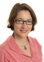
Dr Vanessa Blair - General and Breast Surgeon
Private Service, General Surgery, Breast
Description
Dr Vanessa Blair is an oncoplastic breast surgeon and, when surgery is required, will work with you to reach a carefully considered decision about the best reconstructive options tailored to your particular circumstances.
Breast disorders include:
- fibrocystic disease – benign changes in the breast tissue causes it to become dense or “lumpy”
- fibroadenomas – benign tumours of the breast tissue
- cysts – fluid-filled sacs
- breast infections
- breast cancer.
Many of these conditions do not require surgery and we will work with other specialists to find out the best treatment plan for you.
Consultants
-

Dr Vanessa Blair
General & Breast Surgeon
Referral Expectations
Fees and Charges Description
Dr Vanessa Blair is a Southern Cross Affiliated Provider for a number of procedures. If you are a Southern Cross policy holder you may be eligible for prior approval and a streamlined claims process.
Procedures / Treatments
Simple or Total: all breast tissue, skin and the nipple are surgically removed but the muscles lying under the breast and the lymph nodes are left in place. Modified Radical: all breast tissue, skin and the nipple as well as some lymph tissue are surgically removed. Partial: the breast lump and a portion of other breast tissue (up to one quarter of the breast) as well as lymph tissue are surgically removed. Lumpectomy: the breast lump and surrounding tissue, as well as some lymph tissue, are surgically removed. When combined with radiation treatment, this is known as breast-conserving surgery.
Simple or Total: all breast tissue, skin and the nipple are surgically removed but the muscles lying under the breast and the lymph nodes are left in place. Modified Radical: all breast tissue, skin and the nipple as well as some lymph tissue are surgically removed. Partial: the breast lump and a portion of other breast tissue (up to one quarter of the breast) as well as lymph tissue are surgically removed. Lumpectomy: the breast lump and surrounding tissue, as well as some lymph tissue, are surgically removed. When combined with radiation treatment, this is known as breast-conserving surgery.
Simple or Total: all breast tissue, skin and the nipple are surgically removed but the muscles lying under the breast and the lymph nodes are left in place.
Modified Radical: all breast tissue, skin and the nipple as well as some lymph tissue are surgically removed.
Partial: the breast lump and a portion of other breast tissue (up to one quarter of the breast) as well as lymph tissue are surgically removed.
Lumpectomy: the breast lump and surrounding tissue, as well as some lymph tissue, are surgically removed. When combined with radiation treatment, this is known as breast-conserving surgery.
When a breast has been removed (mastectomy) because of cancer or other disease, it is possible in most cases to reconstruct a breast similar to a natural breast. A breast reconstruction can be performed as part of the breast removal operation or can be performed months or years later. There are two methods of breast reconstruction: one involves using an implant; the other uses tissue taken from another part of your body. There may be medical reasons why one of these methods is more suitable for you or, in other cases, you may be given a choice. Implants A silicone sack filled with either silicone gel or saline (salt water) is inserted underneath the chest muscle and skin. Before being inserted, the skin will sometimes need to be stretched to the required breast size. This is done by placing an empty bag where the implant will finally go, and gradually filling it with saline over weeks or months. The bag is then replaced by the implant in an operation that will probably take 2-3 hours under general anaesthesia (you will sleep through it). You will probably stay in hospital for 2-5 days. Flap reconstruction A flap taken from another part of the body such as your back, stomach or buttocks, is used to reconstruct the breast. This is a more complicated operation than having an implant and may last up to 6 hours and require a 5- to 7-day stay in hospital.
When a breast has been removed (mastectomy) because of cancer or other disease, it is possible in most cases to reconstruct a breast similar to a natural breast. A breast reconstruction can be performed as part of the breast removal operation or can be performed months or years later. There are two methods of breast reconstruction: one involves using an implant; the other uses tissue taken from another part of your body. There may be medical reasons why one of these methods is more suitable for you or, in other cases, you may be given a choice. Implants A silicone sack filled with either silicone gel or saline (salt water) is inserted underneath the chest muscle and skin. Before being inserted, the skin will sometimes need to be stretched to the required breast size. This is done by placing an empty bag where the implant will finally go, and gradually filling it with saline over weeks or months. The bag is then replaced by the implant in an operation that will probably take 2-3 hours under general anaesthesia (you will sleep through it). You will probably stay in hospital for 2-5 days. Flap reconstruction A flap taken from another part of the body such as your back, stomach or buttocks, is used to reconstruct the breast. This is a more complicated operation than having an implant and may last up to 6 hours and require a 5- to 7-day stay in hospital.
When a breast has been removed (mastectomy) because of cancer or other disease, it is possible in most cases to reconstruct a breast similar to a natural breast. A breast reconstruction can be performed as part of the breast removal operation or can be performed months or years later.
There are two methods of breast reconstruction: one involves using an implant; the other uses tissue taken from another part of your body. There may be medical reasons why one of these methods is more suitable for you or, in other cases, you may be given a choice.
Implants
A silicone sack filled with either silicone gel or saline (salt water) is inserted underneath the chest muscle and skin. Before being inserted, the skin will sometimes need to be stretched to the required breast size. This is done by placing an empty bag where the implant will finally go, and gradually filling it with saline over weeks or months. The bag is then replaced by the implant in an operation that will probably take 2-3 hours under general anaesthesia (you will sleep through it). You will probably stay in hospital for 2-5 days.
Flap reconstruction
A flap taken from another part of the body such as your back, stomach or buttocks, is used to reconstruct the breast. This is a more complicated operation than having an implant and may last up to 6 hours and require a 5- to 7-day stay in hospital.
General surgery covers some disorders of the liver and biliary system. The most common of these is pain caused by gallstones. These are formed if the gallbladder is not working properly, and the standard treatment is to remove the gallbladder (cholecystectomy). This procedure is usually performed using a laparoscopic (keyhole) approach.
General surgery covers some disorders of the liver and biliary system. The most common of these is pain caused by gallstones. These are formed if the gallbladder is not working properly, and the standard treatment is to remove the gallbladder (cholecystectomy). This procedure is usually performed using a laparoscopic (keyhole) approach.
General surgery covers some disorders of the liver and biliary system. The most common of these is pain caused by gallstones. These are formed if the gallbladder is not working properly, and the standard treatment is to remove the gallbladder (cholecystectomy). This procedure is usually performed using a laparoscopic (keyhole) approach.
Conditions of the gut dealt with by general surgery include disorders of the oesophagus, stomach, small bowel, large bowel and anus. These range from complex conditions such as ulceration or cancer in the bowel through to fairly minor conditions such as haemorrhoids. Many of the more major conditions such as bowel cancer will require surgery, or sometimes treatment with medication, chemotherapy or radiotherapy. Haemorrhoids are a condition where the veins under the lining of the anus are congested and enlarged. Less severe haemorrhoids can be managed with simple treatments such as injection or banding which can be performed in the clinic while larger ones will require surgery.
Conditions of the gut dealt with by general surgery include disorders of the oesophagus, stomach, small bowel, large bowel and anus. These range from complex conditions such as ulceration or cancer in the bowel through to fairly minor conditions such as haemorrhoids. Many of the more major conditions such as bowel cancer will require surgery, or sometimes treatment with medication, chemotherapy or radiotherapy. Haemorrhoids are a condition where the veins under the lining of the anus are congested and enlarged. Less severe haemorrhoids can be managed with simple treatments such as injection or banding which can be performed in the clinic while larger ones will require surgery.
Conditions of the gut dealt with by general surgery include disorders of the oesophagus, stomach, small bowel, large bowel and anus. These range from complex conditions such as ulceration or cancer in the bowel through to fairly minor conditions such as haemorrhoids. Many of the more major conditions such as bowel cancer will require surgery, or sometimes treatment with medication, chemotherapy or radiotherapy.
Haemorrhoids are a condition where the veins under the lining of the anus are congested and enlarged. Less severe haemorrhoids can be managed with simple treatments such as injection or banding which can be performed in the clinic while larger ones will require surgery.
A hernia exists where part of the abdominal wall is weakened, and the contents of the abdomen push through to the outside. This is most commonly seen in the groin area but can occur in other places. Surgical treatment is usually quite straightforward and involves returning the abdominal contents to the inside and then reinforcing the abdominal wall in some way.
A hernia exists where part of the abdominal wall is weakened, and the contents of the abdomen push through to the outside. This is most commonly seen in the groin area but can occur in other places. Surgical treatment is usually quite straightforward and involves returning the abdominal contents to the inside and then reinforcing the abdominal wall in some way.
A hernia exists where part of the abdominal wall is weakened, and the contents of the abdomen push through to the outside. This is most commonly seen in the groin area but can occur in other places. Surgical treatment is usually quite straightforward and involves returning the abdominal contents to the inside and then reinforcing the abdominal wall in some way.
Skin conditions dealt with by general surgery include lumps, tumours and other lesions of the skin and underlying tissues. These are often fairly simple conditions that can be dealt with by performing minor operations under local anaesthetic (the area of skin being treated is numbed). Often these procedures are performed as outpatient or day case procedures. Skin Cancer New Zealand has a very high rate of skin cancer, when compared to other countries. The most common forms of skin cancer usually appear on areas of skin that have been over-exposed to the sun. Risk factors for developing skin cancer are: prolonged exposure to the sun; people with fair skin; and possibly over-exposure to UV light from sun beds. There are three main types of skin cancers: basal cell carcinoma, squamous cell carcinoma and malignant melanoma. Basal Cell Carcinoma (BCC) This is the most common type and is found on skin surfaces that are exposed to sun. A BCC remains localised and does not usually spread to other areas of the body. Sometimes BCC’s can ulcerate and scab so it is important not to mistake it for a sore. BCCs occur more commonly on the face, back of hands and back. They appear usually as small, red lumps that don’t heal and sometimes bleed or become itchy. They have the tendency to change in size and sometimes in colour. Treatment Often a BCC can be diagnosed just by its appearance. In other cases it will be removed totally and sent for examination and diagnosis, or a biopsy may be taken and just a sample sent for diagnosis. Removal of a BCC will require an appointment with a doctor or surgeon. It will be termed minor surgery and will require a local anaesthetic (numbing of the area) and possibly some stitches. A very small number of BCCs will require a general anaesthetic (you will sleep through the operation) for removal. Squamous Cell Carcinoma (SCC) This type of skin cancer also affects areas of the skin that have exposure to the sun. The most common area is the face, but an SCC can also affect other parts of the body and can spread to other parts of the body. The spreading (metastasising) can potentially be fatal if not successfully treated. A SCC usually begins as a keratosis that looks like an area of thickened scaly skin, it may then develop into a raised, hard lump which enlarges. SCCs can sometimes be painful. Often the edges are irregular and it can appear wart like, the colour can be reddish brown. Sometimes it can appear like a recurring ulcer that does not heal. All SCCs will need to be removed, because of their potential for spread. The removal and diagnosis is the same as for a BCC. Malignant Melanoma This is the most serious form of skin cancer. It can spread to other parts of the body and people can die from this disease. A melanoma usually starts as a pigmented growth on normal skin. They often, but not always, occur on areas that have high sun exposure. In some cases, a melanoma may develop from existing pigmented moles. What to look for: an existing mole that changes colour (it may be black, dark blue or even red and white) the colour pigment may be uneven the edges of the mole/freckle may be irregular and have a spreading edge the surface of the mole/freckle may be flaky/crusted and raised sudden growth of an existing or new mole/freckle inflammation and or itchiness surrounding an existing or new mole/freckle. Treatment It is important that any suspect moles or freckles are checked by a GP or a dermatologist. The sooner a melanoma is treated, there is less chance of it spreading. A biopsy or removal will be carried out depending on the size of the cancer. Tissue samples will be sent for examination, as this will aid in diagnosis and help determine the type of treatment required. If the melanoma has spread more surgery may be required to take more of the affected skin. Samples from lymph nodes that are near to the cancer may be tested for spread, then chemotherapy or radiotherapy may be required to treat this spread. Once a melanoma has been diagnosed, a patient may be referred to an oncologist (a doctor who specialises in cancer). A melanoma that is in the early stages can be treated more successfully and cure rates are much higher than one that has spread.
Skin conditions dealt with by general surgery include lumps, tumours and other lesions of the skin and underlying tissues. These are often fairly simple conditions that can be dealt with by performing minor operations under local anaesthetic (the area of skin being treated is numbed). Often these procedures are performed as outpatient or day case procedures. Skin Cancer New Zealand has a very high rate of skin cancer, when compared to other countries. The most common forms of skin cancer usually appear on areas of skin that have been over-exposed to the sun. Risk factors for developing skin cancer are: prolonged exposure to the sun; people with fair skin; and possibly over-exposure to UV light from sun beds. There are three main types of skin cancers: basal cell carcinoma, squamous cell carcinoma and malignant melanoma. Basal Cell Carcinoma (BCC) This is the most common type and is found on skin surfaces that are exposed to sun. A BCC remains localised and does not usually spread to other areas of the body. Sometimes BCC’s can ulcerate and scab so it is important not to mistake it for a sore. BCCs occur more commonly on the face, back of hands and back. They appear usually as small, red lumps that don’t heal and sometimes bleed or become itchy. They have the tendency to change in size and sometimes in colour. Treatment Often a BCC can be diagnosed just by its appearance. In other cases it will be removed totally and sent for examination and diagnosis, or a biopsy may be taken and just a sample sent for diagnosis. Removal of a BCC will require an appointment with a doctor or surgeon. It will be termed minor surgery and will require a local anaesthetic (numbing of the area) and possibly some stitches. A very small number of BCCs will require a general anaesthetic (you will sleep through the operation) for removal. Squamous Cell Carcinoma (SCC) This type of skin cancer also affects areas of the skin that have exposure to the sun. The most common area is the face, but an SCC can also affect other parts of the body and can spread to other parts of the body. The spreading (metastasising) can potentially be fatal if not successfully treated. A SCC usually begins as a keratosis that looks like an area of thickened scaly skin, it may then develop into a raised, hard lump which enlarges. SCCs can sometimes be painful. Often the edges are irregular and it can appear wart like, the colour can be reddish brown. Sometimes it can appear like a recurring ulcer that does not heal. All SCCs will need to be removed, because of their potential for spread. The removal and diagnosis is the same as for a BCC. Malignant Melanoma This is the most serious form of skin cancer. It can spread to other parts of the body and people can die from this disease. A melanoma usually starts as a pigmented growth on normal skin. They often, but not always, occur on areas that have high sun exposure. In some cases, a melanoma may develop from existing pigmented moles. What to look for: an existing mole that changes colour (it may be black, dark blue or even red and white) the colour pigment may be uneven the edges of the mole/freckle may be irregular and have a spreading edge the surface of the mole/freckle may be flaky/crusted and raised sudden growth of an existing or new mole/freckle inflammation and or itchiness surrounding an existing or new mole/freckle. Treatment It is important that any suspect moles or freckles are checked by a GP or a dermatologist. The sooner a melanoma is treated, there is less chance of it spreading. A biopsy or removal will be carried out depending on the size of the cancer. Tissue samples will be sent for examination, as this will aid in diagnosis and help determine the type of treatment required. If the melanoma has spread more surgery may be required to take more of the affected skin. Samples from lymph nodes that are near to the cancer may be tested for spread, then chemotherapy or radiotherapy may be required to treat this spread. Once a melanoma has been diagnosed, a patient may be referred to an oncologist (a doctor who specialises in cancer). A melanoma that is in the early stages can be treated more successfully and cure rates are much higher than one that has spread.
Skin conditions dealt with by general surgery include lumps, tumours and other lesions of the skin and underlying tissues. These are often fairly simple conditions that can be dealt with by performing minor operations under local anaesthetic (the area of skin being treated is numbed). Often these procedures are performed as outpatient or day case procedures.
Skin Cancer
New Zealand has a very high rate of skin cancer, when compared to other countries. The most common forms of skin cancer usually appear on areas of skin that have been over-exposed to the sun.
Risk factors for developing skin cancer are: prolonged exposure to the sun; people with fair skin; and possibly over-exposure to UV light from sun beds.
There are three main types of skin cancers: basal cell carcinoma, squamous cell carcinoma and malignant melanoma.
Basal Cell Carcinoma (BCC)
This is the most common type and is found on skin surfaces that are exposed to sun. A BCC remains localised and does not usually spread to other areas of the body. Sometimes BCC’s can ulcerate and scab so it is important not to mistake it for a sore.
BCCs occur more commonly on the face, back of hands and back. They appear usually as small, red lumps that don’t heal and sometimes bleed or become itchy. They have the tendency to change in size and sometimes in colour.
Treatment
Often a BCC can be diagnosed just by its appearance. In other cases it will be removed totally and sent for examination and diagnosis, or a biopsy may be taken and just a sample sent for diagnosis.
Removal of a BCC will require an appointment with a doctor or surgeon. It will be termed minor surgery and will require a local anaesthetic (numbing of the area) and possibly some stitches. A very small number of BCCs will require a general anaesthetic (you will sleep through the operation) for removal.
Squamous Cell Carcinoma (SCC)
This type of skin cancer also affects areas of the skin that have exposure to the sun. The most common area is the face, but an SCC can also affect other parts of the body and can spread to other parts of the body. The spreading (metastasising) can potentially be fatal if not successfully treated.
A SCC usually begins as a keratosis that looks like an area of thickened scaly skin, it may then develop into a raised, hard lump which enlarges. SCCs can sometimes be painful. Often the edges are irregular and it can appear wart like, the colour can be reddish brown. Sometimes it can appear like a recurring ulcer that does not heal.
All SCCs will need to be removed, because of their potential for spread. The removal and diagnosis is the same as for a BCC.
Malignant Melanoma
This is the most serious form of skin cancer. It can spread to other parts of the body and people can die from this disease.
A melanoma usually starts as a pigmented growth on normal skin. They often, but not always, occur on areas that have high sun exposure. In some cases, a melanoma may develop from existing pigmented moles.
What to look for:
- an existing mole that changes colour (it may be black, dark blue or even red and white)
- the colour pigment may be uneven
- the edges of the mole/freckle may be irregular and have a spreading edge
- the surface of the mole/freckle may be flaky/crusted and raised
- sudden growth of an existing or new mole/freckle
- inflammation and or itchiness surrounding an existing or new mole/freckle.
Treatment
It is important that any suspect moles or freckles are checked by a GP or a dermatologist. The sooner a melanoma is treated, there is less chance of it spreading.
A biopsy or removal will be carried out depending on the size of the cancer. Tissue samples will be sent for examination, as this will aid in diagnosis and help determine the type of treatment required. If the melanoma has spread more surgery may be required to take more of the affected skin. Samples from lymph nodes that are near to the cancer may be tested for spread, then chemotherapy or radiotherapy may be required to treat this spread.
Once a melanoma has been diagnosed, a patient may be referred to an oncologist (a doctor who specialises in cancer).
A melanoma that is in the early stages can be treated more successfully and cure rates are much higher than one that has spread.
Parking
- Vinery Lane Surgery, Whangārei: Free roadside parking is available at the front of the clinic and paid public parking behind building.
- Kensington Hospital, Whangārei: Dedicated free off street parking is provided.
- St Marks, Newmarket Auckland: Dedicated free parking off MacMurray road.
Website
Contact Details
4 Vinery Lane, Whangārei
Northland
-
Phone
(09) 438 7477
-
Fax
(09) 438 7478
Email
Website
Healthlink EDI: vanblair
Vinery Lane Surgery
Ground Floor
4 Vinery Lane (off Bank Street)
Whangarei
Street Address
Vinery Lane Surgery
Ground Floor
4 Vinery Lane (off Bank Street)
Whangārei
St Marks Breast Centre, 12 Saint Marks Road, Remuera, Auckland
Central Auckland
-
Phone
(09) 520 0389 or 0800 ST MARKS
-
Fax
(09) 520 0589
Website
Was this page helpful?
This page was last updated at 3:28PM on March 15, 2024. This information is reviewed and edited by Dr Vanessa Blair - General and Breast Surgeon.

