
Wellington > Private Hospitals & Specialists >
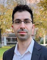
Mr Ahmed Barazanchi - General, Bariatric and Upper GI Surgeon
Private Service, General Surgery, Bariatric (Weight Loss) Surgery
Today
Description
Ahmed Barazanchi is a New Zealand trained Upper GI Surgeon based in Wellington. Ahmed has a private practice, consulting at Wakefield Specialist Centre, where patients can expect timely and exceptional surgical care with a focus on minimally invasive techniques.
Ahmed also provides oesophagogastric, bariatric, and general surgical care at Wellington Public Hospital, where he is currently the lead oesophageal surgeon.
Ahmed offers a range of specialised services, including:
General Surgery
- Laparoscopic cholecystectomy
- Laparoscopic hernia surgery (inguinal, umbilical and incisional)
- Open haemorrhoidectomy
Upper GI Surgery
- Anti-reflux surgery (fundoplication), including revisional hiatal surgery
- Bariatric surgery (Roux-en-Y gastric bypass, mini-bypass, sleeve gastrectomy)
- Revisional bariatric surgery for poor food tolerance, reflux, and weight gain
- Minimally invasive oesophagectomy and gastrectomy for malignant and benign conditions
- Heller’s myotomy for achalasia
Upper GI Endoscopy
- Diagnostic and interventional with a focus on the treatment of surgical disease
Consultants
-
Mr Ahmed Barazanchi
General, Bariatric & Upper GI Surgeon
Ages
Adult / Pakeke, Older adult / Kaumātua, Youth / Rangatahi
How do I access this service?
Referral
Contact us
We welcome patient direct enquiries
Fees and Charges Categorisation
Fees apply
Fees and Charges Description
Southern Cross Affiliated Provider and NIB First Choice member
Hours
| Mon – Fri | 8:30 AM – 5:00 PM |
|---|
Languages Spoken
English, Arabic
Services Provided
Laparoscopic: several small incisions (cuts) are made in the lower right abdomen (stomach) and a narrow tube with a tiny camera attached (laparoscope) in inserted. This allows the surgeon a view of the appendix and, by inserting small surgical instruments through the other cuts, the appendix can be removed. Open: an incision is made in the lower right abdomen and the appendix removed.
Laparoscopic: several small incisions (cuts) are made in the lower right abdomen (stomach) and a narrow tube with a tiny camera attached (laparoscope) in inserted. This allows the surgeon a view of the appendix and, by inserting small surgical instruments through the other cuts, the appendix can be removed. Open: an incision is made in the lower right abdomen and the appendix removed.
Laparoscopic: several small incisions (cuts) are made in the lower right abdomen (stomach) and a narrow tube with a tiny camera attached (laparoscope) in inserted. This allows the surgeon a view of the appendix and, by inserting small surgical instruments through the other cuts, the appendix can be removed.
Open: an incision is made in the lower right abdomen and the appendix removed.
Bariatric or weight loss surgery refers to a number of different procedures that can be performed to treat obesity. Procedures fall into three main types: Malabsorptive - these procedures involve bypassing a section of the small intestine thus reducing the amount of food absorbed into the body. Laparoscopic Gastric Bypass involves bypassing a section of the intestine so that less food is absorbed. Restrictive - these procedures involve reducing the size of the stomach, usually by creating a small pouch at the top of the stomach which limits the amount of food that can be eaten. Laparoscopic Gastric Banding is a surgical treatment for obesity that involves placing an inflatable cuff around the upper stomach. The cuff can be further inflated or deflated to adjust the volume of food that can be consumed. Malabsorptive/Restrictive Combination - these procedures combine both techniques e.g. gastric bypass surgery in which a small stomach pouch is formed and its outlet connected to part of the small intestine.
Bariatric or weight loss surgery refers to a number of different procedures that can be performed to treat obesity. Procedures fall into three main types: Malabsorptive - these procedures involve bypassing a section of the small intestine thus reducing the amount of food absorbed into the body. Laparoscopic Gastric Bypass involves bypassing a section of the intestine so that less food is absorbed. Restrictive - these procedures involve reducing the size of the stomach, usually by creating a small pouch at the top of the stomach which limits the amount of food that can be eaten. Laparoscopic Gastric Banding is a surgical treatment for obesity that involves placing an inflatable cuff around the upper stomach. The cuff can be further inflated or deflated to adjust the volume of food that can be consumed. Malabsorptive/Restrictive Combination - these procedures combine both techniques e.g. gastric bypass surgery in which a small stomach pouch is formed and its outlet connected to part of the small intestine.
Bariatric or weight loss surgery refers to a number of different procedures that can be performed to treat obesity. Procedures fall into three main types:
Malabsorptive - these procedures involve bypassing a section of the small intestine thus reducing the amount of food absorbed into the body.
Laparoscopic Gastric Bypass involves bypassing a section of the intestine so that less food is absorbed.
Restrictive - these procedures involve reducing the size of the stomach, usually by creating a small pouch at the top of the stomach which limits the amount of food that can be eaten.
Laparoscopic Gastric Banding is a surgical treatment for obesity that involves placing an inflatable cuff around the upper stomach. The cuff can be further inflated or deflated to adjust the volume of food that can be consumed.
Malabsorptive/Restrictive Combination - these procedures combine both techniques e.g. gastric bypass surgery in which a small stomach pouch is formed and its outlet connected to part of the small intestine.
Gallstones are formed if the gallbladder is not working properly, and the standard treatment is to remove the gallbladder (cholecystectomy). This procedure is usually performed using a laparoscopic (keyhole) approach. Laparoscopic: several small incisions (cuts) are made in the abdomen (stomach) and a narrow tube with a tiny camera attached (laparoscope) is inserted. This allows the surgeon a view of the gallbladder and, by inserting small surgical instruments through the other cuts, the gallbladder can be removed. Open: an abdominal incision is made and the gallbladder removed.
Gallstones are formed if the gallbladder is not working properly, and the standard treatment is to remove the gallbladder (cholecystectomy). This procedure is usually performed using a laparoscopic (keyhole) approach. Laparoscopic: several small incisions (cuts) are made in the abdomen (stomach) and a narrow tube with a tiny camera attached (laparoscope) is inserted. This allows the surgeon a view of the gallbladder and, by inserting small surgical instruments through the other cuts, the gallbladder can be removed. Open: an abdominal incision is made and the gallbladder removed.
Gallstones are formed if the gallbladder is not working properly, and the standard treatment is to remove the gallbladder (cholecystectomy). This procedure is usually performed using a laparoscopic (keyhole) approach.
Laparoscopic: several small incisions (cuts) are made in the abdomen (stomach) and a narrow tube with a tiny camera attached (laparoscope) is inserted. This allows the surgeon a view of the gallbladder and, by inserting small surgical instruments through the other cuts, the gallbladder can be removed.
Open: an abdominal incision is made and the gallbladder removed.
Partial: the diseased part of the stomach is removed and the remaining section is reattached to the oesophagus (food pipe) or small intestine. Total: all of the stomach is removed and the oesophagus is attached directly to the small intestine.
Partial: the diseased part of the stomach is removed and the remaining section is reattached to the oesophagus (food pipe) or small intestine. Total: all of the stomach is removed and the oesophagus is attached directly to the small intestine.
Partial: the diseased part of the stomach is removed and the remaining section is reattached to the oesophagus (food pipe) or small intestine.
Total: all of the stomach is removed and the oesophagus is attached directly to the small intestine.
Conditions of the gut dealt with by general surgery include disorders of the oesophagus, stomach, small bowel, large bowel and anus. These range from complex conditions such as ulceration or cancer in the bowel through to fairly minor conditions such as haemorrhoids. Many of the more major conditions such as bowel cancer will require surgery, or sometimes treatment with medication, chemotherapy or radiotherapy. Haemorrhoids are a condition where the veins under the lining of the anus are congested and enlarged. Less severe haemorrhoids can be managed with simple treatments such as injection or banding which can be performed in the clinic while larger ones will require surgery.
Conditions of the gut dealt with by general surgery include disorders of the oesophagus, stomach, small bowel, large bowel and anus. These range from complex conditions such as ulceration or cancer in the bowel through to fairly minor conditions such as haemorrhoids. Many of the more major conditions such as bowel cancer will require surgery, or sometimes treatment with medication, chemotherapy or radiotherapy. Haemorrhoids are a condition where the veins under the lining of the anus are congested and enlarged. Less severe haemorrhoids can be managed with simple treatments such as injection or banding which can be performed in the clinic while larger ones will require surgery.
Conditions of the gut dealt with by general surgery include disorders of the oesophagus, stomach, small bowel, large bowel and anus. These range from complex conditions such as ulceration or cancer in the bowel through to fairly minor conditions such as haemorrhoids. Many of the more major conditions such as bowel cancer will require surgery, or sometimes treatment with medication, chemotherapy or radiotherapy.
Haemorrhoids are a condition where the veins under the lining of the anus are congested and enlarged. Less severe haemorrhoids can be managed with simple treatments such as injection or banding which can be performed in the clinic while larger ones will require surgery.
Gastroscopy allows examination of the upper part of your digestive tract i.e. oesophagus (food pipe), stomach and duodenum (top section of the small intestine), by passing a gastroscope (long, flexible tube with a camera on the end) through your mouth and down your digestive tract. Images from the camera are displayed on a television monitor. Sometimes a small tissue sample (biopsy) will need to be taken during the procedure for later examination at a laboratory. Gastroscopy may be used to diagnose peptic ulcers, tumours, gastritis etc. Complications from this procedure are very rare but can occur. They include: bleeding if a biopsy is performed; allergic reaction to the sedative or throat spray; perforation (tearing) of the stomach with the instrument (this is a serious but extremely rare complication). What to expect All endoscopic procedures are viewed as a surgical procedure and generally the same preparation will apply. You will not be able to eat or drink anything for 6 hours before your gastroscopy. When you are ready for the procedure, the back of your throat will be sprayed with anaesthetic. You will also be offered medication (a sedative) to make you go into a light sleep. This will be given by an injection into a vein in your arm or hand. The gastroscopy will take approximately 15 minutes, but you will probably sleep for another 30 minutes. You will spend some time in a recovery unit (probably 1-2 hours) to sleep off the sedative and to allow staff to monitor you (take blood pressure readings etc). Because you have been sedated (given medication to make you sleep) it is important that you arrange for someone else to drive you home. If biopsies are taken for examination, your GP will be sent the results within 2-3 weeks.
Gastroscopy allows examination of the upper part of your digestive tract i.e. oesophagus (food pipe), stomach and duodenum (top section of the small intestine), by passing a gastroscope (long, flexible tube with a camera on the end) through your mouth and down your digestive tract. Images from the camera are displayed on a television monitor. Sometimes a small tissue sample (biopsy) will need to be taken during the procedure for later examination at a laboratory. Gastroscopy may be used to diagnose peptic ulcers, tumours, gastritis etc. Complications from this procedure are very rare but can occur. They include: bleeding if a biopsy is performed; allergic reaction to the sedative or throat spray; perforation (tearing) of the stomach with the instrument (this is a serious but extremely rare complication). What to expect All endoscopic procedures are viewed as a surgical procedure and generally the same preparation will apply. You will not be able to eat or drink anything for 6 hours before your gastroscopy. When you are ready for the procedure, the back of your throat will be sprayed with anaesthetic. You will also be offered medication (a sedative) to make you go into a light sleep. This will be given by an injection into a vein in your arm or hand. The gastroscopy will take approximately 15 minutes, but you will probably sleep for another 30 minutes. You will spend some time in a recovery unit (probably 1-2 hours) to sleep off the sedative and to allow staff to monitor you (take blood pressure readings etc). Because you have been sedated (given medication to make you sleep) it is important that you arrange for someone else to drive you home. If biopsies are taken for examination, your GP will be sent the results within 2-3 weeks.
Gastroscopy allows examination of the upper part of your digestive tract i.e. oesophagus (food pipe), stomach and duodenum (top section of the small intestine), by passing a gastroscope (long, flexible tube with a camera on the end) through your mouth and down your digestive tract. Images from the camera are displayed on a television monitor. Sometimes a small tissue sample (biopsy) will need to be taken during the procedure for later examination at a laboratory.
Gastroscopy may be used to diagnose peptic ulcers, tumours, gastritis etc.
Complications from this procedure are very rare but can occur. They include: bleeding if a biopsy is performed; allergic reaction to the sedative or throat spray; perforation (tearing) of the stomach with the instrument (this is a serious but extremely rare complication).
What to expect
All endoscopic procedures are viewed as a surgical procedure and generally the same preparation will apply. You will not be able to eat or drink anything for 6 hours before your gastroscopy. When you are ready for the procedure, the back of your throat will be sprayed with anaesthetic. You will also be offered medication (a sedative) to make you go into a light sleep. This will be given by an injection into a vein in your arm or hand.
The gastroscopy will take approximately 15 minutes, but you will probably sleep for another 30 minutes. You will spend some time in a recovery unit (probably 1-2 hours) to sleep off the sedative and to allow staff to monitor you (take blood pressure readings etc). Because you have been sedated (given medication to make you sleep) it is important that you arrange for someone else to drive you home.
If biopsies are taken for examination, your GP will be sent the results within 2-3 weeks.
Haemorrhoids are a condition where the veins under the lining of the anus are congested and enlarged. Less severe haemorrhoids can be managed with simple treatments such as injection or banding which can be performed in the clinic while larger ones will require surgery. Haemorrhoid removal: Haemorrhoidectomy: each haemorrhoid or pile is tied off and then cut away. Stapled Haemorrhoidectomy: a circular stapling device is used to pull the haemorrhoid tissue back into its normal position.
Haemorrhoids are a condition where the veins under the lining of the anus are congested and enlarged. Less severe haemorrhoids can be managed with simple treatments such as injection or banding which can be performed in the clinic while larger ones will require surgery. Haemorrhoid removal: Haemorrhoidectomy: each haemorrhoid or pile is tied off and then cut away. Stapled Haemorrhoidectomy: a circular stapling device is used to pull the haemorrhoid tissue back into its normal position.
Haemorrhoids are a condition where the veins under the lining of the anus are congested and enlarged. Less severe haemorrhoids can be managed with simple treatments such as injection or banding which can be performed in the clinic while larger ones will require surgery.
Haemorrhoid removal:
Haemorrhoidectomy: each haemorrhoid or pile is tied off and then cut away.
Stapled Haemorrhoidectomy: a circular stapling device is used to pull the haemorrhoid tissue back into its normal position.
A hernia exists where part of the abdominal wall is weakened, and the contents of the abdomen push through to the outside. This is most commonly seen in the groin area but can occur in other places. Surgical treatment is usually quite straightforward and involves returning the abdominal contents to the inside and then reinforcing the abdominal wall in some way. Hiatus Hernia: Laparoscopic: several small incisions (cuts) are made in the abdomen (stomach) and a narrow tube with a tiny camera attached (laparoscope) is inserted. Small instruments are inserted through the other cuts, allowing the surgeon to push the hernia (part of the stomach and lower oesophagus that is bulging into the chest) back into position in the abdominal cavity. The hiatus (opening) in the diaphragm (a sheet of muscle between the chest and stomach) is tightened and the stomach is stitched into place. Open: an abdominal incision is made over the hernia and the hernia is pushed back into position in the abdominal cavity. The hiatus (opening in the diaphragm) is tightened and the stomach is stitched into place. Fundoplication: during the above procedures, the top part of the stomach (fundus) may be secured in position by wrapping it around the oesophagus. Inguinal Hernia: Laparoscopic: several small incisions are made in the abdomen and a narrow tube with a tiny camera attached (laparoscope) is inserted. Small instruments are inserted through the other cuts, allowing the surgeon to push the hernia (part of the intestine that is bulging through the abdominal wall) back into its original position. The weakness in the abdominal wall is repaired. Open: an abdominal incision is made and the hernia is pushed back into position. The weakness in the abdominal wall is repaired. Umbilical Hernia: An incision is made underneath the navel (tummy button) and the hernia (part of the intestine that is bulging through the abdominal wall) is pushed back into the abdominal cavity. The weakness in the abdominal wall is repaired. Incisional Hernia: Laparoscopic: several small incisions are made in the abdomen and a narrow tube with a tiny camera attached (laparoscope) is inserted. Small instruments are inserted through the other cuts, allowing the surgeon to push the hernia (part of the intestine that is bulging through the abdominal wall) back into its original position. Open: an abdominal incision is made and the hernia is pushed back into position.
A hernia exists where part of the abdominal wall is weakened, and the contents of the abdomen push through to the outside. This is most commonly seen in the groin area but can occur in other places. Surgical treatment is usually quite straightforward and involves returning the abdominal contents to the inside and then reinforcing the abdominal wall in some way. Hiatus Hernia: Laparoscopic: several small incisions (cuts) are made in the abdomen (stomach) and a narrow tube with a tiny camera attached (laparoscope) is inserted. Small instruments are inserted through the other cuts, allowing the surgeon to push the hernia (part of the stomach and lower oesophagus that is bulging into the chest) back into position in the abdominal cavity. The hiatus (opening) in the diaphragm (a sheet of muscle between the chest and stomach) is tightened and the stomach is stitched into place. Open: an abdominal incision is made over the hernia and the hernia is pushed back into position in the abdominal cavity. The hiatus (opening in the diaphragm) is tightened and the stomach is stitched into place. Fundoplication: during the above procedures, the top part of the stomach (fundus) may be secured in position by wrapping it around the oesophagus. Inguinal Hernia: Laparoscopic: several small incisions are made in the abdomen and a narrow tube with a tiny camera attached (laparoscope) is inserted. Small instruments are inserted through the other cuts, allowing the surgeon to push the hernia (part of the intestine that is bulging through the abdominal wall) back into its original position. The weakness in the abdominal wall is repaired. Open: an abdominal incision is made and the hernia is pushed back into position. The weakness in the abdominal wall is repaired. Umbilical Hernia: An incision is made underneath the navel (tummy button) and the hernia (part of the intestine that is bulging through the abdominal wall) is pushed back into the abdominal cavity. The weakness in the abdominal wall is repaired. Incisional Hernia: Laparoscopic: several small incisions are made in the abdomen and a narrow tube with a tiny camera attached (laparoscope) is inserted. Small instruments are inserted through the other cuts, allowing the surgeon to push the hernia (part of the intestine that is bulging through the abdominal wall) back into its original position. Open: an abdominal incision is made and the hernia is pushed back into position.
A hernia exists where part of the abdominal wall is weakened, and the contents of the abdomen push through to the outside. This is most commonly seen in the groin area but can occur in other places. Surgical treatment is usually quite straightforward and involves returning the abdominal contents to the inside and then reinforcing the abdominal wall in some way.
Hiatus Hernia:
Laparoscopic: several small incisions (cuts) are made in the abdomen (stomach) and a narrow tube with a tiny camera attached (laparoscope) is inserted. Small instruments are inserted through the other cuts, allowing the surgeon to push the hernia (part of the stomach and lower oesophagus that is bulging into the chest) back into position in the abdominal cavity. The hiatus (opening) in the diaphragm (a sheet of muscle between the chest and stomach) is tightened and the stomach is stitched into place.
Open: an abdominal incision is made over the hernia and the hernia is pushed back into position in the abdominal cavity. The hiatus (opening in the diaphragm) is tightened and the stomach is stitched into place.
Fundoplication: during the above procedures, the top part of the stomach (fundus) may be secured in position by wrapping it around the oesophagus.
Inguinal Hernia:
Laparoscopic: several small incisions are made in the abdomen and a narrow tube with a tiny camera attached (laparoscope) is inserted. Small instruments are inserted through the other cuts, allowing the surgeon to push the hernia (part of the intestine that is bulging through the abdominal wall) back into its original position. The weakness in the abdominal wall is repaired.
Open: an abdominal incision is made and the hernia is pushed back into position. The weakness in the abdominal wall is repaired.
Umbilical Hernia:
An incision is made underneath the navel (tummy button) and the hernia (part of the intestine that is bulging through the abdominal wall) is pushed back into the abdominal cavity. The weakness in the abdominal wall is repaired.
Incisional Hernia:
Laparoscopic: several small incisions are made in the abdomen and a narrow tube with a tiny camera attached (laparoscope) is inserted. Small instruments are inserted through the other cuts, allowing the surgeon to push the hernia (part of the intestine that is bulging through the abdominal wall) back into its original position.
Open: an abdominal incision is made and the hernia is pushed back into position.
Disability Assistance
Wheelchair access, Wheelchair accessible toilet, Mobility parking space
Parking
Free patient parking is provided at the clinic
Pharmacy
Contact Details
-
Phone
(04) 381 8100
Healthlink EDI
evohcare
Email
Website
99 Rintoul Street
Newtown
Wellington 6021
Street Address
99 Rintoul Street
Newtown
Wellington 6021
Was this page helpful?
This page was last updated at 4:35PM on March 26, 2026. This information is reviewed and edited by Mr Ahmed Barazanchi - General, Bariatric and Upper GI Surgeon.