
Waikato > Private Hospitals & Specialists >
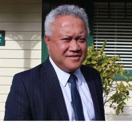
Simi Lolohea - Colorectal, General & Laparoscopic Surgeon
Private Service, General Surgery
Today
8:00 AM to 5:00 PM.
Description
- Colon and rectal conditions
- Perianal conditions
- Laparoscopic colectomy
- Laparoscopic cholecystectomy
- Laparoscopic umbilical/inguinal/incisional hernias
- Pelvic floor disorders
- Colonoscopy and gastroscopy
- Pseudomyxoma peritonei
The colon and the rectum are part of the digestive tract that processes the food we eat. Together they make up the large intestine or large bowel and are located in the abdomen between the small intestine and the anus. The colon is about 1.8m long and absorbs water and nutrients from food. The rectum is the last segment of the large intestine and is about 20 -25cm long. This is where waste material is stored before it passes out of the body through the anus.
A colorectal surgeon is a general surgeon who has had further training and specializes in the diagnosis and treatment of diseases of the colon, rectum, and anus.
What is Laparoscopic Surgery?
Laparoscopic (or keyhole) surgical procedures are performed through several minor cuts (incisions), usually only 5-10mm long, rather than through one large incision.
A long, narrow surgical telescope (laparoscope) with a tiny camera and light source attached is inserted through one of the incisions so that the surgeon can view the inside of the body on a TV monitor.
The surgeon then passes specially designed surgical instruments through the other incisions and carries out the procedure using the TV monitor to guide the instruments.
Laparoscopic surgery is usually associated with less blood loss during surgery and less pain and scarring following surgery. In most cases, time spent in the hospital is less, and overall recovery time from the operation is less than with conventional open surgery.
Consultants
-
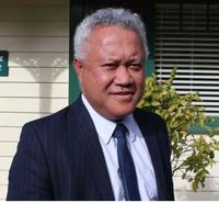
Mr Simi Lolohea
Colorectal, General & Laparoscopic Surgeon
Referral Expectations
Fees and Charges Description
Mr Lolohea is a Southern Cross Affiliated Provider for consultations and:
- Colonoscopy, flexible sigmoidoscopy, gastroscopy.
- Laparoscopic cholecystectomy with an intraoperative cholangiogram, open or laparoscopic cholecystectomy,
- Open or laparoscopic repair of inguinal hernia, open repair of epigastric hernia, open repair of incisional hernia, open repair of inguinal hernia, open repair of umbilical hernia.
Mr Lolohea is an NIB Health Affiliated Provider
Hours
8:00 AM to 5:00 PM.
| Mon – Fri | 8:00 AM – 5:00 PM |
|---|
Procedures / Treatments
Peritoneal surface malignancy can be divided into primary and secondary. Indication for referral includes Pseudomyxoma peritonei (PMP), peritoneal mesothelioma and adenocarcinoma of the appendix and colon. Prior to the development of peritonectomy, patients with no-gynaecological peritoneal carcinomatosis had a dismal prognosis. Patients treated by surgery have a median survival of 2 - 8 months. Quality of life for these patients is almost always very poor. Peritonectomy is a complex and challenging multi-modal therapy. Treatments offered at Braemar Hospital include peritonectomy and heated intraperitoneal chemotherapy (HIPEC). These procedures offer selected patients with the above diseases a good chance of long-term survival together with effective local control of their disease. The prognosis is even worse for those whose disease could not be removed with surgery or controlled by intravenous chemotherapy. These patients have been found to have a better quality of life and long term survival if they are treated with PIPAC. PIPAC (Pressurised Intra-peritoneal Chemotherapy) is the delivery of gaseous chemotherapy intraperitoneally at high concentrations using keyhole surgery. Usually, patients are discharged the next day with minimal discomfort.
Peritoneal surface malignancy can be divided into primary and secondary. Indication for referral includes Pseudomyxoma peritonei (PMP), peritoneal mesothelioma and adenocarcinoma of the appendix and colon. Prior to the development of peritonectomy, patients with no-gynaecological peritoneal carcinomatosis had a dismal prognosis. Patients treated by surgery have a median survival of 2 - 8 months. Quality of life for these patients is almost always very poor. Peritonectomy is a complex and challenging multi-modal therapy. Treatments offered at Braemar Hospital include peritonectomy and heated intraperitoneal chemotherapy (HIPEC). These procedures offer selected patients with the above diseases a good chance of long-term survival together with effective local control of their disease. The prognosis is even worse for those whose disease could not be removed with surgery or controlled by intravenous chemotherapy. These patients have been found to have a better quality of life and long term survival if they are treated with PIPAC. PIPAC (Pressurised Intra-peritoneal Chemotherapy) is the delivery of gaseous chemotherapy intraperitoneally at high concentrations using keyhole surgery. Usually, patients are discharged the next day with minimal discomfort.
Peritoneal surface malignancy can be divided into primary and secondary. Indication for referral includes Pseudomyxoma peritonei (PMP), peritoneal mesothelioma and adenocarcinoma of the appendix and colon.
Prior to the development of peritonectomy, patients with no-gynaecological peritoneal carcinomatosis had a dismal prognosis. Patients treated by surgery have a median survival of 2 - 8 months. Quality of life for these patients is almost always very poor.
Peritonectomy is a complex and challenging multi-modal therapy. Treatments offered at Braemar Hospital include peritonectomy and heated intraperitoneal chemotherapy (HIPEC). These procedures offer selected patients with the above diseases a good chance of long-term survival together with effective local control of their disease.
The prognosis is even worse for those whose disease could not be removed with surgery or controlled by intravenous chemotherapy. These patients have been found to have a better quality of life and long term survival if they are treated with PIPAC. PIPAC (Pressurised Intra-peritoneal Chemotherapy) is the delivery of gaseous chemotherapy intraperitoneally at high concentrations using keyhole surgery. Usually, patients are discharged the next day with minimal discomfort.
Conditions of the gut dealt with by general surgery include disorders of the oesophagus, stomach, small bowel, large bowel and anus. These range from complex conditions such as ulceration or cancer in the bowel through to fairly minor conditions such as haemorrhoids. Many of the more major conditions such as bowel cancer will require surgery, or sometimes treatment with medication, chemotherapy or radiotherapy. Haemorrhoids are a condition where the veins under the lining of the anus are congested and enlarged. Less severe haemorrhoids can be managed with simple treatments such as injection or banding which can be performed in the clinic while larger ones will require surgery.
Conditions of the gut dealt with by general surgery include disorders of the oesophagus, stomach, small bowel, large bowel and anus. These range from complex conditions such as ulceration or cancer in the bowel through to fairly minor conditions such as haemorrhoids. Many of the more major conditions such as bowel cancer will require surgery, or sometimes treatment with medication, chemotherapy or radiotherapy. Haemorrhoids are a condition where the veins under the lining of the anus are congested and enlarged. Less severe haemorrhoids can be managed with simple treatments such as injection or banding which can be performed in the clinic while larger ones will require surgery.
Conditions of the gut dealt with by general surgery include disorders of the oesophagus, stomach, small bowel, large bowel and anus. These range from complex conditions such as ulceration or cancer in the bowel through to fairly minor conditions such as haemorrhoids. Many of the more major conditions such as bowel cancer will require surgery, or sometimes treatment with medication, chemotherapy or radiotherapy.
Haemorrhoids are a condition where the veins under the lining of the anus are congested and enlarged. Less severe haemorrhoids can be managed with simple treatments such as injection or banding which can be performed in the clinic while larger ones will require surgery.
Gastroscopy allows examination of the upper part of your digestive tract i.e. oesophagus (food pipe), stomach and duodenum (top section of the small intestine), by passing a gastroscope (long, flexible tube with a camera on the end) through your mouth and down your digestive tract. Images from the camera are displayed on a television monitor. Sometimes a small tissue sample (biopsy) will need to be taken during the procedure for later examination at a laboratory. Gastroscopy may be used to diagnose peptic ulcers, tumours, gastritis etc. Complications from this procedure are very rare but can occur. They include: bleeding if a biopsy is performed; allergic reaction to the sedative or throat spray; perforation (tearing) of the stomach with the instrument (this is a serious but extremely rare complication). What to expect All endoscopic procedures are viewed as a surgical procedure and generally the same preparation will apply. You will not be able to eat or drink anything for 6 hours before your gastroscopy. When you are ready for the procedure, the back of your throat will be sprayed with anaesthetic. You will also be offered medication (a sedative) to make you go into a light sleep. This will be given by an injection into a vein in your arm or hand. The gastroscopy will take approximately 15 minutes, but you will probably sleep for another 30 minutes. You will spend some time in a recovery unit (probably 1-2 hours) to sleep off the sedative and to allow staff to monitor you (take blood pressure readings etc). Because you have been sedated (given medication to make you sleep) it is important that you arrange for someone else to drive you home. If biopsies are taken for examination, your GP will be sent the results within 2-3 weeks.
Gastroscopy allows examination of the upper part of your digestive tract i.e. oesophagus (food pipe), stomach and duodenum (top section of the small intestine), by passing a gastroscope (long, flexible tube with a camera on the end) through your mouth and down your digestive tract. Images from the camera are displayed on a television monitor. Sometimes a small tissue sample (biopsy) will need to be taken during the procedure for later examination at a laboratory. Gastroscopy may be used to diagnose peptic ulcers, tumours, gastritis etc. Complications from this procedure are very rare but can occur. They include: bleeding if a biopsy is performed; allergic reaction to the sedative or throat spray; perforation (tearing) of the stomach with the instrument (this is a serious but extremely rare complication). What to expect All endoscopic procedures are viewed as a surgical procedure and generally the same preparation will apply. You will not be able to eat or drink anything for 6 hours before your gastroscopy. When you are ready for the procedure, the back of your throat will be sprayed with anaesthetic. You will also be offered medication (a sedative) to make you go into a light sleep. This will be given by an injection into a vein in your arm or hand. The gastroscopy will take approximately 15 minutes, but you will probably sleep for another 30 minutes. You will spend some time in a recovery unit (probably 1-2 hours) to sleep off the sedative and to allow staff to monitor you (take blood pressure readings etc). Because you have been sedated (given medication to make you sleep) it is important that you arrange for someone else to drive you home. If biopsies are taken for examination, your GP will be sent the results within 2-3 weeks.
Gastroscopy allows examination of the upper part of your digestive tract i.e. oesophagus (food pipe), stomach and duodenum (top section of the small intestine), by passing a gastroscope (long, flexible tube with a camera on the end) through your mouth and down your digestive tract. Images from the camera are displayed on a television monitor. Sometimes a small tissue sample (biopsy) will need to be taken during the procedure for later examination at a laboratory.
Gastroscopy may be used to diagnose peptic ulcers, tumours, gastritis etc.
Complications from this procedure are very rare but can occur. They include: bleeding if a biopsy is performed; allergic reaction to the sedative or throat spray; perforation (tearing) of the stomach with the instrument (this is a serious but extremely rare complication).
What to expect
All endoscopic procedures are viewed as a surgical procedure and generally the same preparation will apply. You will not be able to eat or drink anything for 6 hours before your gastroscopy. When you are ready for the procedure, the back of your throat will be sprayed with anaesthetic. You will also be offered medication (a sedative) to make you go into a light sleep. This will be given by an injection into a vein in your arm or hand.
The gastroscopy will take approximately 15 minutes, but you will probably sleep for another 30 minutes. You will spend some time in a recovery unit (probably 1-2 hours) to sleep off the sedative and to allow staff to monitor you (take blood pressure readings etc). Because you have been sedated (given medication to make you sleep) it is important that you arrange for someone else to drive you home.
If biopsies are taken for examination, your GP will be sent the results within 2-3 weeks.
Colonoscopy is the examination of your colon (large bowel) using a colonoscope (long, flexible tube with a camera on the end). The colonoscope is passed into your rectum (bottom) and then moved slowly along the entire colon, while images from the camera are displayed on a television monitor. The procedure takes from 10 minutes to an hour. Sometimes a small tissue sample (biopsy) will need to be taken during the procedure for later examination at a laboratory. A colonoscopy may help diagnose conditions such as polyps (small growths of tissue projecting into the bowel), tumours, ulcerative colitis (inflammation of the colon) and diverticulitis (inflammation of sacs that form on the walls of the colon). Colonoscopy may also be used to remove polyps in the colon. Risks of a colonoscopy are rare but include: bleeding if a biopsy is performed; allergic reaction to the sedative; perforation (tearing) of the bowel wall. What to expect It is important that the bowel is completely empty before the procedure takes place. This means that you will only be able to have liquids on the day before, and will probably have to take some oral laxative medication (to make you go to the toilet more). When you are ready for the procedure, you will be given medication (a sedative) to make you go into a light sleep. This will be given by an injection into a vein in your arm or hand. The colonoscopy will usually take 15 – 30 minutes, but you will probably sleep for another 30 minutes. Because you have been sedated (given medication to make you sleep) it is important that you arrange for someone else to drive you home. Some patients may experience discomfort after the procedure, due to air remaining in the colon.
Colonoscopy is the examination of your colon (large bowel) using a colonoscope (long, flexible tube with a camera on the end). The colonoscope is passed into your rectum (bottom) and then moved slowly along the entire colon, while images from the camera are displayed on a television monitor. The procedure takes from 10 minutes to an hour. Sometimes a small tissue sample (biopsy) will need to be taken during the procedure for later examination at a laboratory. A colonoscopy may help diagnose conditions such as polyps (small growths of tissue projecting into the bowel), tumours, ulcerative colitis (inflammation of the colon) and diverticulitis (inflammation of sacs that form on the walls of the colon). Colonoscopy may also be used to remove polyps in the colon. Risks of a colonoscopy are rare but include: bleeding if a biopsy is performed; allergic reaction to the sedative; perforation (tearing) of the bowel wall. What to expect It is important that the bowel is completely empty before the procedure takes place. This means that you will only be able to have liquids on the day before, and will probably have to take some oral laxative medication (to make you go to the toilet more). When you are ready for the procedure, you will be given medication (a sedative) to make you go into a light sleep. This will be given by an injection into a vein in your arm or hand. The colonoscopy will usually take 15 – 30 minutes, but you will probably sleep for another 30 minutes. Because you have been sedated (given medication to make you sleep) it is important that you arrange for someone else to drive you home. Some patients may experience discomfort after the procedure, due to air remaining in the colon.
Colonoscopy is the examination of your colon (large bowel) using a colonoscope (long, flexible tube with a camera on the end). The colonoscope is passed into your rectum (bottom) and then moved slowly along the entire colon, while images from the camera are displayed on a television monitor.
The procedure takes from 10 minutes to an hour. Sometimes a small tissue sample (biopsy) will need to be taken during the procedure for later examination at a laboratory.
A colonoscopy may help diagnose conditions such as polyps (small growths of tissue projecting into the bowel), tumours, ulcerative colitis (inflammation of the colon) and diverticulitis (inflammation of sacs that form on the walls of the colon).
Colonoscopy may also be used to remove polyps in the colon.
Risks of a colonoscopy are rare but include: bleeding if a biopsy is performed; allergic reaction to the sedative; perforation (tearing) of the bowel wall.
What to expect
It is important that the bowel is completely empty before the procedure takes place. This means that you will only be able to have liquids on the day before, and will probably have to take some oral laxative medication (to make you go to the toilet more).
When you are ready for the procedure, you will be given medication (a sedative) to make you go into a light sleep. This will be given by an injection into a vein in your arm or hand.
The colonoscopy will usually take 15 – 30 minutes, but you will probably sleep for another 30 minutes. Because you have been sedated (given medication to make you sleep) it is important that you arrange for someone else to drive you home.
Some patients may experience discomfort after the procedure, due to air remaining in the colon.
A long, narrow tube with a tiny camera attached (sigmoidoscope) is inserted into your anus and moved through your lower large intestine (bowel). This allows the surgeon a view of the lining of the lower large intestine (sigmoid colon). If necessary, a biopsy (small piece of tissue) may be taken for examination in the laboratory.
A long, narrow tube with a tiny camera attached (sigmoidoscope) is inserted into your anus and moved through your lower large intestine (bowel). This allows the surgeon a view of the lining of the lower large intestine (sigmoid colon). If necessary, a biopsy (small piece of tissue) may be taken for examination in the laboratory.
A long, narrow tube with a tiny camera attached (sigmoidoscope) is inserted into your anus and moved through your lower large intestine (bowel). This allows the surgeon a view of the lining of the lower large intestine (sigmoid colon). If necessary, a biopsy (small piece of tissue) may be taken for examination in the laboratory.
There are two types of IBD, ulcerative colitis and Crohn’s disease. In these conditions, the immune system attacks the lining of the colon causing inflammation and ulceration, bleeding and diarrhoea. In ulcerative colitis this only involves the large intestine, whereas in Crohn’s disease areas within the entire intestine can be involved. Both diseases are chronic (long term) with symptoms coming (relapse) and going (remission) over a number of years. Symptoms depend on what part of the intestine is involved but include: abdominal pain diarrhoea with bleeding tiredness fevers infections around the anus (bottom) weight loss can occur if the condition has been present for some time. Diagnosis is made when the symptoms, examination and blood tests suggest inflammatory bowel disease, infection is ruled out, and you undergo a colonoscopy with biopsy. Treatment depends on the severity of the symptoms and what part of the intestine is affected. Medication is aimed at suppressing the immune system, which is harming the lining of the bowel. This is done via oral or intravenous medication as well as medication given as an enema (via the bottom). Other treatments include changes in the diet to optimise nutrition and health. Treatment in some cases requires surgery to remove affected parts of the bowel. For more information see https://crohnsandcolitis.org.nz/
There are two types of IBD, ulcerative colitis and Crohn’s disease. In these conditions, the immune system attacks the lining of the colon causing inflammation and ulceration, bleeding and diarrhoea. In ulcerative colitis this only involves the large intestine, whereas in Crohn’s disease areas within the entire intestine can be involved. Both diseases are chronic (long term) with symptoms coming (relapse) and going (remission) over a number of years. Symptoms depend on what part of the intestine is involved but include: abdominal pain diarrhoea with bleeding tiredness fevers infections around the anus (bottom) weight loss can occur if the condition has been present for some time. Diagnosis is made when the symptoms, examination and blood tests suggest inflammatory bowel disease, infection is ruled out, and you undergo a colonoscopy with biopsy. Treatment depends on the severity of the symptoms and what part of the intestine is affected. Medication is aimed at suppressing the immune system, which is harming the lining of the bowel. This is done via oral or intravenous medication as well as medication given as an enema (via the bottom). Other treatments include changes in the diet to optimise nutrition and health. Treatment in some cases requires surgery to remove affected parts of the bowel. For more information see https://crohnsandcolitis.org.nz/
There are two types of IBD, ulcerative colitis and Crohn’s disease. In these conditions, the immune system attacks the lining of the colon causing inflammation and ulceration, bleeding and diarrhoea. In ulcerative colitis this only involves the large intestine, whereas in Crohn’s disease areas within the entire intestine can be involved. Both diseases are chronic (long term) with symptoms coming (relapse) and going (remission) over a number of years.
Symptoms depend on what part of the intestine is involved but include:
- abdominal pain
- diarrhoea with bleeding
- tiredness
- fevers
- infections around the anus (bottom)
- weight loss can occur if the condition has been present for some time.
Diagnosis is made when the symptoms, examination and blood tests suggest inflammatory bowel disease, infection is ruled out, and you undergo a colonoscopy with biopsy.
Treatment depends on the severity of the symptoms and what part of the intestine is affected. Medication is aimed at suppressing the immune system, which is harming the lining of the bowel. This is done via oral or intravenous medication as well as medication given as an enema (via the bottom). Other treatments include changes in the diet to optimise nutrition and health. Treatment in some cases requires surgery to remove affected parts of the bowel. For more information see https://crohnsandcolitis.org.nz/
Haemorrhoids are a condition where the veins under the lining of the anus are congested and enlarged. Less severe haemorrhoids can be managed with simple treatments such as injection or banding which can be performed in the clinic while larger ones will require surgery. Haemorrhoid removal: Haemorrhoidectomy: each haemorrhoid or pile is tied off and then cut away. Stapled Haemorrhoidectomy: a circular stapling device is used to pull the haemorrhoid tissue back into its normal position.
Haemorrhoids are a condition where the veins under the lining of the anus are congested and enlarged. Less severe haemorrhoids can be managed with simple treatments such as injection or banding which can be performed in the clinic while larger ones will require surgery. Haemorrhoid removal: Haemorrhoidectomy: each haemorrhoid or pile is tied off and then cut away. Stapled Haemorrhoidectomy: a circular stapling device is used to pull the haemorrhoid tissue back into its normal position.
Haemorrhoids are a condition where the veins under the lining of the anus are congested and enlarged. Less severe haemorrhoids can be managed with simple treatments such as injection or banding which can be performed in the clinic while larger ones will require surgery.
Haemorrhoid removal:
Haemorrhoidectomy: each haemorrhoid or pile is tied off and then cut away.
Stapled Haemorrhoidectomy: a circular stapling device is used to pull the haemorrhoid tissue back into its normal position.
This is cancer that begins in your colon or rectum. Often, it may start as a polyp which is a growth of abnormal tissue on the lining of the colon or rectum. Most people will not have symptoms of colorectal cancer until the disease is at a fairly advanced stage. Then they may experience symptoms such as: change in bowel habit that lasts for more than a few days blood in the stool stomach pain. Tests used to confirm a diagnosis of colorectal cancer include: stool blood test – a sample of stool is tested for traces of blood sigmoidoscopy colonoscopy barium enema – a chalky white substance (barium) and air are pumped into the colon and x-rays are taken biopsy – a small piece of tissue is removed for examination under a microscope Stool blood tests, sigmoidoscopy, colonoscopy and barium enemas are also used as screening tests to look for colorectal cancer in people without symptoms. If these tests find cancers at an early stage, the chances of successful treatment are much higher than when the cancers are further advanced. Screening tests can also involve the removal of polyps that may become cancerous in the future. Treatment The choice of treatment depends on your overall health as well as how far advanced the cancer is. This is determined in a process known as ‘staging’ in which the tumour size, lymph node involvement and spread to other organs is assessed. The three main forms of treatment for colorectal cancer are: Surgery – the most common treatment. Surgery may involve ‘Open Surgery’ in which a large incision (cut) is made in your abdomen or ‘Laparoscopic Surgery’ in which several much smaller incisions are made. The section of the colon or rectum with the cancer is removed and the two ends are reconnected. In some cases, a temporary or permanent colostomy may be required to remove body wastes. Chemotherapy – anticancer medicines, either taken by mouth (oral) or injected into a vein (intravenous), can destroy cancer cells and slow tumour growth. Chemotherapy is useful to treat cancers that have spread to other parts of the body and may also be used before or after surgery or in combination with radiation therapy. Radiation Therapy – high energy x-rays are used to destroy cancer cells or shrink tumours. It is often used together with surgery, in some cases to shrink the tumour before surgery, or to destroy any cells that may be left behind after surgery.
This is cancer that begins in your colon or rectum. Often, it may start as a polyp which is a growth of abnormal tissue on the lining of the colon or rectum. Most people will not have symptoms of colorectal cancer until the disease is at a fairly advanced stage. Then they may experience symptoms such as: change in bowel habit that lasts for more than a few days blood in the stool stomach pain. Tests used to confirm a diagnosis of colorectal cancer include: stool blood test – a sample of stool is tested for traces of blood sigmoidoscopy colonoscopy barium enema – a chalky white substance (barium) and air are pumped into the colon and x-rays are taken biopsy – a small piece of tissue is removed for examination under a microscope Stool blood tests, sigmoidoscopy, colonoscopy and barium enemas are also used as screening tests to look for colorectal cancer in people without symptoms. If these tests find cancers at an early stage, the chances of successful treatment are much higher than when the cancers are further advanced. Screening tests can also involve the removal of polyps that may become cancerous in the future. Treatment The choice of treatment depends on your overall health as well as how far advanced the cancer is. This is determined in a process known as ‘staging’ in which the tumour size, lymph node involvement and spread to other organs is assessed. The three main forms of treatment for colorectal cancer are: Surgery – the most common treatment. Surgery may involve ‘Open Surgery’ in which a large incision (cut) is made in your abdomen or ‘Laparoscopic Surgery’ in which several much smaller incisions are made. The section of the colon or rectum with the cancer is removed and the two ends are reconnected. In some cases, a temporary or permanent colostomy may be required to remove body wastes. Chemotherapy – anticancer medicines, either taken by mouth (oral) or injected into a vein (intravenous), can destroy cancer cells and slow tumour growth. Chemotherapy is useful to treat cancers that have spread to other parts of the body and may also be used before or after surgery or in combination with radiation therapy. Radiation Therapy – high energy x-rays are used to destroy cancer cells or shrink tumours. It is often used together with surgery, in some cases to shrink the tumour before surgery, or to destroy any cells that may be left behind after surgery.
This is cancer that begins in your colon or rectum. Often, it may start as a polyp which is a growth of abnormal tissue on the lining of the colon or rectum.
Most people will not have symptoms of colorectal cancer until the disease is at a fairly advanced stage. Then they may experience symptoms such as:
- change in bowel habit that lasts for more than a few days
- blood in the stool
- stomach pain.
Tests used to confirm a diagnosis of colorectal cancer include:
- stool blood test – a sample of stool is tested for traces of blood
- sigmoidoscopy
- colonoscopy
- barium enema – a chalky white substance (barium) and air are pumped into the colon and x-rays are taken
- biopsy – a small piece of tissue is removed for examination under a microscope
Stool blood tests, sigmoidoscopy, colonoscopy and barium enemas are also used as screening tests to look for colorectal cancer in people without symptoms. If these tests find cancers at an early stage, the chances of successful treatment are much higher than when the cancers are further advanced. Screening tests can also involve the removal of polyps that may become cancerous in the future.
Treatment
The choice of treatment depends on your overall health as well as how far advanced the cancer is. This is determined in a process known as ‘staging’ in which the tumour size, lymph node involvement and spread to other organs is assessed.
The three main forms of treatment for colorectal cancer are:
Surgery – the most common treatment. Surgery may involve ‘Open Surgery’ in which a large incision (cut) is made in your abdomen or ‘Laparoscopic Surgery’ in which several much smaller incisions are made. The section of the colon or rectum with the cancer is removed and the two ends are reconnected. In some cases, a temporary or permanent colostomy may be required to remove body wastes.
Chemotherapy – anticancer medicines, either taken by mouth (oral) or injected into a vein (intravenous), can destroy cancer cells and slow tumour growth. Chemotherapy is useful to treat cancers that have spread to other parts of the body and may also be used before or after surgery or in combination with radiation therapy.
Radiation Therapy – high energy x-rays are used to destroy cancer cells or shrink tumours. It is often used together with surgery, in some cases to shrink the tumour before surgery, or to destroy any cells that may be left behind after surgery.
Laparoscopic: several small incisions (cuts) are made in the abdomen and a narrow tube with a tiny camera attached (laparoscope) is inserted. This allows the surgeon a view of the colon and, by inserting small surgical instruments through the other cuts, part or all of the colon can be removed. Open: an abdominal incision is made and part or all of the colon is removed.
Laparoscopic: several small incisions (cuts) are made in the abdomen and a narrow tube with a tiny camera attached (laparoscope) is inserted. This allows the surgeon a view of the colon and, by inserting small surgical instruments through the other cuts, part or all of the colon can be removed. Open: an abdominal incision is made and part or all of the colon is removed.
Laparoscopic: several small incisions (cuts) are made in the abdomen and a narrow tube with a tiny camera attached (laparoscope) is inserted. This allows the surgeon a view of the colon and, by inserting small surgical instruments through the other cuts, part or all of the colon can be removed.
Open: an abdominal incision is made and part or all of the colon is removed.
An opening is made in the skin of the abdomen (stomach) to allow drainage of stools (faeces) from the colon into a collection bag on the outside. This may be temporary to allow time for healing of the colon or, if the entire colon has been removed, it may be permanent.
An opening is made in the skin of the abdomen (stomach) to allow drainage of stools (faeces) from the colon into a collection bag on the outside. This may be temporary to allow time for healing of the colon or, if the entire colon has been removed, it may be permanent.
An opening is made in the skin of the abdomen (stomach) to allow drainage of stools (faeces) from the colon into a collection bag on the outside. This may be temporary to allow time for healing of the colon or, if the entire colon has been removed, it may be permanent.
Laparoscopic: several small incisions (cuts) are made in the abdomen (stomach) and a narrow tube with a tiny camera attached (laparoscope) is inserted. This allows the surgeon to view the rectum and, by inserting small surgical instruments through the other cuts, part or all of the rectum can be removed. Open: an abdominal incision is made and part or all of the rectum removed.
Laparoscopic: several small incisions (cuts) are made in the abdomen (stomach) and a narrow tube with a tiny camera attached (laparoscope) is inserted. This allows the surgeon to view the rectum and, by inserting small surgical instruments through the other cuts, part or all of the rectum can be removed. Open: an abdominal incision is made and part or all of the rectum removed.
Laparoscopic: several small incisions (cuts) are made in the abdomen (stomach) and a narrow tube with a tiny camera attached (laparoscope) is inserted. This allows the surgeon to view the rectum and, by inserting small surgical instruments through the other cuts, part or all of the rectum can be removed.
Open: an abdominal incision is made and part or all of the rectum removed.
General surgery covers some disorders of the liver and biliary system. The most common of these is pain caused by gallstones. These are formed if the gallbladder is not working properly, and the standard treatment is to remove the gallbladder (cholecystectomy). This procedure is usually performed using a laparoscopic (keyhole) approach. Laparoscopic: several small incisions (cuts) are made in the abdomen (stomach) and a narrow tube with a tiny camera attached (laparoscope) is inserted. This allows the surgeon a view of the gallbladder and, by inserting small surgical instruments through the other cuts, the gallbladder can be removed. Open: an abdominal incision is made and the gallbladder removed.
General surgery covers some disorders of the liver and biliary system. The most common of these is pain caused by gallstones. These are formed if the gallbladder is not working properly, and the standard treatment is to remove the gallbladder (cholecystectomy). This procedure is usually performed using a laparoscopic (keyhole) approach. Laparoscopic: several small incisions (cuts) are made in the abdomen (stomach) and a narrow tube with a tiny camera attached (laparoscope) is inserted. This allows the surgeon a view of the gallbladder and, by inserting small surgical instruments through the other cuts, the gallbladder can be removed. Open: an abdominal incision is made and the gallbladder removed.
General surgery covers some disorders of the liver and biliary system. The most common of these is pain caused by gallstones. These are formed if the gallbladder is not working properly, and the standard treatment is to remove the gallbladder (cholecystectomy). This procedure is usually performed using a laparoscopic (keyhole) approach.
Laparoscopic: several small incisions (cuts) are made in the abdomen (stomach) and a narrow tube with a tiny camera attached (laparoscope) is inserted. This allows the surgeon a view of the gallbladder and, by inserting small surgical instruments through the other cuts, the gallbladder can be removed.
Open: an abdominal incision is made and the gallbladder removed.
A hernia exists where part of the abdominal wall is weakened, and the contents of the abdomen push through to the outside. This is most commonly seen in the groin area but can occur in other places. Surgical treatment is usually quite straightforward and involves returning the abdominal contents to the inside and then reinforcing the abdominal wall in some way.
A hernia exists where part of the abdominal wall is weakened, and the contents of the abdomen push through to the outside. This is most commonly seen in the groin area but can occur in other places. Surgical treatment is usually quite straightforward and involves returning the abdominal contents to the inside and then reinforcing the abdominal wall in some way.
A hernia exists where part of the abdominal wall is weakened, and the contents of the abdomen push through to the outside. This is most commonly seen in the groin area but can occur in other places. Surgical treatment is usually quite straightforward and involves returning the abdominal contents to the inside and then reinforcing the abdominal wall in some way.
Parking
Patient parking is provided at the practice.
Pharmacy
Find your nearest pharmacy here
Website
Contact Details
107 Clarence Street, Hamilton
Waikato
8:00 AM to 5:00 PM.
-
Phone
(07) 858 0771
Healthlink EDI
thesurgc
Email
Website
Urgent appointments available
107 Clarence Street
Hamilton Lake
Hamilton
Waikato 3204
Street Address
107 Clarence Street
Hamilton Lake
Hamilton
Waikato 3204
Was this page helpful?
This page was last updated at 12:06PM on July 8, 2025. This information is reviewed and edited by Simi Lolohea - Colorectal, General & Laparoscopic Surgeon.