
Central Auckland, East Auckland, North Auckland, South Auckland, West Auckland > Private Hospitals & Specialists >
The Specialists Takapuna - General, Bariatric, Breast, Skin Cancer & Thyroid Surgery
Private Surgical Service, General Surgery, Breast
Today
Description
We are a new Surgical Centre offering state-of-the-art facilities in the heart of Takapuna.
Consultants
-
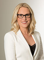
Dr Katherine Gale
Oncoplastic Breast Surgeon
-
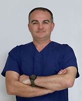
Associate Professor Richard Martin
General Surgeon
-

Mr Jason Robertson
Upper GI and Bariatric Surgeon
Hours
| Mon – Fri | 8:00 AM – 4:30 PM |
|---|
Procedures / Treatments
Shave Biopsy: the top layers of skin in the area being investigated are shaved off with a scalpel (surgical knife) for investigation under a microscope. Punch Biopsy: a small cylindrical core of tissue is taken from the area being investigated for examination under a microscope. Excision Biopsy: all of the lesion or area being investigated is cut out with a scalpel for examination under a microscope. Incision Biopsy: part of the lesion is cut out with a scalpel for examination under a microscope.
Shave Biopsy: the top layers of skin in the area being investigated are shaved off with a scalpel (surgical knife) for investigation under a microscope. Punch Biopsy: a small cylindrical core of tissue is taken from the area being investigated for examination under a microscope. Excision Biopsy: all of the lesion or area being investigated is cut out with a scalpel for examination under a microscope. Incision Biopsy: part of the lesion is cut out with a scalpel for examination under a microscope.
Shave Biopsy: the top layers of skin in the area being investigated are shaved off with a scalpel (surgical knife) for investigation under a microscope.
Punch Biopsy: a small cylindrical core of tissue is taken from the area being investigated for examination under a microscope.
Excision Biopsy: all of the lesion or area being investigated is cut out with a scalpel for examination under a microscope.
Incision Biopsy: part of the lesion is cut out with a scalpel for examination under a microscope.
Skin lesions can be divided into two groups: Benign (non-cancerous): e.g. moles, cysts, warts, tags. These may be removed to prevent spreading (warts), stop discomfort if the lesion is being irritated by clothing/jewellery or to improve appearance. Malignant (cancerous): basal cell and squamous cell carcinomas are generally slow growing and unlikely to spread to other parts of the body. Melanoma is a serious skin cancer that can spread to other parts of the body. Urgent removal is recommended. Surgery to remove skin lesions usually involves an office or outpatient visit, local anaesthesia (the area around the scar is numbed by injecting a local anaesthetic) and stitches. You may or may not have a dressing put on the wound and it is important to keep the area dry for 24 hours. Stitches may be removed in 1-2 weeks. You may need to take a few days off work after the surgery.
Skin lesions can be divided into two groups: Benign (non-cancerous): e.g. moles, cysts, warts, tags. These may be removed to prevent spreading (warts), stop discomfort if the lesion is being irritated by clothing/jewellery or to improve appearance. Malignant (cancerous): basal cell and squamous cell carcinomas are generally slow growing and unlikely to spread to other parts of the body. Melanoma is a serious skin cancer that can spread to other parts of the body. Urgent removal is recommended. Surgery to remove skin lesions usually involves an office or outpatient visit, local anaesthesia (the area around the scar is numbed by injecting a local anaesthetic) and stitches. You may or may not have a dressing put on the wound and it is important to keep the area dry for 24 hours. Stitches may be removed in 1-2 weeks. You may need to take a few days off work after the surgery.
Skin lesions can be divided into two groups:
- Benign (non-cancerous): e.g. moles, cysts, warts, tags. These may be removed to prevent spreading (warts), stop discomfort if the lesion is being irritated by clothing/jewellery or to improve appearance.
- Malignant (cancerous): basal cell and squamous cell carcinomas are generally slow growing and unlikely to spread to other parts of the body. Melanoma is a serious skin cancer that can spread to other parts of the body. Urgent removal is recommended.
Surgery to remove skin lesions usually involves an office or outpatient visit, local anaesthesia (the area around the scar is numbed by injecting a local anaesthetic) and stitches. You may or may not have a dressing put on the wound and it is important to keep the area dry for 24 hours. Stitches may be removed in 1-2 weeks. You may need to take a few days off work after the surgery.
A hernia exists where part of the abdominal wall is weakened, and the contents of the abdomen push through to the outside. This is most commonly seen in the groin area but can occur in other places. Surgical treatment is usually quite straightforward and involves returning the abdominal contents to the inside and then reinforcing the abdominal wall in some way. Hiatus Hernia: Laparoscopic: several small incisions (cuts) are made in the abdomen (stomach) and a narrow tube with a tiny camera attached (laparoscope) is inserted. Small instruments are inserted through the other cuts, allowing the surgeon to push the hernia (part of the stomach and lower oesophagus that is bulging into the chest) back into position in the abdominal cavity. The hiatus (opening) in the diaphragm (a sheet of muscle between the chest and stomach) is tightened and the stomach is stitched into place. Open: an abdominal incision is made over the hernia and the hernia is pushed back into position in the abdominal cavity. The hiatus (opening in the diaphragm) is tightened and the stomach is stitched into place. Fundoplication: during the above procedures, the top part of the stomach (fundus) may be secured in position by wrapping it around the oesophagus. Inguinal Hernia: Laparoscopic: several small incisions are made in the abdomen and a narrow tube with a tiny camera attached (laparoscope) is inserted. Small instruments are inserted through the other cuts, allowing the surgeon to push the hernia (part of the intestine that is bulging through the abdominal wall) back into its original position. The weakness in the abdominal wall is repaired. Open: an abdominal incision is made and the hernia is pushed back into position. The weakness in the abdominal wall is repaired. Umbilical Hernia: An incision is made underneath the navel (tummy button) and the hernia (part of the intestine that is bulging through the abdominal wall) is pushed back into the abdominal cavity. The weakness in the abdominal wall is repaired. Incisional Hernia: Laparoscopic: several small incisions are made in the abdomen and a narrow tube with a tiny camera attached (laparoscope) is inserted. Small instruments are inserted through the other cuts, allowing the surgeon to push the hernia (part of the intestine that is bulging through the abdominal wall) back into its original position. Open: an abdominal incision is made and the hernia is pushed back into position.
A hernia exists where part of the abdominal wall is weakened, and the contents of the abdomen push through to the outside. This is most commonly seen in the groin area but can occur in other places. Surgical treatment is usually quite straightforward and involves returning the abdominal contents to the inside and then reinforcing the abdominal wall in some way. Hiatus Hernia: Laparoscopic: several small incisions (cuts) are made in the abdomen (stomach) and a narrow tube with a tiny camera attached (laparoscope) is inserted. Small instruments are inserted through the other cuts, allowing the surgeon to push the hernia (part of the stomach and lower oesophagus that is bulging into the chest) back into position in the abdominal cavity. The hiatus (opening) in the diaphragm (a sheet of muscle between the chest and stomach) is tightened and the stomach is stitched into place. Open: an abdominal incision is made over the hernia and the hernia is pushed back into position in the abdominal cavity. The hiatus (opening in the diaphragm) is tightened and the stomach is stitched into place. Fundoplication: during the above procedures, the top part of the stomach (fundus) may be secured in position by wrapping it around the oesophagus. Inguinal Hernia: Laparoscopic: several small incisions are made in the abdomen and a narrow tube with a tiny camera attached (laparoscope) is inserted. Small instruments are inserted through the other cuts, allowing the surgeon to push the hernia (part of the intestine that is bulging through the abdominal wall) back into its original position. The weakness in the abdominal wall is repaired. Open: an abdominal incision is made and the hernia is pushed back into position. The weakness in the abdominal wall is repaired. Umbilical Hernia: An incision is made underneath the navel (tummy button) and the hernia (part of the intestine that is bulging through the abdominal wall) is pushed back into the abdominal cavity. The weakness in the abdominal wall is repaired. Incisional Hernia: Laparoscopic: several small incisions are made in the abdomen and a narrow tube with a tiny camera attached (laparoscope) is inserted. Small instruments are inserted through the other cuts, allowing the surgeon to push the hernia (part of the intestine that is bulging through the abdominal wall) back into its original position. Open: an abdominal incision is made and the hernia is pushed back into position.
A hernia exists where part of the abdominal wall is weakened, and the contents of the abdomen push through to the outside. This is most commonly seen in the groin area but can occur in other places. Surgical treatment is usually quite straightforward and involves returning the abdominal contents to the inside and then reinforcing the abdominal wall in some way.
Hiatus Hernia:
Laparoscopic: several small incisions (cuts) are made in the abdomen (stomach) and a narrow tube with a tiny camera attached (laparoscope) is inserted. Small instruments are inserted through the other cuts, allowing the surgeon to push the hernia (part of the stomach and lower oesophagus that is bulging into the chest) back into position in the abdominal cavity. The hiatus (opening) in the diaphragm (a sheet of muscle between the chest and stomach) is tightened and the stomach is stitched into place.
Open: an abdominal incision is made over the hernia and the hernia is pushed back into position in the abdominal cavity. The hiatus (opening in the diaphragm) is tightened and the stomach is stitched into place.
Fundoplication: during the above procedures, the top part of the stomach (fundus) may be secured in position by wrapping it around the oesophagus.
Inguinal Hernia:
Laparoscopic: several small incisions are made in the abdomen and a narrow tube with a tiny camera attached (laparoscope) is inserted. Small instruments are inserted through the other cuts, allowing the surgeon to push the hernia (part of the intestine that is bulging through the abdominal wall) back into its original position. The weakness in the abdominal wall is repaired.
Open: an abdominal incision is made and the hernia is pushed back into position. The weakness in the abdominal wall is repaired.
Umbilical Hernia:
An incision is made underneath the navel (tummy button) and the hernia (part of the intestine that is bulging through the abdominal wall) is pushed back into the abdominal cavity. The weakness in the abdominal wall is repaired.
Incisional Hernia:
Laparoscopic: several small incisions are made in the abdomen and a narrow tube with a tiny camera attached (laparoscope) is inserted. Small instruments are inserted through the other cuts, allowing the surgeon to push the hernia (part of the intestine that is bulging through the abdominal wall) back into its original position.
Open: an abdominal incision is made and the hernia is pushed back into position.
Laparoscopic: several small incisions (cuts) are made in the lower right abdomen (stomach) and a narrow tube with a tiny camera attached (laparoscope) in inserted. This allows the surgeon a view of the appendix and, by inserting small surgical instruments through the other cuts, the appendix can be removed. Open: an incision is made in the lower right abdomen and the appendix removed.
Laparoscopic: several small incisions (cuts) are made in the lower right abdomen (stomach) and a narrow tube with a tiny camera attached (laparoscope) in inserted. This allows the surgeon a view of the appendix and, by inserting small surgical instruments through the other cuts, the appendix can be removed. Open: an incision is made in the lower right abdomen and the appendix removed.
Laparoscopic: several small incisions (cuts) are made in the lower right abdomen (stomach) and a narrow tube with a tiny camera attached (laparoscope) in inserted. This allows the surgeon a view of the appendix and, by inserting small surgical instruments through the other cuts, the appendix can be removed.
Open: an incision is made in the lower right abdomen and the appendix removed.
Gallstones are formed if the gallbladder is not working properly, and the standard treatment is to remove the gallbladder (cholecystectomy). This procedure is usually performed using a laparoscopic (keyhole) approach. Laparoscopic: several small incisions (cuts) are made in the abdomen (stomach) and a narrow tube with a tiny camera attached (laparoscope) is inserted. This allows the surgeon a view of the gallbladder and, by inserting small surgical instruments through the other cuts, the gallbladder can be removed. Open: an abdominal incision is made and the gallbladder removed.
Gallstones are formed if the gallbladder is not working properly, and the standard treatment is to remove the gallbladder (cholecystectomy). This procedure is usually performed using a laparoscopic (keyhole) approach. Laparoscopic: several small incisions (cuts) are made in the abdomen (stomach) and a narrow tube with a tiny camera attached (laparoscope) is inserted. This allows the surgeon a view of the gallbladder and, by inserting small surgical instruments through the other cuts, the gallbladder can be removed. Open: an abdominal incision is made and the gallbladder removed.
Gallstones are formed if the gallbladder is not working properly, and the standard treatment is to remove the gallbladder (cholecystectomy). This procedure is usually performed using a laparoscopic (keyhole) approach.
Laparoscopic: several small incisions (cuts) are made in the abdomen (stomach) and a narrow tube with a tiny camera attached (laparoscope) is inserted. This allows the surgeon a view of the gallbladder and, by inserting small surgical instruments through the other cuts, the gallbladder can be removed.
Open: an abdominal incision is made and the gallbladder removed.
Partial: the diseased part of the stomach is removed and the remaining section is reattached to the oesophagus (food pipe) or small intestine. Total: all of the stomach is removed and the oesophagus is attached directly to the small intestine.
Partial: the diseased part of the stomach is removed and the remaining section is reattached to the oesophagus (food pipe) or small intestine. Total: all of the stomach is removed and the oesophagus is attached directly to the small intestine.
Partial: the diseased part of the stomach is removed and the remaining section is reattached to the oesophagus (food pipe) or small intestine.
Total: all of the stomach is removed and the oesophagus is attached directly to the small intestine.
Gastroscopy allows examination of the upper part of your digestive tract i.e. oesophagus (food pipe), stomach and duodenum (top section of the small intestine), by passing a gastroscope (long, flexible tube with a camera on the end) through your mouth and down your digestive tract. Images from the camera are displayed on a television monitor. Sometimes a small tissue sample (biopsy) will need to be taken during the procedure for later examination at a laboratory. Gastroscopy may be used to diagnose peptic ulcers, tumours, gastritis etc. Complications from this procedure are very rare but can occur. They include: bleeding if a biopsy is performed; allergic reaction to the sedative or throat spray; perforation (tearing) of the stomach with the instrument (this is a serious but extremely rare complication). What to expect All endoscopic procedures are viewed as a surgical procedure and generally the same preparation will apply. You will not be able to eat or drink anything for 6 hours before your gastroscopy. When you are ready for the procedure, the back of your throat will be sprayed with anaesthetic. You will also be offered medication (a sedative) to make you go into a light sleep. This will be given by an injection into a vein in your arm or hand. The gastroscopy will take approximately 15 minutes, but you will probably sleep for another 30 minutes. You will spend some time in a recovery unit (probably 1-2 hours) to sleep off the sedative and to allow staff to monitor you (take blood pressure readings etc). Because you have been sedated (given medication to make you sleep) it is important that you arrange for someone else to drive you home. If biopsies are taken for examination, your GP will be sent the results within 2-3 weeks.
Gastroscopy allows examination of the upper part of your digestive tract i.e. oesophagus (food pipe), stomach and duodenum (top section of the small intestine), by passing a gastroscope (long, flexible tube with a camera on the end) through your mouth and down your digestive tract. Images from the camera are displayed on a television monitor. Sometimes a small tissue sample (biopsy) will need to be taken during the procedure for later examination at a laboratory. Gastroscopy may be used to diagnose peptic ulcers, tumours, gastritis etc. Complications from this procedure are very rare but can occur. They include: bleeding if a biopsy is performed; allergic reaction to the sedative or throat spray; perforation (tearing) of the stomach with the instrument (this is a serious but extremely rare complication). What to expect All endoscopic procedures are viewed as a surgical procedure and generally the same preparation will apply. You will not be able to eat or drink anything for 6 hours before your gastroscopy. When you are ready for the procedure, the back of your throat will be sprayed with anaesthetic. You will also be offered medication (a sedative) to make you go into a light sleep. This will be given by an injection into a vein in your arm or hand. The gastroscopy will take approximately 15 minutes, but you will probably sleep for another 30 minutes. You will spend some time in a recovery unit (probably 1-2 hours) to sleep off the sedative and to allow staff to monitor you (take blood pressure readings etc). Because you have been sedated (given medication to make you sleep) it is important that you arrange for someone else to drive you home. If biopsies are taken for examination, your GP will be sent the results within 2-3 weeks.
Gastroscopy allows examination of the upper part of your digestive tract i.e. oesophagus (food pipe), stomach and duodenum (top section of the small intestine), by passing a gastroscope (long, flexible tube with a camera on the end) through your mouth and down your digestive tract. Images from the camera are displayed on a television monitor. Sometimes a small tissue sample (biopsy) will need to be taken during the procedure for later examination at a laboratory.
Gastroscopy may be used to diagnose peptic ulcers, tumours, gastritis etc.
Complications from this procedure are very rare but can occur. They include: bleeding if a biopsy is performed; allergic reaction to the sedative or throat spray; perforation (tearing) of the stomach with the instrument (this is a serious but extremely rare complication).
What to expect
All endoscopic procedures are viewed as a surgical procedure and generally the same preparation will apply. You will not be able to eat or drink anything for 6 hours before your gastroscopy. When you are ready for the procedure, the back of your throat will be sprayed with anaesthetic. You will also be offered medication (a sedative) to make you go into a light sleep. This will be given by an injection into a vein in your arm or hand.
The gastroscopy will take approximately 15 minutes, but you will probably sleep for another 30 minutes. You will spend some time in a recovery unit (probably 1-2 hours) to sleep off the sedative and to allow staff to monitor you (take blood pressure readings etc). Because you have been sedated (given medication to make you sleep) it is important that you arrange for someone else to drive you home.
If biopsies are taken for examination, your GP will be sent the results within 2-3 weeks.
The parathyroid glands are four small glands located in the neck which produce parathyroid hormone, a hormone involved in the regulation of calcium and phosphate levels. Overactivity of one or more of the glands (hyperparathyroidism) results in excessive parathyroid hormone production. Parathyroidectomy is a surgical procedure to remove one or more of the parathyroid glands through an incision (cut) in the front of and at the base of the neck.
The parathyroid glands are four small glands located in the neck which produce parathyroid hormone, a hormone involved in the regulation of calcium and phosphate levels. Overactivity of one or more of the glands (hyperparathyroidism) results in excessive parathyroid hormone production. Parathyroidectomy is a surgical procedure to remove one or more of the parathyroid glands through an incision (cut) in the front of and at the base of the neck.
The parathyroid glands are four small glands located in the neck which produce parathyroid hormone, a hormone involved in the regulation of calcium and phosphate levels. Overactivity of one or more of the glands (hyperparathyroidism) results in excessive parathyroid hormone production.
Parathyroidectomy is a surgical procedure to remove one or more of the parathyroid glands through an incision (cut) in the front of and at the base of the neck.
This is a surgical procedure to remove part or all of the parotid gland, which is the largest of the salivary glands and is located in front of and just below the ear. This surgery is most commonly done to remove tumours, which can be benign (non-cancerous) or malignant (cancerous). It may also be performed for chronic infections or other gland problems. Special care is taken during the surgery to protect the facial nerve, which runs through the parotid gland and controls movement of the face.
This is a surgical procedure to remove part or all of the parotid gland, which is the largest of the salivary glands and is located in front of and just below the ear. This surgery is most commonly done to remove tumours, which can be benign (non-cancerous) or malignant (cancerous). It may also be performed for chronic infections or other gland problems. Special care is taken during the surgery to protect the facial nerve, which runs through the parotid gland and controls movement of the face.
This is a surgical procedure to remove part or all of the parotid gland, which is the largest of the salivary glands and is located in front of and just below the ear.
This surgery is most commonly done to remove tumours, which can be benign (non-cancerous) or malignant (cancerous). It may also be performed for chronic infections or other gland problems.
Special care is taken during the surgery to protect the facial nerve, which runs through the parotid gland and controls movement of the face.
The thyroid is a gland that sits in the front, and towards the bottom of, your neck. It is responsible for producing a hormone called thyroxin that affects many organs including the heart, muscles and bones. Thyroidectomy is a surgical procedure to remove all or part of the thyroid gland for reasons such as thyroid cancer, goitre (enlarged thyroid), thyroid nodules or overactive thyroid (hyperthyroidism) that doesn't respond to other treatments. A thyroidectomy may be total (removal of the entire thyroid gland) or partial or lobectomy (removal of part of the gland).
The thyroid is a gland that sits in the front, and towards the bottom of, your neck. It is responsible for producing a hormone called thyroxin that affects many organs including the heart, muscles and bones. Thyroidectomy is a surgical procedure to remove all or part of the thyroid gland for reasons such as thyroid cancer, goitre (enlarged thyroid), thyroid nodules or overactive thyroid (hyperthyroidism) that doesn't respond to other treatments. A thyroidectomy may be total (removal of the entire thyroid gland) or partial or lobectomy (removal of part of the gland).
The thyroid is a gland that sits in the front, and towards the bottom of, your neck. It is responsible for producing a hormone called thyroxin that affects many organs including the heart, muscles and bones.
Thyroidectomy is a surgical procedure to remove all or part of the thyroid gland for reasons such as thyroid cancer, goitre (enlarged thyroid), thyroid nodules or overactive thyroid (hyperthyroidism) that doesn't respond to other treatments.
A thyroidectomy may be total (removal of the entire thyroid gland) or partial or lobectomy (removal of part of the gland).
Breast Biopsy Open Excisional: a small incision (cut) is made as close as possible to the lump and the lump, together with a surrounding margin of tissue, is removed for examination. If the lump is large, only a portion of it may be removed. Fine Needle Aspiration and Core Needle Biopsy: both these procedures involve inserting a needle through your skin into the breast lump and removing a sample of tissue for examination. Mastectomy Simple or Total: all breast tissue, skin and the nipple are surgically removed but the muscles lying under the breast and the lymph nodes are left in place. Modified Radical: all breast tissue, skin and the nipple as well as some lymph tissue are surgically removed. Partial: the breast lump and a portion of other breast tissue (up to one quarter of the breast) as well as lymph tissue are surgically removed. Lumpectomy: the breast lump and surrounding tissue, as well as some lymph tissue, are surgically removed. When combined with radiation treatment, this is known as breast-conserving surgery. Breast Reconstruction A silicone sack filled with either silicone gel or saline (salt water) is inserted underneath your chest muscle and skin. Before being inserted, the skin will sometimes need to be stretched to the required breast size. This is done by placing an empty bag where the implant will finally go, and gradually filling it with saline over weeks or months. The bag is then replaced by the implant in another operation. Breast Enlargement Surgery to increase breast size involves inserting silicone sacks (implants) filled with silicone gel or salt water (saline) under the chest muscle and skin. The procedure involves making a cut (incision) in the armpit, under the breast or around the areola (the dark area around the nipple) from where the implant is inserted. Breast Lift This is an operation that can lift and reshape sagging breasts. The procedure usually involves removing skin from an area below the nipple and reshaping the breast. Breast Reduction Surgery to reduce breast size involves making a cut (incision) around the areola (the dark area around the nipple) straight downwards and along the crease beneath the breast. Glandular tissue, fat and skin are removed and the breast reshaped.
Breast Biopsy Open Excisional: a small incision (cut) is made as close as possible to the lump and the lump, together with a surrounding margin of tissue, is removed for examination. If the lump is large, only a portion of it may be removed. Fine Needle Aspiration and Core Needle Biopsy: both these procedures involve inserting a needle through your skin into the breast lump and removing a sample of tissue for examination. Mastectomy Simple or Total: all breast tissue, skin and the nipple are surgically removed but the muscles lying under the breast and the lymph nodes are left in place. Modified Radical: all breast tissue, skin and the nipple as well as some lymph tissue are surgically removed. Partial: the breast lump and a portion of other breast tissue (up to one quarter of the breast) as well as lymph tissue are surgically removed. Lumpectomy: the breast lump and surrounding tissue, as well as some lymph tissue, are surgically removed. When combined with radiation treatment, this is known as breast-conserving surgery. Breast Reconstruction A silicone sack filled with either silicone gel or saline (salt water) is inserted underneath your chest muscle and skin. Before being inserted, the skin will sometimes need to be stretched to the required breast size. This is done by placing an empty bag where the implant will finally go, and gradually filling it with saline over weeks or months. The bag is then replaced by the implant in another operation. Breast Enlargement Surgery to increase breast size involves inserting silicone sacks (implants) filled with silicone gel or salt water (saline) under the chest muscle and skin. The procedure involves making a cut (incision) in the armpit, under the breast or around the areola (the dark area around the nipple) from where the implant is inserted. Breast Lift This is an operation that can lift and reshape sagging breasts. The procedure usually involves removing skin from an area below the nipple and reshaping the breast. Breast Reduction Surgery to reduce breast size involves making a cut (incision) around the areola (the dark area around the nipple) straight downwards and along the crease beneath the breast. Glandular tissue, fat and skin are removed and the breast reshaped.
Service types: Breast biopsy, Breast cancer surgery (mastectomy), Breast reconstruction, Breast enlargement surgery (augmentation), Breast lift surgery, Breast reduction surgery.
Breast Biopsy
Open Excisional: a small incision (cut) is made as close as possible to the lump and the lump, together with a surrounding margin of tissue, is removed for examination. If the lump is large, only a portion of it may be removed.
Fine Needle Aspiration and Core Needle Biopsy: both these procedures involve inserting a needle through your skin into the breast lump and removing a sample of tissue for examination.
Mastectomy
Simple or Total: all breast tissue, skin and the nipple are surgically removed but the muscles lying under the breast and the lymph nodes are left in place.
Modified Radical: all breast tissue, skin and the nipple as well as some lymph tissue are surgically removed.
Partial: the breast lump and a portion of other breast tissue (up to one quarter of the breast) as well as lymph tissue are surgically removed.
Lumpectomy: the breast lump and surrounding tissue, as well as some lymph tissue, are surgically removed. When combined with radiation treatment, this is known as breast-conserving surgery.
Breast Reconstruction
A silicone sack filled with either silicone gel or saline (salt water) is inserted underneath your chest muscle and skin. Before being inserted, the skin will sometimes need to be stretched to the required breast size. This is done by placing an empty bag where the implant will finally go, and gradually filling it with saline over weeks or months. The bag is then replaced by the implant in another operation.
Breast Enlargement
Surgery to increase breast size involves inserting silicone sacks (implants) filled with silicone gel or salt water (saline) under the chest muscle and skin. The procedure involves making a cut (incision) in the armpit, under the breast or around the areola (the dark area around the nipple) from where the implant is inserted.
Breast Lift
This is an operation that can lift and reshape sagging breasts. The procedure usually involves removing skin from an area below the nipple and reshaping the breast.
Breast Reduction
Surgery to reduce breast size involves making a cut (incision) around the areola (the dark area around the nipple) straight downwards and along the crease beneath the breast. Glandular tissue, fat and skin are removed and the breast reshaped.
Service types: Skin cancer, Skin biopsy, Skin lesion excision. Malignant Melanoma This is the most serious form of skin cancer. It can spread to other parts of the body and people can die from this disease. A melanoma usually starts as a pigmented growth on normal skin. They often, but not always, occur on areas that have high sun exposure. In some cases, a melanoma may develop from existing pigmented moles. What to look for: an existing mole that changes colour (it may be black, dark blue or even red and white) the colour pigment may be uneven the edges of the mole/freckle may be irregular and have a spreading edge the surface of the mole/freckle may be flaky/crusted and raised sudden growth of an existing or new mole/freckle inflammation and or itchiness surrounding an existing or new mole/freckle. Treatment: It is important that any suspect moles or freckles are checked by a GP or a dermatologist. The sooner a melanoma is treated, there is less chance of it spreading. A biopsy or removal will be carried out depending on the size of the cancer. Tissue samples will be sent for examination, as this will aid in diagnosis and help determine the type of treatment required. If the melanoma has spread, more surgery may be required to take more of the affected skin. Samples from lymph nodes that are near to the cancer may be tested for spread, then chemotherapy or radiotherapy may be required to treat this spread. A melanoma that is in the early stages can be treated more successfully and cure rates are much higher than one that has spread.
Service types: Skin cancer, Skin biopsy, Skin lesion excision. Malignant Melanoma This is the most serious form of skin cancer. It can spread to other parts of the body and people can die from this disease. A melanoma usually starts as a pigmented growth on normal skin. They often, but not always, occur on areas that have high sun exposure. In some cases, a melanoma may develop from existing pigmented moles. What to look for: an existing mole that changes colour (it may be black, dark blue or even red and white) the colour pigment may be uneven the edges of the mole/freckle may be irregular and have a spreading edge the surface of the mole/freckle may be flaky/crusted and raised sudden growth of an existing or new mole/freckle inflammation and or itchiness surrounding an existing or new mole/freckle. Treatment: It is important that any suspect moles or freckles are checked by a GP or a dermatologist. The sooner a melanoma is treated, there is less chance of it spreading. A biopsy or removal will be carried out depending on the size of the cancer. Tissue samples will be sent for examination, as this will aid in diagnosis and help determine the type of treatment required. If the melanoma has spread, more surgery may be required to take more of the affected skin. Samples from lymph nodes that are near to the cancer may be tested for spread, then chemotherapy or radiotherapy may be required to treat this spread. A melanoma that is in the early stages can be treated more successfully and cure rates are much higher than one that has spread.
Service types: Skin cancer, Skin biopsy, Skin lesion excision.
Malignant Melanoma
This is the most serious form of skin cancer. It can spread to other parts of the body and people can die from this disease.
A melanoma usually starts as a pigmented growth on normal skin. They often, but not always, occur on areas that have high sun exposure. In some cases, a melanoma may develop from existing pigmented moles.
What to look for:
- an existing mole that changes colour (it may be black, dark blue or even red and white)
- the colour pigment may be uneven
- the edges of the mole/freckle may be irregular and have a spreading edge
- the surface of the mole/freckle may be flaky/crusted and raised
- sudden growth of an existing or new mole/freckle
- inflammation and or itchiness surrounding an existing or new mole/freckle.
Treatment:
It is important that any suspect moles or freckles are checked by a GP or a dermatologist. The sooner a melanoma is treated, there is less chance of it spreading.
A biopsy or removal will be carried out depending on the size of the cancer. Tissue samples will be sent for examination, as this will aid in diagnosis and help determine the type of treatment required. If the melanoma has spread, more surgery may be required to take more of the affected skin. Samples from lymph nodes that are near to the cancer may be tested for spread, then chemotherapy or radiotherapy may be required to treat this spread.
A melanoma that is in the early stages can be treated more successfully and cure rates are much higher than one that has spread.
Bariatric or weight loss surgery refers to a number of different procedures that can be performed to treat obesity. Procedures fall into three main types: Malabsorptive - these procedures involve bypassing a section of the small intestine thus reducing the amount of food absorbed into the body. Laparoscopic Gastric Bypass involves bypassing a section of the intestine so that less food is absorbed. Restrictive - these procedures involve reducing the size of the stomach, usually by creating a small pouch at the top of the stomach which limits the amount of food that can be eaten. Malabsorptive/Restrictive Combination - these procedures combine both techniques e.g. gastric bypass surgery in which a small stomach pouch is formed and its outlet connected to part of the small intestine.
Bariatric or weight loss surgery refers to a number of different procedures that can be performed to treat obesity. Procedures fall into three main types: Malabsorptive - these procedures involve bypassing a section of the small intestine thus reducing the amount of food absorbed into the body. Laparoscopic Gastric Bypass involves bypassing a section of the intestine so that less food is absorbed. Restrictive - these procedures involve reducing the size of the stomach, usually by creating a small pouch at the top of the stomach which limits the amount of food that can be eaten. Malabsorptive/Restrictive Combination - these procedures combine both techniques e.g. gastric bypass surgery in which a small stomach pouch is formed and its outlet connected to part of the small intestine.
Bariatric or weight loss surgery refers to a number of different procedures that can be performed to treat obesity. Procedures fall into three main types:
-
Malabsorptive - these procedures involve bypassing a section of the small intestine thus reducing the amount of food absorbed into the body.
-
Laparoscopic Gastric Bypass involves bypassing a section of the intestine so that less food is absorbed.
-
Restrictive - these procedures involve reducing the size of the stomach, usually by creating a small pouch at the top of the stomach which limits the amount of food that can be eaten.
- Malabsorptive/Restrictive Combination - these procedures combine both techniques e.g. gastric bypass surgery in which a small stomach pouch is formed and its outlet connected to part of the small intestine.
Gastro-oesophageal Reflux Disease (GORD) occurs when the valve between the stomach and the lower end of the oesophagus is not working properly. Laparoscopic Nissen Fundoplication is a surgical procedure for GORD that involves wrapping the top part of the stomach (fundus) around the lower end of the oesophagus. The valve between the stomach and the oesophagus is also replaced or repaired.
Gastro-oesophageal Reflux Disease (GORD) occurs when the valve between the stomach and the lower end of the oesophagus is not working properly. Laparoscopic Nissen Fundoplication is a surgical procedure for GORD that involves wrapping the top part of the stomach (fundus) around the lower end of the oesophagus. The valve between the stomach and the oesophagus is also replaced or repaired.
Gastro-oesophageal Reflux Disease (GORD) occurs when the valve between the stomach and the lower end of the oesophagus is not working properly.
Laparoscopic Nissen Fundoplication is a surgical procedure for GORD that involves wrapping the top part of the stomach (fundus) around the lower end of the oesophagus. The valve between the stomach and the oesophagus is also replaced or repaired.
New Zealand has a very high rate of skin cancer, when compared to other countries. The most common forms of skin cancer usually appear on areas of skin that have been over-exposed to the sun. Risk factors for developing skin cancer are: prolonged exposure to the sun; people with fair skin; and possibly over-exposure to UV light from sun beds. There are three main types of skin cancers: basal cell carcinoma, squamous cell carcinoma and malignant melanoma. Basal Cell Carcinoma (BCC): This is the most common type and is found on skin surfaces that are exposed to sun. A BCC remains localised and does not usually spread to other areas of the body. Sometimes BCCs can ulcerate and scab so it is important not to mistake it for a sore. BCCs occur more commonly on the face, back of hands and back. They appear usually as small, red lumps that don’t heal and sometimes bleed or become itchy. They have the tendency to change in size and sometimes in colour. Treatment: Often a BCC can be diagnosed just by its appearance. In other cases it will be removed totally and sent for examination and diagnosis, or a biopsy may be taken and just a sample sent for diagnosis. Removal of a BCC will require an appointment with a doctor or surgeon. It will be termed minor surgery and will require a local anaesthetic (numbing of the area) and possibly some stitches. A very small number of BCCs will require a general anaesthetic (you will sleep through the operation) for removal. Squamous Cell Carcinoma (SCC): This type of skin cancer also affects areas of the skin that have exposure to the sun. The most common area is the face, but an SCC can also affect other parts of the body and can spread to other parts of the body. The spreading (metastasising) can potentially be fatal if not successfully treated. A SCC usually begins as a keratosis that looks like an area of thickened scaly skin, it may then develop into a raised, hard lump which enlarges. SCCs can sometimes be painful. Often the edges are irregular and it can appear wart like, the colour can be reddish brown. Sometimes it can appear like a recurring ulcer that does not heal. All SCCs will need to be removed, because of their potential for spread. The removal and diagnosis is the same as for a BCC. Malignant Melanoma: This is the most serious form of skin cancer. It can spread to other parts of the body and people can die from this disease. A melanoma usually starts as a pigmented growth on normal skin. They often, but not always, occur on areas that have high sun exposure. In some cases, a melanoma may develop from existing pigmented moles. What to look for: an existing mole that changes colour (it may be black, dark blue or even red and white) the colour pigment may be uneven the edges of the mole/freckle may be irregular and have a spreading edge the surface of the mole/freckle may be flaky/crusted and raised sudden growth of an existing or new mole/freckle inflammation and or itchiness surrounding an existing or new mole/freckle. Treatment: It is important that any suspect moles or freckles are checked by a GP or a dermatologist. The sooner a melanoma is treated, there is less chance of it spreading. A biopsy or removal will be carried out depending on the size of the cancer. Tissue samples will be sent for examination, as this will aid in diagnosis and help determine the type of treatment required. If the melanoma has spread more surgery may be required to take more of the affected skin. Samples from lymph nodes that are near to the cancer may be tested for spread, then chemotherapy or radiotherapy may be required to treat this spread. Once a melanoma has been diagnosed, a patient may be referred to an oncologist (a doctor who specialises in cancer). A melanoma that is in the early stages can be treated more successfully and cure rates are much higher than one that has spread.
New Zealand has a very high rate of skin cancer, when compared to other countries. The most common forms of skin cancer usually appear on areas of skin that have been over-exposed to the sun. Risk factors for developing skin cancer are: prolonged exposure to the sun; people with fair skin; and possibly over-exposure to UV light from sun beds. There are three main types of skin cancers: basal cell carcinoma, squamous cell carcinoma and malignant melanoma. Basal Cell Carcinoma (BCC): This is the most common type and is found on skin surfaces that are exposed to sun. A BCC remains localised and does not usually spread to other areas of the body. Sometimes BCCs can ulcerate and scab so it is important not to mistake it for a sore. BCCs occur more commonly on the face, back of hands and back. They appear usually as small, red lumps that don’t heal and sometimes bleed or become itchy. They have the tendency to change in size and sometimes in colour. Treatment: Often a BCC can be diagnosed just by its appearance. In other cases it will be removed totally and sent for examination and diagnosis, or a biopsy may be taken and just a sample sent for diagnosis. Removal of a BCC will require an appointment with a doctor or surgeon. It will be termed minor surgery and will require a local anaesthetic (numbing of the area) and possibly some stitches. A very small number of BCCs will require a general anaesthetic (you will sleep through the operation) for removal. Squamous Cell Carcinoma (SCC): This type of skin cancer also affects areas of the skin that have exposure to the sun. The most common area is the face, but an SCC can also affect other parts of the body and can spread to other parts of the body. The spreading (metastasising) can potentially be fatal if not successfully treated. A SCC usually begins as a keratosis that looks like an area of thickened scaly skin, it may then develop into a raised, hard lump which enlarges. SCCs can sometimes be painful. Often the edges are irregular and it can appear wart like, the colour can be reddish brown. Sometimes it can appear like a recurring ulcer that does not heal. All SCCs will need to be removed, because of their potential for spread. The removal and diagnosis is the same as for a BCC. Malignant Melanoma: This is the most serious form of skin cancer. It can spread to other parts of the body and people can die from this disease. A melanoma usually starts as a pigmented growth on normal skin. They often, but not always, occur on areas that have high sun exposure. In some cases, a melanoma may develop from existing pigmented moles. What to look for: an existing mole that changes colour (it may be black, dark blue or even red and white) the colour pigment may be uneven the edges of the mole/freckle may be irregular and have a spreading edge the surface of the mole/freckle may be flaky/crusted and raised sudden growth of an existing or new mole/freckle inflammation and or itchiness surrounding an existing or new mole/freckle. Treatment: It is important that any suspect moles or freckles are checked by a GP or a dermatologist. The sooner a melanoma is treated, there is less chance of it spreading. A biopsy or removal will be carried out depending on the size of the cancer. Tissue samples will be sent for examination, as this will aid in diagnosis and help determine the type of treatment required. If the melanoma has spread more surgery may be required to take more of the affected skin. Samples from lymph nodes that are near to the cancer may be tested for spread, then chemotherapy or radiotherapy may be required to treat this spread. Once a melanoma has been diagnosed, a patient may be referred to an oncologist (a doctor who specialises in cancer). A melanoma that is in the early stages can be treated more successfully and cure rates are much higher than one that has spread.
New Zealand has a very high rate of skin cancer, when compared to other countries. The most common forms of skin cancer usually appear on areas of skin that have been over-exposed to the sun.
Risk factors for developing skin cancer are: prolonged exposure to the sun; people with fair skin; and possibly over-exposure to UV light from sun beds.
There are three main types of skin cancers: basal cell carcinoma, squamous cell carcinoma and malignant melanoma.
Basal Cell Carcinoma (BCC):
This is the most common type and is found on skin surfaces that are exposed to sun. A BCC remains localised and does not usually spread to other areas of the body. Sometimes BCCs can ulcerate and scab so it is important not to mistake it for a sore.
BCCs occur more commonly on the face, back of hands and back. They appear usually as small, red lumps that don’t heal and sometimes bleed or become itchy. They have the tendency to change in size and sometimes in colour.
Treatment:
Often a BCC can be diagnosed just by its appearance. In other cases it will be removed totally and sent for examination and diagnosis, or a biopsy may be taken and just a sample sent for diagnosis.
Removal of a BCC will require an appointment with a doctor or surgeon. It will be termed minor surgery and will require a local anaesthetic (numbing of the area) and possibly some stitches. A very small number of BCCs will require a general anaesthetic (you will sleep through the operation) for removal.
Squamous Cell Carcinoma (SCC):
This type of skin cancer also affects areas of the skin that have exposure to the sun. The most common area is the face, but an SCC can also affect other parts of the body and can spread to other parts of the body. The spreading (metastasising) can potentially be fatal if not successfully treated.
A SCC usually begins as a keratosis that looks like an area of thickened scaly skin, it may then develop into a raised, hard lump which enlarges. SCCs can sometimes be painful. Often the edges are irregular and it can appear wart like, the colour can be reddish brown. Sometimes it can appear like a recurring ulcer that does not heal.
All SCCs will need to be removed, because of their potential for spread. The removal and diagnosis is the same as for a BCC.
Malignant Melanoma:
This is the most serious form of skin cancer. It can spread to other parts of the body and people can die from this disease.
A melanoma usually starts as a pigmented growth on normal skin. They often, but not always, occur on areas that have high sun exposure. In some cases, a melanoma may develop from existing pigmented moles.
What to look for:
- an existing mole that changes colour (it may be black, dark blue or even red and white)
- the colour pigment may be uneven
- the edges of the mole/freckle may be irregular and have a spreading edge
- the surface of the mole/freckle may be flaky/crusted and raised
- sudden growth of an existing or new mole/freckle
- inflammation and or itchiness surrounding an existing or new mole/freckle.
Treatment:
It is important that any suspect moles or freckles are checked by a GP or a dermatologist. The sooner a melanoma is treated, there is less chance of it spreading.
A biopsy or removal will be carried out depending on the size of the cancer. Tissue samples will be sent for examination, as this will aid in diagnosis and help determine the type of treatment required. If the melanoma has spread more surgery may be required to take more of the affected skin. Samples from lymph nodes that are near to the cancer may be tested for spread, then chemotherapy or radiotherapy may be required to treat this spread.
Once a melanoma has been diagnosed, a patient may be referred to an oncologist (a doctor who specialises in cancer).
A melanoma that is in the early stages can be treated more successfully and cure rates are much higher than one that has spread.
Disability Assistance
Wheelchair access, Wheelchair accessible toilet, Mobility parking space
Parking
Free patient parking is available.
Pharmacy
Find your nearest pharmacy here
Website
Contact Details
-
Phone
09 869 3080
Healthlink EDI
tstanzac
Email
Website
Level 2, 3 Anzac Street
Takapuna
Auckland
Auckland 0622
Street Address
Level 2, 3 Anzac Street
Takapuna
Auckland
Auckland 0622
Postal Address
Level 2
3 Anzac Street
Takapuna
Auckland
0622
Was this page helpful?
This page was last updated at 3:28PM on July 15, 2025. This information is reviewed and edited by The Specialists Takapuna - General, Bariatric, Breast, Skin Cancer & Thyroid Surgery.