
Waikato > Private Hospitals & Specialists >
Bridgewater Day Surgery - Ophthalmology
Private Surgical Service, Ophthalmology
Today
8:00 AM to 5:00 PM.
Description
We are experts in cataract surgery and cover all aspects of eye surgery and ophthalmic care. This ensures our patients receive the highest standard of treatment and personalized care.
We have been providing comprehensive ophthalmic care to the greater Waikato region for over 30 years.
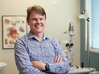
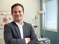
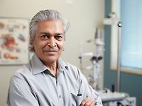
Consultants
-
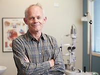
Dr John Dickson
Ophthalmologist
-
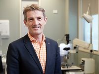
Dr Reid Ferguson
Ophthalmologist
-

Dr Stephen Guest
Ophthalmologist
-
Dr Benjamin Hoy
Ophthalmologist
-

Dr Thiyagaraj Krishnan
Ophthalmologist
-
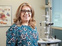
Dr Selma Matloob
Ophthalmologist
-
Dr Michael Merriman
Ophthalmologist
-
Dr Bheema Patil
Ophthalmologist
-
Dr Rohan Weerekoon
Ophthalmologist
-
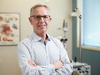
Dr David Worsley
Ophthalmologist
Ages
Adult / Pakeke, Older adult / Kaumātua
How do I access this service?
Make an appointment
Following consultation with the Specialist, your appointment will be arranged by the Specialist's Receptionist. You will be contacted with the details.
Find more details here
Fees and Charges Categorisation
Fees apply
Fees and Charges Description
Depending on your health insurance policy, a prior approval number can be gained before surgery.
If you do not have insurance cover, please discuss with the Bridgewater Day Surgery receptionist your preferred method of payment. Find out more here.
Hours
8:00 AM to 5:00 PM.
| Mon – Fri | 8:00 AM – 5:00 PM |
|---|
Public Holidays: Closed Waitangi Day (6 Feb), Good Friday (3 Apr), Easter Sunday (5 Apr), Easter Monday (6 Apr), ANZAC Day (observed) (27 Apr), King's Birthday (1 Jun), Matariki (10 Jul), Labour Day (26 Oct).
Languages Spoken
English
Procedures / Treatments
Cataract Surgery Modern cataract surgery has evolved to become exceptionally safe and effective, thanks to advances in technology that have made the procedure less invasive. As a result, the outcomes are more predictable, with minimal side effects, and the eye typically recovers swiftly. Individuals usually opt for modern cataract surgery when their diminishing eyesight impedes their day-to-day activities. If a cataract is in its early stages, vision improvement may be achievable with glasses, as advised by your ophthalmologist or optometrist. However, once the cataract becomes dense, glasses become ineffective, necessitating cataract surgery. Various eye conditions such as glaucoma, age-related macular degeneration, prior injuries, or surgeries can also impact vision. Your ophthalmologist will assess your eye and provide guidance on how these conditions may influence cataract surgery. In certain instances, the presence of other eye conditions may influence the timing of surgery; for instance, surgery may be postponed if the cataract isn't the sole contributor to vision loss. Eventually the cataract will prevent normal vision being possible. This is when surgery is considered. Usually occurring when people are in their 70’s or older, cataracts can, however, occur at any age, even at birth. Some illnesses such as diabetes, and some medications such as steroids like prednisone, can contribute to cataracts developing at an earlier age. Cataract surgery, as carried out by Hamilton Eye Clinic’s Ophthalmologists, uses techniques which enable the process to be painless and stress free. Usually spending less than 2 hours at a hospital, like Bridgewater Day Surgery, most people have the 20 minute procedure with local anaesthetic. The cloudy lens is removed and a new one replaces it. The replacement lens is manufactured in different strengths to enable a lens to be selected for your eye. Often the recovery is quick - many people will see clearly the next day. To read more click on the link
Cataract Surgery Modern cataract surgery has evolved to become exceptionally safe and effective, thanks to advances in technology that have made the procedure less invasive. As a result, the outcomes are more predictable, with minimal side effects, and the eye typically recovers swiftly. Individuals usually opt for modern cataract surgery when their diminishing eyesight impedes their day-to-day activities. If a cataract is in its early stages, vision improvement may be achievable with glasses, as advised by your ophthalmologist or optometrist. However, once the cataract becomes dense, glasses become ineffective, necessitating cataract surgery. Various eye conditions such as glaucoma, age-related macular degeneration, prior injuries, or surgeries can also impact vision. Your ophthalmologist will assess your eye and provide guidance on how these conditions may influence cataract surgery. In certain instances, the presence of other eye conditions may influence the timing of surgery; for instance, surgery may be postponed if the cataract isn't the sole contributor to vision loss. Eventually the cataract will prevent normal vision being possible. This is when surgery is considered. Usually occurring when people are in their 70’s or older, cataracts can, however, occur at any age, even at birth. Some illnesses such as diabetes, and some medications such as steroids like prednisone, can contribute to cataracts developing at an earlier age. Cataract surgery, as carried out by Hamilton Eye Clinic’s Ophthalmologists, uses techniques which enable the process to be painless and stress free. Usually spending less than 2 hours at a hospital, like Bridgewater Day Surgery, most people have the 20 minute procedure with local anaesthetic. The cloudy lens is removed and a new one replaces it. The replacement lens is manufactured in different strengths to enable a lens to be selected for your eye. Often the recovery is quick - many people will see clearly the next day. To read more click on the link
Cataract Surgery
Modern cataract surgery has evolved to become exceptionally safe and effective, thanks to advances in technology that have made the procedure less invasive. As a result, the outcomes are more predictable, with minimal side effects, and the eye typically recovers swiftly. Individuals usually opt for modern cataract surgery when their diminishing eyesight impedes their day-to-day activities. If a cataract is in its early stages, vision improvement may be achievable with glasses, as advised by your ophthalmologist or optometrist. However, once the cataract becomes dense, glasses become ineffective, necessitating cataract surgery.
Various eye conditions such as glaucoma, age-related macular degeneration, prior injuries, or surgeries can also impact vision. Your ophthalmologist will assess your eye and provide guidance on how these conditions may influence cataract surgery. In certain instances, the presence of other eye conditions may influence the timing of surgery; for instance, surgery may be postponed if the cataract isn't the sole contributor to vision loss.
Eventually the cataract will prevent normal vision being possible. This is when surgery is considered. Usually occurring when people are in their 70’s or older, cataracts can, however, occur at any age, even at birth.
Some illnesses such as diabetes, and some medications such as steroids like prednisone, can contribute to cataracts developing at an earlier age. Cataract surgery, as carried out by Hamilton Eye Clinic’s Ophthalmologists, uses techniques which enable the process to be painless and stress free.
Usually spending less than 2 hours at a hospital, like Bridgewater Day Surgery, most people have the 20 minute procedure with local anaesthetic.
The cloudy lens is removed and a new one replaces it. The replacement lens is manufactured in different strengths to enable a lens to be selected for your eye.
Often the recovery is quick - many people will see clearly the next day.
To read more click on the link
Glaucoma (often associated with raised eye pressure) Glaucoma Treatment Glaucoma is a group of conditions that damage the optic nerve, usually gradually and often without early symptoms. Eye pressure is one of the most important risk factors, but glaucoma can occur even when pressure is within the normal range. While there is more to glaucoma than pressure alone, currently the only proven way to slow or prevent further damage is to lower the eye pressure. If eye pressure is reduced to an appropriate level for the individual patient, glaucoma can often be stabilised or slowed significantly, helping to preserve vision throughout a patient’s lifetime. Because every eye is different, we set an individual target pressure based on the type and severity of glaucoma, the amount of optic nerve damage, and other risk factors. Treatment Options Eye drops Many patients use medicated eye drops to lower pressure. Some people need one drop, while others may require a combination of treatments. Laser treatment Selective Laser Trabeculoplasty (SLT) is a safe and commonly used treatment for certain types of glaucoma. It can reduce eye pressure, may decrease the need for drops, and is increasingly considered as an early treatment option for suitable patients. Its effect can last for years, although repeat treatment or additional therapy may sometimes be needed. Surgery Surgery may be recommended when glaucoma is progressing despite drops or laser treatment, when lower pressures are required, or when drops are not tolerated. There are several surgical options, and the most suitable procedure depends on the individual patient. Ongoing Care Glaucoma is usually a lifelong condition. Although damage already done cannot be reversed, ongoing treatment and regular monitoring can help protect remaining vision. Follow-up schedules vary, but many patients are reviewed every 3 to 12 months depending on their needs. To read more click on the link
Glaucoma (often associated with raised eye pressure) Glaucoma Treatment Glaucoma is a group of conditions that damage the optic nerve, usually gradually and often without early symptoms. Eye pressure is one of the most important risk factors, but glaucoma can occur even when pressure is within the normal range. While there is more to glaucoma than pressure alone, currently the only proven way to slow or prevent further damage is to lower the eye pressure. If eye pressure is reduced to an appropriate level for the individual patient, glaucoma can often be stabilised or slowed significantly, helping to preserve vision throughout a patient’s lifetime. Because every eye is different, we set an individual target pressure based on the type and severity of glaucoma, the amount of optic nerve damage, and other risk factors. Treatment Options Eye drops Many patients use medicated eye drops to lower pressure. Some people need one drop, while others may require a combination of treatments. Laser treatment Selective Laser Trabeculoplasty (SLT) is a safe and commonly used treatment for certain types of glaucoma. It can reduce eye pressure, may decrease the need for drops, and is increasingly considered as an early treatment option for suitable patients. Its effect can last for years, although repeat treatment or additional therapy may sometimes be needed. Surgery Surgery may be recommended when glaucoma is progressing despite drops or laser treatment, when lower pressures are required, or when drops are not tolerated. There are several surgical options, and the most suitable procedure depends on the individual patient. Ongoing Care Glaucoma is usually a lifelong condition. Although damage already done cannot be reversed, ongoing treatment and regular monitoring can help protect remaining vision. Follow-up schedules vary, but many patients are reviewed every 3 to 12 months depending on their needs. To read more click on the link
Glaucoma (often associated with raised eye pressure)
Glaucoma Treatment
Glaucoma is a group of conditions that damage the optic nerve, usually gradually and often without early symptoms. Eye pressure is one of the most important risk factors, but glaucoma can occur even when pressure is within the normal range. While there is more to glaucoma than pressure alone, currently the only proven way to slow or prevent further damage is to lower the eye pressure.
If eye pressure is reduced to an appropriate level for the individual patient, glaucoma can often be stabilised or slowed significantly, helping to preserve vision throughout a patient’s lifetime. Because every eye is different, we set an individual target pressure based on the type and severity of glaucoma, the amount of optic nerve damage, and other risk factors.
Treatment Options
Eye drops
Many patients use medicated eye drops to lower pressure. Some people need one drop, while others may require a combination of treatments.
Laser treatment
Selective Laser Trabeculoplasty (SLT) is a safe and commonly used treatment for certain types of glaucoma. It can reduce eye pressure, may decrease the need for drops, and is increasingly considered as an early treatment option for suitable patients. Its effect can last for years, although repeat treatment or additional therapy may sometimes be needed.
Surgery
Surgery may be recommended when glaucoma is progressing despite drops or laser treatment, when lower pressures are required, or when drops are not tolerated. There are several surgical options, and the most suitable procedure depends on the individual patient.
Ongoing Care
Glaucoma is usually a lifelong condition. Although damage already done cannot be reversed, ongoing treatment and regular monitoring can help protect remaining vision. Follow-up schedules vary, but many patients are reviewed every 3 to 12 months depending on their needs.
To read more click on the link
Oculoplastic and lacrimal surgery is a sub-specialty of ophthalmology which focuses on disorders of the eyelids, tear-drainage system and the bones behind the eye, commonly known as the orbit. Oculoplastic surgery also includes cosmetic surgery of the eyes. Common problems that require Oculoplastic, lacrimal or orbital surgery include: Facial fractures and injuries Tumors Droopy eyelids Blocked tear ducts Skin cancer Birth deformities Thyroid eye disease Excessive watering of the eye Hamilton Eye Clinic has specialists who are experts in oculoplastic and lacrimal surgery. To read more click on the link
Oculoplastic and lacrimal surgery is a sub-specialty of ophthalmology which focuses on disorders of the eyelids, tear-drainage system and the bones behind the eye, commonly known as the orbit. Oculoplastic surgery also includes cosmetic surgery of the eyes. Common problems that require Oculoplastic, lacrimal or orbital surgery include: Facial fractures and injuries Tumors Droopy eyelids Blocked tear ducts Skin cancer Birth deformities Thyroid eye disease Excessive watering of the eye Hamilton Eye Clinic has specialists who are experts in oculoplastic and lacrimal surgery. To read more click on the link
Oculoplastic and lacrimal surgery is a sub-specialty of ophthalmology which focuses on disorders of the eyelids, tear-drainage system and the bones behind the eye, commonly known as the orbit.
Oculoplastic surgery also includes cosmetic surgery of the eyes.
Common problems that require Oculoplastic, lacrimal or orbital surgery include:
- Facial fractures and injuries
- Tumors
- Droopy eyelids
- Blocked tear ducts
- Skin cancer
- Birth deformities
- Thyroid eye disease
- Excessive watering of the eye
Hamilton Eye Clinic has specialists who are experts in oculoplastic and lacrimal surgery. To read more click on the link
Strabismus (Squint) Treatment Strabismus can be treated surgically or non-surgically. This condition is ideally treated as early as possible (particularly in children) so that it does not progress to require extensive corrections. There are a number of non-surgical treatments for strabismus, all of which put the weaker eye to use in order to strengthen it. These treatments include: Eyeglasses or contact lenses, which can be used to help people who have crossed eyes due to an uncorrected farsightedness. Botox, which can be used to relax the contracted muscles in the eyes, making it easier for the eyes to focus where they need to. Patching or covering the stronger eye, to strengthen the weakened eye. Surgery In order to surgically correct strabismus, the surgeon shortens or lengthens the muscles that control the eye. First, a small incision into the eye is made in order to reach the muscle that requires strengthening or weakening. To strengthen or shorten a muscle, a small section from one end of the muscle is removed and the two ends are reattached at the same location. To weaken or add length to a muscle, a partial cut is usually made across the muscle, allowing the eye to turn further than was previously allowed. In adults, this surgery is usually performed under local anaesthetic and the patient can often go home on the same day. Occasionally the patient may experience double vision after surgery as the brain adjusts to the new way of seeing, but this should correct itself over the following weeks. To read more click on the link
Strabismus (Squint) Treatment Strabismus can be treated surgically or non-surgically. This condition is ideally treated as early as possible (particularly in children) so that it does not progress to require extensive corrections. There are a number of non-surgical treatments for strabismus, all of which put the weaker eye to use in order to strengthen it. These treatments include: Eyeglasses or contact lenses, which can be used to help people who have crossed eyes due to an uncorrected farsightedness. Botox, which can be used to relax the contracted muscles in the eyes, making it easier for the eyes to focus where they need to. Patching or covering the stronger eye, to strengthen the weakened eye. Surgery In order to surgically correct strabismus, the surgeon shortens or lengthens the muscles that control the eye. First, a small incision into the eye is made in order to reach the muscle that requires strengthening or weakening. To strengthen or shorten a muscle, a small section from one end of the muscle is removed and the two ends are reattached at the same location. To weaken or add length to a muscle, a partial cut is usually made across the muscle, allowing the eye to turn further than was previously allowed. In adults, this surgery is usually performed under local anaesthetic and the patient can often go home on the same day. Occasionally the patient may experience double vision after surgery as the brain adjusts to the new way of seeing, but this should correct itself over the following weeks. To read more click on the link
Strabismus (Squint) Treatment
Strabismus can be treated surgically or non-surgically. This condition is ideally treated as early as possible (particularly in children) so that it does not progress to require extensive corrections. There are a number of non-surgical treatments for strabismus, all of which put the weaker eye to use in order to strengthen it. These treatments include:
- Eyeglasses or contact lenses, which can be used to help people who have crossed eyes due to an uncorrected farsightedness.
- Botox, which can be used to relax the contracted muscles in the eyes, making it easier for the eyes to focus where they need to.
- Patching or covering the stronger eye, to strengthen the weakened eye.
- Surgery
In order to surgically correct strabismus, the surgeon shortens or lengthens the muscles that control the eye. First, a small incision into the eye is made in order to reach the muscle that requires strengthening or weakening. To strengthen or shorten a muscle, a small section from one end of the muscle is removed and the two ends are reattached at the same location. To weaken or add length to a muscle, a partial cut is usually made across the muscle, allowing the eye to turn further than was previously allowed.
In adults, this surgery is usually performed under local anaesthetic and the patient can often go home on the same day. Occasionally the patient may experience double vision after surgery as the brain adjusts to the new way of seeing, but this should correct itself over the following weeks.
To read more click on the link
Retinal Tears If a tear of the retina has occurred, laser treatment or cryotherapy may be used to seal the tear, so that a retinal detachment doesn’t occur. Laser treatment (photocoagulation) – A tiny laser beam is directed through your pupil using special lenses, and creates multiple “spot welds” around the retinal tear to seal it. Cryotherapy (freezing treatment) – A small probe is applied to the outside of the eye overlying the tear and freezes through to the tear, creating a scar that seals the hole. Both treatments are usually performed under local anaesthesia and can be associated with a little discomfort. The eye may be red and a little sore for a few days after cryotherapy. Types of surgery repair retinal detachment Pneumatic Retinopexy Cryotherapy (freezing) or laser treatment is applied to the retinal tear. A gas bubble is injected into the vitreous gel to push the detached retina against the back of the eye and allow the tear to seal. You are asked to maintain your head in a certain position for several days. The gas bubble may take several weeks to disappear, during which time it is unsafe to fly in an aeroplane. Vitrectomy Tiny incisions are made in the wall of the eye and fine instruments are positioned inside the eye to remove the vitreous gel and replace it with a gas bubble. Laser or cryotherapy is applied to the retinal tear to seal it, and positioning of the head for several days is required. Over time, the gas is absorbed and replaced by the eye’s own clear fluid, but while you have a gas bubble in the eye, it is unsafe to fly. Scleral buckling A cryotherapy probe is initially used to seal the retinal tear from the outside of the eye. A strip of silicone or plastic called a “buckle” is then sewn onto the white outside wall of the eye over the site of the retinal tear. This pushes the wall of the eye in closer to the underlying retinal tear and encourages the retina to reattach. The buckle is left on the eye permanently, but is hidden away under the tissue behind the eyelids. Your surgeon will discuss with you which is the most appropriate operation for your eye. Treatment of retinal detachment When part of the retina has detached, vision will be lost unless reattachment surgery is performed. Fortunately, almost all retinal detachments can be repaired, although sometimes more than one operation is required. Reattaching the retina usually removes the shadow in vision caused by retinal detachment. If, however, the shadow extended into the centre of the vision, a degree of permanent damage may have occurred, and it is unlikely that your reading vision will be fully restored, even when surgery to reattach the retina is successful. To read more click on the link
Retinal Tears If a tear of the retina has occurred, laser treatment or cryotherapy may be used to seal the tear, so that a retinal detachment doesn’t occur. Laser treatment (photocoagulation) – A tiny laser beam is directed through your pupil using special lenses, and creates multiple “spot welds” around the retinal tear to seal it. Cryotherapy (freezing treatment) – A small probe is applied to the outside of the eye overlying the tear and freezes through to the tear, creating a scar that seals the hole. Both treatments are usually performed under local anaesthesia and can be associated with a little discomfort. The eye may be red and a little sore for a few days after cryotherapy. Types of surgery repair retinal detachment Pneumatic Retinopexy Cryotherapy (freezing) or laser treatment is applied to the retinal tear. A gas bubble is injected into the vitreous gel to push the detached retina against the back of the eye and allow the tear to seal. You are asked to maintain your head in a certain position for several days. The gas bubble may take several weeks to disappear, during which time it is unsafe to fly in an aeroplane. Vitrectomy Tiny incisions are made in the wall of the eye and fine instruments are positioned inside the eye to remove the vitreous gel and replace it with a gas bubble. Laser or cryotherapy is applied to the retinal tear to seal it, and positioning of the head for several days is required. Over time, the gas is absorbed and replaced by the eye’s own clear fluid, but while you have a gas bubble in the eye, it is unsafe to fly. Scleral buckling A cryotherapy probe is initially used to seal the retinal tear from the outside of the eye. A strip of silicone or plastic called a “buckle” is then sewn onto the white outside wall of the eye over the site of the retinal tear. This pushes the wall of the eye in closer to the underlying retinal tear and encourages the retina to reattach. The buckle is left on the eye permanently, but is hidden away under the tissue behind the eyelids. Your surgeon will discuss with you which is the most appropriate operation for your eye. Treatment of retinal detachment When part of the retina has detached, vision will be lost unless reattachment surgery is performed. Fortunately, almost all retinal detachments can be repaired, although sometimes more than one operation is required. Reattaching the retina usually removes the shadow in vision caused by retinal detachment. If, however, the shadow extended into the centre of the vision, a degree of permanent damage may have occurred, and it is unlikely that your reading vision will be fully restored, even when surgery to reattach the retina is successful. To read more click on the link
If a tear of the retina has occurred, laser treatment or cryotherapy may be used to seal the tear, so that a retinal detachment doesn’t occur.
Laser treatment (photocoagulation) – A tiny laser beam is directed through your pupil using special lenses, and creates multiple “spot welds” around the retinal tear to seal it.
Cryotherapy (freezing treatment) – A small probe is applied to the outside of the eye overlying the tear and freezes through to the tear, creating a scar that seals the hole. Both treatments are usually performed under local anaesthesia and can be associated with a little discomfort. The eye may be red and a little sore for a few days after cryotherapy.
-
Pneumatic RetinopexyCryotherapy (freezing) or laser treatment is applied to the retinal tear. A gas bubble is injected into the vitreous gel to push the detached retina against the back of the eye and allow the tear to seal. You are asked to maintain your head in a certain position for several days. The gas bubble may take several weeks to disappear, during which time it is unsafe to fly in an aeroplane.
-
VitrectomyTiny incisions are made in the wall of the eye and fine instruments are positioned inside the eye to remove the vitreous gel and replace it with a gas bubble. Laser or cryotherapy is applied to the retinal tear to seal it, and positioning of the head for several days is required. Over time, the gas is absorbed and replaced by the eye’s own clear fluid, but while you have a gas bubble in the eye, it is unsafe to fly.
-
Scleral bucklingA cryotherapy probe is initially used to seal the retinal tear from the outside of the eye. A strip of silicone or plastic called a “buckle” is then sewn onto the white outside wall of the eye over the site of the retinal tear. This pushes the wall of the eye in closer to the underlying retinal tear and encourages the retina to reattach. The buckle is left on the eye permanently, but is hidden away under the tissue behind the eyelids. Your surgeon will discuss with you which is the most appropriate operation for your eye.
When part of the retina has detached, vision will be lost unless reattachment surgery is performed. Fortunately, almost all retinal detachments can be repaired, although sometimes more than one operation is required. Reattaching the retina usually removes the shadow in vision caused by retinal detachment. If, however, the shadow extended into the centre of the vision, a degree of permanent damage may have occurred, and it is unlikely that your reading vision will be fully restored, even when surgery to reattach the retina is successful.
To read more click on the link
Intravitreal injections are a procedure in which medication is injected directly into the vitreous humor - the gel-like substance inside the eye. These injections are commonly used to treat various conditions such as age-related macular degeneration (AMD), diabetic retinopathy and retinal vein occlusion and are also used to deliver antibiotic, antifungal and antiviral medications in patients with eye infections. To learn more click on the link
Intravitreal injections are a procedure in which medication is injected directly into the vitreous humor - the gel-like substance inside the eye. These injections are commonly used to treat various conditions such as age-related macular degeneration (AMD), diabetic retinopathy and retinal vein occlusion and are also used to deliver antibiotic, antifungal and antiviral medications in patients with eye infections. To learn more click on the link
Intravitreal injections are a procedure in which medication is injected directly into the vitreous humor - the gel-like substance inside the eye. These injections are commonly used to treat various conditions such as age-related macular degeneration (AMD), diabetic retinopathy and retinal vein occlusion and are also used to deliver antibiotic, antifungal and antiviral medications in patients with eye infections.
To learn more click on the link
Oculoplastic surgery focuses on surgical treatment of eyelid and tear duct problems, including both functional and cosmetic issues. Common procedures are: Blepharoplasty: removal of excess skin and/or fat from the upper and/or lower eyelids Ptosis (droopy eyelid) repair Skin cancer surgery Tear duct surgery To learn more please click on the link
Oculoplastic surgery focuses on surgical treatment of eyelid and tear duct problems, including both functional and cosmetic issues. Common procedures are: Blepharoplasty: removal of excess skin and/or fat from the upper and/or lower eyelids Ptosis (droopy eyelid) repair Skin cancer surgery Tear duct surgery To learn more please click on the link
Oculoplastic surgery focuses on surgical treatment of eyelid and tear duct problems, including both functional and cosmetic issues.
Common procedures are:
- Blepharoplasty: removal of excess skin and/or fat from the upper and/or lower eyelids
- Ptosis (droopy eyelid) repair
- Skin cancer surgery
- Tear duct surgery
To learn more please click on the link
There are two kinds of growths over the eye: a pterygium and a pinguecula. Both are benign growths over the cornea and the conjunctiva and are fairly common. Pterygia and pingueculae often present with a feeling that something is in the eye, known as the “foreign body sensation”. Although benign, pterygia contain blood vessels and can form scar tissue that can permanently disfigure the eye and should be checked by an eye specialist when noticed. Pterygium A pterygium is a wing-shaped extension of thickened tissue on the surface (conjunctiva) of the white of the eye, which grows onto the adjacent cornea (the window into the eye). Pterygia are benign growths (not cancers), which can continue to grow across the eye and eventually seriously affect sight. It is a fairly common condition. It may occur in one or both eyes. Pterygia generally start in susceptible young adults and gradually increase in size over the years. In some people, growth may cease after a period, and particularly in the elderly, they may become inactive. To read more click on the link
There are two kinds of growths over the eye: a pterygium and a pinguecula. Both are benign growths over the cornea and the conjunctiva and are fairly common. Pterygia and pingueculae often present with a feeling that something is in the eye, known as the “foreign body sensation”. Although benign, pterygia contain blood vessels and can form scar tissue that can permanently disfigure the eye and should be checked by an eye specialist when noticed. Pterygium A pterygium is a wing-shaped extension of thickened tissue on the surface (conjunctiva) of the white of the eye, which grows onto the adjacent cornea (the window into the eye). Pterygia are benign growths (not cancers), which can continue to grow across the eye and eventually seriously affect sight. It is a fairly common condition. It may occur in one or both eyes. Pterygia generally start in susceptible young adults and gradually increase in size over the years. In some people, growth may cease after a period, and particularly in the elderly, they may become inactive. To read more click on the link
There are two kinds of growths over the eye: a pterygium and a pinguecula. Both are benign growths over the cornea and the conjunctiva and are fairly common.
Pterygia and pingueculae often present with a feeling that something is in the eye, known as the “foreign body sensation”. Although benign, pterygia contain blood vessels and can form scar tissue that can permanently disfigure the eye and should be checked by an eye specialist when noticed.
A pterygium is a wing-shaped extension of thickened tissue on the surface (conjunctiva) of the white of the eye, which grows onto the adjacent cornea (the window into the eye). Pterygia are benign growths (not cancers), which can continue to grow across the eye and eventually seriously affect sight.
It is a fairly common condition. It may occur in one or both eyes.
Pterygia generally start in susceptible young adults and gradually increase in size over the years. In some people, growth may cease after a period, and particularly in the elderly, they may become inactive.
To read more click on the link
Service types: Retinal surgery, Retinal detachment.
Service types: Retinal surgery, Retinal detachment.
Service types: Retinal surgery, Retinal detachment.
Online Booking URL
Refreshments
After surgery a light refreshment is provided. Please specify if any special dietary requirements
(e.g gluten free, vegetarian etc).
Travel Directions
Please see our useful map to find our location on Grantham Street.
Parking
Please arrange transport to and from your surgery appointment as you are NOT permitted to drive following surgery until advised by your specialist. Find out more here
Accommodation
We can suggest nearby accommodation options if you have a need for an overnight stay. Please ask the secretary for your doctor to help with this.
Pharmacy
Find your nearest pharmacy here
Other
Contact Details
Bridgewater Day Surgery
Waikato
8:00 AM to 5:00 PM.
-
Phone
(07) 834 0006
Healthlink EDI
hecanbds
Email
Website
Bridgewater Building, 130 Grantham Street
Hamilton East
Hamilton
Waikato 3247
Street Address
Bridgewater Building, 130 Grantham Street
Hamilton East
Hamilton
Waikato 3247
Was this page helpful?
This page was last updated at 10:59AM on May 21, 2026. This information is reviewed and edited by Bridgewater Day Surgery - Ophthalmology.