Central Auckland, East Auckland, North Auckland, South Auckland, West Auckland > Private Hospitals & Specialists >
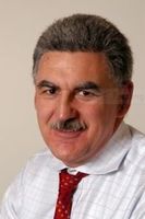
Dr Lance West - Oral & Maxillofacial Surgeon
Private Service, Oral & Maxillofacial Surgery
Today
8:30 AM to 3:30 PM.
Description
Lance West graduated BDS at the University of Otago in 1976. He trained in Oral and Maxillofacial Surgery at the Royal London Hospital and the Royal Dental Hospital, London, gaining Fellowship of the Royal College of Surgeons of Edinburgh in 1982 and Fellowship of the Royal College of Surgeons in Ireland in 1983. He gained Fellowship of the Royal Australasian College of Dental Surgeons (General Stream) in 1986 and Fellowship of the Royal Australian College of Dental Surgeons in Oral and Maxillofacial Surgery in 1987.
He has been in private oral and maxillofacial surgery practice in Auckland since 1987. He has wide experience in the surgical aspects of dental implants including bone and soft tissue reconstruction in cases where implants may otherwise not be possible. He also specialises in surgical correction of jaw and facial deformities, wisdom tooth extraction and orthodontically related procedures such as exposures of impacted and unerupted teeth.
He is a keen rugby follower and has many years experience of treating facial injuries including fractures and injuries of the jaw/facial bones.
Dental Team
-

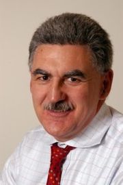
Dr Lance West
Oral & Maxillofacial Surgeon
Referral Expectations
Lance is happy to receive referrals from GPs, orthodontists and dentists but also welcomes enquires directly from patients.
You can use our online referral form
Fees and Charges Description
Initial consultation costs vary dependent upon the procedure. Please contact us for further detail.
We are happy to receive private, ACC and insurance funded patients.
More information can be found here
Hours
8:30 AM to 3:30 PM.
| Mon – Fri | 8:30 AM – 3:30 PM |
|---|
Public Holidays: Closed Good Friday (18 Apr), Easter Sunday (20 Apr), Easter Monday (21 Apr), ANZAC Day (25 Apr), King's Birthday (2 Jun), Matariki (20 Jun), Labour Day (27 Oct), Auckland Anniversary (26 Jan), Waitangi Day (6 Feb).
Services Provided
Gum tissue at the site of the implant is opened up to expose the bone. The bone is drilled and a titanium implant is inserted where the root of your tooth had been. Once the bone and gum has healed (3-6 months), the post is attached to the implant and the crown is placed over the post and cemented into place.
Gum tissue at the site of the implant is opened up to expose the bone. The bone is drilled and a titanium implant is inserted where the root of your tooth had been. Once the bone and gum has healed (3-6 months), the post is attached to the implant and the crown is placed over the post and cemented into place.
Gum tissue at the site of the implant is opened up to expose the bone. The bone is drilled and a titanium implant is inserted where the root of your tooth had been. Once the bone and gum has healed (3-6 months), the post is attached to the implant and the crown is placed over the post and cemented into place.
Orthognathic surgery corrects malpositioning of the upper and or lower jaws. This is usually done in combination with orthodontic treatment carried out with a specialist orthodontist. Surgery is usually carried out through an incision inside the mouth. The bones are cut and the jaws mobilised and following repositioning are held with titanium plates and screws which maintain the position of the bones while they heal. Common deformities suitable for orthognathic surgical correction are: Mandibular retrognathia (lower jaw too short) Mandibular prognathism (lower jaw too far forward) Maxillary retrognathia (upper jaw too far back) Vertical maxillary excess (upper jaw too tall, often resulting in a gummy smile or difficulty getting the upper and lower lips together) Transverse maxillary deficiency (upper jaw too narrow, often resulting in cross bite) Anterior open bite (vertical gap between upper and lower front teeth) Jaw and facial asymmetries Orthognathic surgery will help restore a normal occlusion and may improve a person's ability to chew and speak. Breathing may become easier for some people. The surgery may also improve facial appearance.
Orthognathic surgery corrects malpositioning of the upper and or lower jaws. This is usually done in combination with orthodontic treatment carried out with a specialist orthodontist. Surgery is usually carried out through an incision inside the mouth. The bones are cut and the jaws mobilised and following repositioning are held with titanium plates and screws which maintain the position of the bones while they heal. Common deformities suitable for orthognathic surgical correction are: Mandibular retrognathia (lower jaw too short) Mandibular prognathism (lower jaw too far forward) Maxillary retrognathia (upper jaw too far back) Vertical maxillary excess (upper jaw too tall, often resulting in a gummy smile or difficulty getting the upper and lower lips together) Transverse maxillary deficiency (upper jaw too narrow, often resulting in cross bite) Anterior open bite (vertical gap between upper and lower front teeth) Jaw and facial asymmetries Orthognathic surgery will help restore a normal occlusion and may improve a person's ability to chew and speak. Breathing may become easier for some people. The surgery may also improve facial appearance.
Orthognathic surgery corrects malpositioning of the upper and or lower jaws. This is usually done in combination with orthodontic treatment carried out with a specialist orthodontist. Surgery is usually carried out through an incision inside the mouth. The bones are cut and the jaws mobilised and following repositioning are held with titanium plates and screws which maintain the position of the bones while they heal.
Common deformities suitable for orthognathic surgical correction are:
- Mandibular retrognathia (lower jaw too short)
- Mandibular prognathism (lower jaw too far forward)
- Maxillary retrognathia (upper jaw too far back)
- Vertical maxillary excess (upper jaw too tall, often resulting in a gummy smile or difficulty getting the upper and lower lips together)
- Transverse maxillary deficiency (upper jaw too narrow, often resulting in cross bite)
- Anterior open bite (vertical gap between upper and lower front teeth)
- Jaw and facial asymmetries
Orthognathic surgery will help restore a normal occlusion and may improve a person's ability to chew and speak. Breathing may become easier for some people. The surgery may also improve facial appearance.
Parotidectomy: an incision (cut) is made in front of the ear and runs down below the jaw line. Part or all of the parotid gland is removed. Superficial parotidectomy: an incision is made in front of the ear and runs down beneath the ear lobe. The superficial (top) lobe of the parotid gland is removed. Submandibular gland surgery: an incision is made just below the jaw bone and the submandibular gland removed.
Parotidectomy: an incision (cut) is made in front of the ear and runs down below the jaw line. Part or all of the parotid gland is removed. Superficial parotidectomy: an incision is made in front of the ear and runs down beneath the ear lobe. The superficial (top) lobe of the parotid gland is removed. Submandibular gland surgery: an incision is made just below the jaw bone and the submandibular gland removed.
Parotidectomy: an incision (cut) is made in front of the ear and runs down below the jaw line. Part or all of the parotid gland is removed.
Superficial parotidectomy: an incision is made in front of the ear and runs down beneath the ear lobe. The superficial (top) lobe of the parotid gland is removed.
Submandibular gland surgery: an incision is made just below the jaw bone and the submandibular gland removed.
Arthroscopic: several small incisions (cuts) are made over the joint in front of the ear. A small telescopic instrument with a tiny camera attached (arthroscope) is inserted, allowing the surgeon a view of the joint. Small instruments can be inserted into the other cuts to free up the joint by e.g. removing adhesions and scarring, or repositioning a disc. Arthroplasty (open surgery): an incision is made in front of the ear, giving the surgeon access to reconstruct the joint by e.g. smoothing joint surfaces, repairing discs or removing diseased tissue. If a joint replacement is necessary, a second incision under the angle of the jaw may be required.
Arthroscopic: several small incisions (cuts) are made over the joint in front of the ear. A small telescopic instrument with a tiny camera attached (arthroscope) is inserted, allowing the surgeon a view of the joint. Small instruments can be inserted into the other cuts to free up the joint by e.g. removing adhesions and scarring, or repositioning a disc. Arthroplasty (open surgery): an incision is made in front of the ear, giving the surgeon access to reconstruct the joint by e.g. smoothing joint surfaces, repairing discs or removing diseased tissue. If a joint replacement is necessary, a second incision under the angle of the jaw may be required.
Arthroscopic: several small incisions (cuts) are made over the joint in front of the ear. A small telescopic instrument with a tiny camera attached (arthroscope) is inserted, allowing the surgeon a view of the joint. Small instruments can be inserted into the other cuts to free up the joint by e.g. removing adhesions and scarring, or repositioning a disc.
Arthroplasty (open surgery): an incision is made in front of the ear, giving the surgeon access to reconstruct the joint by e.g. smoothing joint surfaces, repairing discs or removing diseased tissue. If a joint replacement is necessary, a second incision under the angle of the jaw may be required.
Wisdom teeth are the third molars right at the back of your mouth. They usually appear during your late teens or early twenties. If there is not enough room in your mouth they may partially erupt through the gum or not at all. This is referred to as an impacted wisdom tooth. Due to their location wisdom teeth can be difficult to clean and are more susceptible to decay, gum disease and recurrent infections. They can cause crowding of teeth and, on rare occasions, cysts and tumours develop around them. Your dentist will advise if some or all of your wisdom teeth need to be removed. Wisdom teeth will usually only be removed if your dentist believes they will be a significant compromise to your oral health. Impacted tooth extraction Your dentist may recommend extraction if you are at significantly greater risk of infection or tooth decay. Impacted teeth may be removed by your dentist or they may refer you to an oral & maxillofacial surgeon. An incision (cut) is made in your gum and access to the impacted tooth cleared by pushing aside gum tissue and, if necessary, removing some bone. The tooth is removed whole or in pieces and the gum stitched together over the hole.
Wisdom teeth are the third molars right at the back of your mouth. They usually appear during your late teens or early twenties. If there is not enough room in your mouth they may partially erupt through the gum or not at all. This is referred to as an impacted wisdom tooth. Due to their location wisdom teeth can be difficult to clean and are more susceptible to decay, gum disease and recurrent infections. They can cause crowding of teeth and, on rare occasions, cysts and tumours develop around them. Your dentist will advise if some or all of your wisdom teeth need to be removed. Wisdom teeth will usually only be removed if your dentist believes they will be a significant compromise to your oral health. Impacted tooth extraction Your dentist may recommend extraction if you are at significantly greater risk of infection or tooth decay. Impacted teeth may be removed by your dentist or they may refer you to an oral & maxillofacial surgeon. An incision (cut) is made in your gum and access to the impacted tooth cleared by pushing aside gum tissue and, if necessary, removing some bone. The tooth is removed whole or in pieces and the gum stitched together over the hole.
Wisdom teeth are the third molars right at the back of your mouth. They usually appear during your late teens or early twenties. If there is not enough room in your mouth they may partially erupt through the gum or not at all. This is referred to as an impacted wisdom tooth.
Due to their location wisdom teeth can be difficult to clean and are more susceptible to decay, gum disease and recurrent infections. They can cause crowding of teeth and, on rare occasions, cysts and tumours develop around them.
Your dentist will advise if some or all of your wisdom teeth need to be removed. Wisdom teeth will usually only be removed if your dentist believes they will be a significant compromise to your oral health.
Impacted tooth extraction
Your dentist may recommend extraction if you are at significantly greater risk of infection or tooth decay. Impacted teeth may be removed by your dentist or they may refer you to an oral & maxillofacial surgeon.
An incision (cut) is made in your gum and access to the impacted tooth cleared by pushing aside gum tissue and, if necessary, removing some bone. The tooth is removed whole or in pieces and the gum stitched together over the hole.
Parking
There are three allocated off street parking spaces marked in green and numbered 21, 22 and 26 for Lance West's patients at 5 St Marks Road.
Website
Contact Details
5 St Mark's Road, Remuera
Central Auckland
8:30 AM to 3:30 PM.
-
Phone
(09) 520 6223
-
Fax
(09) 520 6233
Email
Website
Use our online contact form
Street Address
5 St Mark's Road
Remuera
Auckland
Was this page helpful?
This page was last updated at 1:35PM on April 8, 2025. This information is reviewed and edited by Dr Lance West - Oral & Maxillofacial Surgeon.


