Central Auckland, East Auckland, North Auckland, South Auckland, West Auckland > Private Hospitals & Specialists >
OMS Specialists | Mr Richard Cobb & Mr Ryan Smit - Oral & Maxillofacial Surgeons
Private Service, Oral & Maxillofacial Surgery
Today
8:00 AM to 5:00 PM.
Description
OMS Specialists are specialist oral, jaw and facial surgeons based in a modern and purpose designed surgical clinic in Auckland. Our surgeons have degrees in both Medicine and Dentistry and are trained to the highest international standards in Oral and Maxillofacial Surgery.
We pride ourselves on the use of minimally invasive techniques and can provide treatment under local anaesthetic, intravenous sedation or general anaesthesia as required.
Procedures offered cover the following specialty areas:
Consultants
-

Mr Richard Cobb
Specialist Oral & Maxillofacial Surgeon
-

Mr Simon Roberts
Specialist Oral & Maxillofacial Surgeon
-

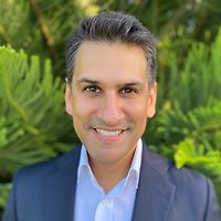
Mr Ryan Smit
Specialist Oral & Maxillofacial Surgeon
Ages
Child / Tamariki, Youth / Rangatahi, Adult / Pakeke, Older adult / Kaumātua
How do I access this service?
Referral Expectations
Fees and Charges Categorisation
Fees apply
Fees and Charges Description
We are a Southern Cross Affiliated Provider and NIB First Choice Health Partner. All health insurers accepted.
Hours
8:00 AM to 5:00 PM.
| Mon | 8:00 AM – 5:00 PM |
|---|---|
| Tue | 8:00 AM – 3:00 PM |
| Wed | 8:00 AM – 5:00 PM |
| Thu | 8:00 AM – 3:00 PM |
| Fri | 8:00 AM – 5:00 PM |
Services Provided
Wisdom teeth are the third molars right at the back of your mouth. They usually appear during your late teens or early twenties. If there is not enough room in your mouth they may partially erupt through the gum or not at all. This is referred to as an impacted wisdom tooth. Due to their location wisdom teeth can be difficult to clean and are more susceptible to decay, gum disease and recurrent infections. They can cause crowding of teeth and, on rare occasions, cysts and tumours develop around them. Your dentist will advise if some or all of your wisdom teeth need to be removed. Wisdom teeth will usually only be removed if your dentist believes they will be a significant compromise to your oral health. Impacted tooth extraction Your dentist may recommend extraction if you are at significantly greater risk of infection or tooth decay. Impacted teeth may be removed by your dentist or they may refer you to an oral & maxillofacial surgeon. An incision (cut) is made in your gum and access to the impacted tooth cleared by pushing aside gum tissue and, if necessary, removing some bone. The tooth is removed whole or in pieces and the gum stitched together over the hole.
Wisdom teeth are the third molars right at the back of your mouth. They usually appear during your late teens or early twenties. If there is not enough room in your mouth they may partially erupt through the gum or not at all. This is referred to as an impacted wisdom tooth. Due to their location wisdom teeth can be difficult to clean and are more susceptible to decay, gum disease and recurrent infections. They can cause crowding of teeth and, on rare occasions, cysts and tumours develop around them. Your dentist will advise if some or all of your wisdom teeth need to be removed. Wisdom teeth will usually only be removed if your dentist believes they will be a significant compromise to your oral health. Impacted tooth extraction Your dentist may recommend extraction if you are at significantly greater risk of infection or tooth decay. Impacted teeth may be removed by your dentist or they may refer you to an oral & maxillofacial surgeon. An incision (cut) is made in your gum and access to the impacted tooth cleared by pushing aside gum tissue and, if necessary, removing some bone. The tooth is removed whole or in pieces and the gum stitched together over the hole.
Wisdom teeth are the third molars right at the back of your mouth. They usually appear during your late teens or early twenties. If there is not enough room in your mouth they may partially erupt through the gum or not at all. This is referred to as an impacted wisdom tooth.
Due to their location wisdom teeth can be difficult to clean and are more susceptible to decay, gum disease and recurrent infections. They can cause crowding of teeth and, on rare occasions, cysts and tumours develop around them.
Your dentist will advise if some or all of your wisdom teeth need to be removed. Wisdom teeth will usually only be removed if your dentist believes they will be a significant compromise to your oral health.
Impacted tooth extraction
Your dentist may recommend extraction if you are at significantly greater risk of infection or tooth decay. Impacted teeth may be removed by your dentist or they may refer you to an oral & maxillofacial surgeon.
An incision (cut) is made in your gum and access to the impacted tooth cleared by pushing aside gum tissue and, if necessary, removing some bone. The tooth is removed whole or in pieces and the gum stitched together over the hole.
Gum tissue at the site of the implant is opened up to expose the bone. The bone is drilled and a titanium implant is inserted where the root of your tooth had been. Once the bone and gum has healed (3-6 months), the post is attached to the implant and the crown is placed over the post and cemented into place.
Gum tissue at the site of the implant is opened up to expose the bone. The bone is drilled and a titanium implant is inserted where the root of your tooth had been. Once the bone and gum has healed (3-6 months), the post is attached to the implant and the crown is placed over the post and cemented into place.
Gum tissue at the site of the implant is opened up to expose the bone. The bone is drilled and a titanium implant is inserted where the root of your tooth had been. Once the bone and gum has healed (3-6 months), the post is attached to the implant and the crown is placed over the post and cemented into place.
There are three large pairs of glands (parotid, sublingual and submandibular) in your mouth that produce saliva which helps break down food as part of the digestion process. Salivary gland surgery involves the removal of one or more of the salivary glands for reasons including: tumours (benign or cancerous), chronic infections or blockages, salivary stones or injuries or cysts. Care is taken to avoid damaging nearby nerves, especially those that control facial movement.
There are three large pairs of glands (parotid, sublingual and submandibular) in your mouth that produce saliva which helps break down food as part of the digestion process. Salivary gland surgery involves the removal of one or more of the salivary glands for reasons including: tumours (benign or cancerous), chronic infections or blockages, salivary stones or injuries or cysts. Care is taken to avoid damaging nearby nerves, especially those that control facial movement.
There are three large pairs of glands (parotid, sublingual and submandibular) in your mouth that produce saliva which helps break down food as part of the digestion process.
Salivary gland surgery involves the removal of one or more of the salivary glands for reasons including: tumours (benign or cancerous), chronic infections or blockages, salivary stones or injuries or cysts.
Care is taken to avoid damaging nearby nerves, especially those that control facial movement.
Arthroscopic: several small incisions (cuts) are made over the joint in front of the ear. A small telescopic instrument with a tiny camera attached (arthroscope) is inserted, allowing the surgeon a view of the joint. Small instruments can be inserted into the other cuts to free up the joint by e.g. removing adhesions and scarring, or repositioning a disc. Arthroplasty (open surgery): an incision is made in front of the ear, giving the surgeon access to reconstruct the joint by e.g. smoothing joint surfaces, repairing discs or removing diseased tissue. If a joint replacement is necessary, a second incision under the angle of the jaw may be required.
Arthroscopic: several small incisions (cuts) are made over the joint in front of the ear. A small telescopic instrument with a tiny camera attached (arthroscope) is inserted, allowing the surgeon a view of the joint. Small instruments can be inserted into the other cuts to free up the joint by e.g. removing adhesions and scarring, or repositioning a disc. Arthroplasty (open surgery): an incision is made in front of the ear, giving the surgeon access to reconstruct the joint by e.g. smoothing joint surfaces, repairing discs or removing diseased tissue. If a joint replacement is necessary, a second incision under the angle of the jaw may be required.
Arthroscopic: several small incisions (cuts) are made over the joint in front of the ear. A small telescopic instrument with a tiny camera attached (arthroscope) is inserted, allowing the surgeon a view of the joint. Small instruments can be inserted into the other cuts to free up the joint by e.g. removing adhesions and scarring, or repositioning a disc.
Arthroplasty (open surgery): an incision is made in front of the ear, giving the surgeon access to reconstruct the joint by e.g. smoothing joint surfaces, repairing discs or removing diseased tissue. If a joint replacement is necessary, a second incision under the angle of the jaw may be required.
Scar appearance can be improved by various methods including a surgical procedure known as scar revision. This usually involves cutting out the old scar, closing the wound with stitches and, in some cases, moving the scar so that it is hidden by natural features of the body. Scar revision is usually performed under local anaesthesia (the area around the scar is numbed by injecting a local anaesthetic). Sometimes you may also be given steroid injections at the time of surgery. Immediately following the procedure, you will need to remain at the clinic for about an hour, during which you will be encouraged to walk around. You may or may not have a dressing put on the wound and it is important to keep the area dry for 24 hours. Stitches may be removed in 1-2 weeks. You may need to take a few days off work after the surgery.
Scar appearance can be improved by various methods including a surgical procedure known as scar revision. This usually involves cutting out the old scar, closing the wound with stitches and, in some cases, moving the scar so that it is hidden by natural features of the body. Scar revision is usually performed under local anaesthesia (the area around the scar is numbed by injecting a local anaesthetic). Sometimes you may also be given steroid injections at the time of surgery. Immediately following the procedure, you will need to remain at the clinic for about an hour, during which you will be encouraged to walk around. You may or may not have a dressing put on the wound and it is important to keep the area dry for 24 hours. Stitches may be removed in 1-2 weeks. You may need to take a few days off work after the surgery.
Scar appearance can be improved by various methods including a surgical procedure known as scar revision. This usually involves cutting out the old scar, closing the wound with stitches and, in some cases, moving the scar so that it is hidden by natural features of the body.
Scar revision is usually performed under local anaesthesia (the area around the scar is numbed by injecting a local anaesthetic). Sometimes you may also be given steroid injections at the time of surgery. Immediately following the procedure, you will need to remain at the clinic for about an hour, during which you will be encouraged to walk around. You may or may not have a dressing put on the wound and it is important to keep the area dry for 24 hours. Stitches may be removed in 1-2 weeks. You may need to take a few days off work after the surgery.
Skin lesions can be divided into two groups: benign (non-cancerous): e.g. moles, cysts, warts, tags. These may be removed to prevent spreading (warts), stop discomfort if the lesion is being irritated by clothing/jewellery or to improve appearance. malignant (cancerous): basal cell and squamous cell carcinomas are generally slow growing and unlikely to spread to other parts of the body. Melanoma is a serious skin cancer that can spread to other parts of the body. Urgent removal is recommended. Surgery to remove skin lesions usually involves an office or outpatient visit, local anaesthesia (the area around the scar is numbed by injecting a local anaesthetic) and stitches. You may or may not have a dressing put on the wound and it is important to keep the area dry for 24 hours. Stitches may be removed in 1-2 weeks. You may need to take a few days off work after the surgery.
Skin lesions can be divided into two groups: benign (non-cancerous): e.g. moles, cysts, warts, tags. These may be removed to prevent spreading (warts), stop discomfort if the lesion is being irritated by clothing/jewellery or to improve appearance. malignant (cancerous): basal cell and squamous cell carcinomas are generally slow growing and unlikely to spread to other parts of the body. Melanoma is a serious skin cancer that can spread to other parts of the body. Urgent removal is recommended. Surgery to remove skin lesions usually involves an office or outpatient visit, local anaesthesia (the area around the scar is numbed by injecting a local anaesthetic) and stitches. You may or may not have a dressing put on the wound and it is important to keep the area dry for 24 hours. Stitches may be removed in 1-2 weeks. You may need to take a few days off work after the surgery.
Skin lesions can be divided into two groups:
- benign (non-cancerous): e.g. moles, cysts, warts, tags. These may be removed to prevent spreading (warts), stop discomfort if the lesion is being irritated by clothing/jewellery or to improve appearance.
- malignant (cancerous): basal cell and squamous cell carcinomas are generally slow growing and unlikely to spread to other parts of the body. Melanoma is a serious skin cancer that can spread to other parts of the body. Urgent removal is recommended.
Surgery to remove skin lesions usually involves an office or outpatient visit, local anaesthesia (the area around the scar is numbed by injecting a local anaesthetic) and stitches. You may or may not have a dressing put on the wound and it is important to keep the area dry for 24 hours. Stitches may be removed in 1-2 weeks. You may need to take a few days off work after the surgery.
Online Booking URL
Public Transport
The Auckland Transport website is a good resource to plan your public transport options.
Parking
Free parking is available at the clinic
Pharmacy
Find your pharmacy here
Website
Contact Details
123 Carlton Gore Road, Newmarket, Auckland
Central Auckland
8:00 AM to 5:00 PM.
-
Phone
(09) 477 0058
Healthlink EDI
implants
Email
Website
123 Carlton Gore Road
Newmarket
Auckland
Auckland 1052
Street Address
123 Carlton Gore Road
Newmarket
Auckland
Auckland 1052
Was this page helpful?
This page was last updated at 9:04AM on January 8, 2026. This information is reviewed and edited by OMS Specialists | Mr Richard Cobb & Mr Ryan Smit - Oral & Maxillofacial Surgeons.

