
Central Auckland, East Auckland, North Auckland, South Auckland, West Auckland > Private Hospitals & Specialists >
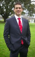
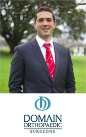
Chris Fougere - Hip, Knee, Ankle & Foot Orthopaedic Surgeon
Private Service, Orthopaedics
Today
8:30 AM to 5:00 PM.
Description
Chris Fougere is a New Zealand trained orthopaedic surgeon. He completed undergraduate training at the Auckland University School of Medicine, then entered the NZ Advanced Orthopaedic training programme, attaining FRACS (Ortho) in 2009.
Further fellowship training followed in 2010 in Adult Joint Replacement and Revision in Sydney, Adult Deformity and Shoulder Reconstruction in Melbourne concluding with Adult Deformity in 2011 in Florida with Dr Dror Paley.
Chris has a consultant appointment at Auckland City Hospital, focusing on Fracture, Deformity Surgery and Joint Reconstruction. His private practice is with Domain Orthopaedic Surgeons.
Chris Fougere's main interests are:
- Joint Preservation (Arthroscopy, Osteotomy) and Replacement
- Fracture
- Injury and Deformity Surgery of the:
- Hip
- Knee
- Foot.
Consultants
-
Mr Chris Fougere
Orthopaedic Surgeon
Referral Expectations
You need to bring with you:
- any letters or reports from your doctor or hospital
- any x-rays, CT or MRI films and reports
- all medicines you are taking including herbal and natural remedies
- your pharmaceutical entitlement card
- your ACC number, if you have one.
Hours
8:30 AM to 5:00 PM.
| Mon – Fri | 8:30 AM – 5:00 PM |
|---|
Procedures / Treatments
For elderly patients joint replacement surgery is commonly required to treat damaged joints from wearing out, arthritis or other forms of joint disease including rheumatoid arthritis. In these procedures the damaged joint surface is removed and replaced with artificial surfaces normally made from metal (chromium cobalt alloy, titanium), plastic (high density polyethelene) or ceramic which act as alternate bearing surfaces for the damaged joint. These operations are major procedures which require the patient to be in hospital for several days and followed by a significant period of rehabilitation. The hospital has several ways of approaching the procedure for replacement and the specifics for the procedure will be covered at the time of assessment and booking of surgery. Occasionally blood transfusions are required; if you have some concerns raise this with your surgeon during consultation.
For elderly patients joint replacement surgery is commonly required to treat damaged joints from wearing out, arthritis or other forms of joint disease including rheumatoid arthritis. In these procedures the damaged joint surface is removed and replaced with artificial surfaces normally made from metal (chromium cobalt alloy, titanium), plastic (high density polyethelene) or ceramic which act as alternate bearing surfaces for the damaged joint. These operations are major procedures which require the patient to be in hospital for several days and followed by a significant period of rehabilitation. The hospital has several ways of approaching the procedure for replacement and the specifics for the procedure will be covered at the time of assessment and booking of surgery. Occasionally blood transfusions are required; if you have some concerns raise this with your surgeon during consultation.
Over the last 30 years a large number of orthopaedic procedures on joints have been performed using an arthroscope, where a fiber optic telescope is used to look inside the joint. Through this type of keyhole surgery, fine instruments can be introduced through small incisions (portals) to allow surgery to be performed without the need for large cuts. This allows many procedures to be performed as a day stay and allows quicker return to normal function of the joint. Arthroscopic surgery is less painful than open surgery and decreases the risk of healing problems. Arthroscopy allows access to parts of the joints which can not be accessed by other types of surgery.
Over the last 30 years a large number of orthopaedic procedures on joints have been performed using an arthroscope, where a fiber optic telescope is used to look inside the joint. Through this type of keyhole surgery, fine instruments can be introduced through small incisions (portals) to allow surgery to be performed without the need for large cuts. This allows many procedures to be performed as a day stay and allows quicker return to normal function of the joint. Arthroscopic surgery is less painful than open surgery and decreases the risk of healing problems. Arthroscopy allows access to parts of the joints which can not be accessed by other types of surgery.
In many cases tendons will be lengthened to improve the muscle balance around a joint or tendons will be transferred to give overall better joint function. This occurs in children with neuromuscular conditions but also applies to a number of other conditions. Most of these procedures involve some sort of splintage after the surgery followed by a period of rehabilitation, normally supervised by a physiotherapist.
In many cases tendons will be lengthened to improve the muscle balance around a joint or tendons will be transferred to give overall better joint function. This occurs in children with neuromuscular conditions but also applies to a number of other conditions. Most of these procedures involve some sort of splintage after the surgery followed by a period of rehabilitation, normally supervised by a physiotherapist.
Hip Arthroscopy Small incisions (cuts) are made in the hip area and a small telescopic instrument with a tiny camera attached (arthroscope) is inserted. This allows the surgeon to look inside the joint, identify problems and, in some cases, operate. Tiny instruments can be passed through the arthroscope to remove loose, damaged or inflamed tissue. Hip Replacement An incision (cut) is made on the side of the thigh to allow the surgeon access to the hip joint. The diseased and damaged parts of the hip joint are removed and replaced with smooth, artificial metal ‘ball’ and plastic ‘socket’ parts.
Hip Arthroscopy Small incisions (cuts) are made in the hip area and a small telescopic instrument with a tiny camera attached (arthroscope) is inserted. This allows the surgeon to look inside the joint, identify problems and, in some cases, operate. Tiny instruments can be passed through the arthroscope to remove loose, damaged or inflamed tissue. Hip Replacement An incision (cut) is made on the side of the thigh to allow the surgeon access to the hip joint. The diseased and damaged parts of the hip joint are removed and replaced with smooth, artificial metal ‘ball’ and plastic ‘socket’ parts.
Hip Arthroscopy
Small incisions (cuts) are made in the hip area and a small telescopic instrument with a tiny camera attached (arthroscope) is inserted. This allows the surgeon to look inside the joint, identify problems and, in some cases, operate. Tiny instruments can be passed through the arthroscope to remove loose, damaged or inflamed tissue.
Hip Replacement
An incision (cut) is made on the side of the thigh to allow the surgeon access to the hip joint. The diseased and damaged parts of the hip joint are removed and replaced with smooth, artificial metal ‘ball’ and plastic ‘socket’ parts.
Knee Arthroscopy Several small incisions (cuts) are made on the knee through which is inserted a small telescopic instrument with a tiny camera attached (arthroscope). This allows the surgeon to look inside the joint, identify problems and, in some cases, make repairs to damaged tissue. Knee Replacement An incision (cut) is made on the front of the knee to allow the surgeon access to the knee joint. The damaged and painful areas of the thigh bone (femur) and lower leg bone (tibia), including the knee joint, are removed and replaced with metal and plastic parts.
Knee Arthroscopy Several small incisions (cuts) are made on the knee through which is inserted a small telescopic instrument with a tiny camera attached (arthroscope). This allows the surgeon to look inside the joint, identify problems and, in some cases, make repairs to damaged tissue. Knee Replacement An incision (cut) is made on the front of the knee to allow the surgeon access to the knee joint. The damaged and painful areas of the thigh bone (femur) and lower leg bone (tibia), including the knee joint, are removed and replaced with metal and plastic parts.
Knee Arthroscopy
Several small incisions (cuts) are made on the knee through which is inserted a small telescopic instrument with a tiny camera attached (arthroscope). This allows the surgeon to look inside the joint, identify problems and, in some cases, make repairs to damaged tissue.
Knee Replacement
An incision (cut) is made on the front of the knee to allow the surgeon access to the knee joint. The damaged and painful areas of the thigh bone (femur) and lower leg bone (tibia), including the knee joint, are removed and replaced with metal and plastic parts.
Ankle Arthroscopy Two or three small incisions (cuts) are made in the ankle and a small telescopic instrument with a tiny camera attached (arthroscope) is inserted. This allows the surgeon to look inside the joint, identify problems and, in some cases, operate. Tiny instruments can be passed through the arthroscope to remove bony spurs, damaged cartilage or inflamed tissue. Ankle Replacement An incision (cut) is made in the front of, and several smaller cuts on the outside of, the ankle. The damaged ankle joint is replaced with a metal and plastic implant.
Ankle Arthroscopy Two or three small incisions (cuts) are made in the ankle and a small telescopic instrument with a tiny camera attached (arthroscope) is inserted. This allows the surgeon to look inside the joint, identify problems and, in some cases, operate. Tiny instruments can be passed through the arthroscope to remove bony spurs, damaged cartilage or inflamed tissue. Ankle Replacement An incision (cut) is made in the front of, and several smaller cuts on the outside of, the ankle. The damaged ankle joint is replaced with a metal and plastic implant.
Ankle Arthroscopy
Two or three small incisions (cuts) are made in the ankle and a small telescopic instrument with a tiny camera attached (arthroscope) is inserted. This allows the surgeon to look inside the joint, identify problems and, in some cases, operate. Tiny instruments can be passed through the arthroscope to remove bony spurs, damaged cartilage or inflamed tissue.
Ankle Replacement
An incision (cut) is made in the front of, and several smaller cuts on the outside of, the ankle. The damaged ankle joint is replaced with a metal and plastic implant.
A bunion is a lump of bone and soft tissue that forms where the big toe joins the foot. Typically caused by ill-fitting shoes, bunions may require surgery to relieve pain and allow a return to normal activities. Click here for more information.
A bunion is a lump of bone and soft tissue that forms where the big toe joins the foot. Typically caused by ill-fitting shoes, bunions may require surgery to relieve pain and allow a return to normal activities. Click here for more information.
A bunion is a lump of bone and soft tissue that forms where the big toe joins the foot. Typically caused by ill-fitting shoes, bunions may require surgery to relieve pain and allow a return to normal activities.
Click here for more information.
Orthopaedic surgeons have expertise in the treatment of fractured (broken) bones, particularly in the assessment of damage that may have occurred around the fracture. Follow-up of a fracture may involve monitoring the progress of the healing bone, checking the position of the bone in a cast and deciding when other steps in management such as re-manipulation of the fracture or removal of a cast is required. Click here for more information about fractures.
Orthopaedic surgeons have expertise in the treatment of fractured (broken) bones, particularly in the assessment of damage that may have occurred around the fracture. Follow-up of a fracture may involve monitoring the progress of the healing bone, checking the position of the bone in a cast and deciding when other steps in management such as re-manipulation of the fracture or removal of a cast is required. Click here for more information about fractures.
Orthopaedic surgeons have expertise in the treatment of fractured (broken) bones, particularly in the assessment of damage that may have occurred around the fracture.
Follow-up of a fracture may involve monitoring the progress of the healing bone, checking the position of the bone in a cast and deciding when other steps in management such as re-manipulation of the fracture or removal of a cast is required.
Click here for more information about fractures.
Orthopaedic deformities can be congenital or acquired as the result of injury, infection or tumour. Resulting in crooked limbs or discrepancies in limb length, such deformities can affect appearance and function and can often cause significant pain. Osteotomy is the division of a crooked or bent bone to improve alignment of the limb. These procedures normally involve some form of internal fixation, such as rods or plates, or external fixation which involves external wires and pins to hold the bone. The type of procedure for fixation will be explained when the surgery is planned. Some of the more common orthopaedic deformities are: Intoeing Bow Legs (Genu Varum) Club Foot (Talipes) Developmental Dislocation of the Hip Bunions Limb Length Discrepancy
Orthopaedic deformities can be congenital or acquired as the result of injury, infection or tumour. Resulting in crooked limbs or discrepancies in limb length, such deformities can affect appearance and function and can often cause significant pain. Osteotomy is the division of a crooked or bent bone to improve alignment of the limb. These procedures normally involve some form of internal fixation, such as rods or plates, or external fixation which involves external wires and pins to hold the bone. The type of procedure for fixation will be explained when the surgery is planned. Some of the more common orthopaedic deformities are: Intoeing Bow Legs (Genu Varum) Club Foot (Talipes) Developmental Dislocation of the Hip Bunions Limb Length Discrepancy
Orthopaedic deformities can be congenital or acquired as the result of injury, infection or tumour. Resulting in crooked limbs or discrepancies in limb length, such deformities can affect appearance and function and can often cause significant pain.
Some of the more common orthopaedic deformities are:
Public Transport
The Auckland Transport Journey Planner will help you to plan your journey.
Parking
Parking is available in designated spaces in the lower ground car park at the Titoki Street practice. Please enter the carpark to the right hand side of the building.
Website
Contact Details
20 Titoki Street, Parnell, Auckland
Central Auckland
8:30 AM to 5:00 PM.
-
Phone
(09) 307 4285
-
Fax
(09) 307 4280
Healthlink EDI
domainor
Email
Website
20 Titoki Street
Parnell
Auckland 1052
Street Address
20 Titoki Street
Parnell
Auckland 1052
Was this page helpful?
This page was last updated at 9:46AM on November 23, 2022. This information is reviewed and edited by Chris Fougere - Hip, Knee, Ankle & Foot Orthopaedic Surgeon.