
Central Auckland, East Auckland, North Auckland, South Auckland, West Auckland > Private Hospitals & Specialists >
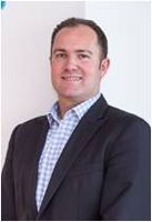
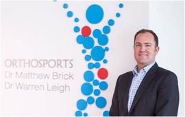
Dr Warren Leigh - Knee, Hip, Shoulder & Spine Orthopaedic Surgeon
Private Service, Orthopaedics, Spinal
Description
Dr Warren Leigh is a New Zealand trained orthopaedic surgeon who specializes in the management of sports injuries. His practice includes arthroscopic surgery of the hip, knee and shoulder, spine surgery and knee arthroplasty.
Dr Leigh is based at the Millennium Institute of Sport and Health and operates at Southern Cross North Harbour.
Warren grew up in Tauranga before moving to study medicine at the University of Auckland. He then completed the NZ Orthopaedic Association's advanced surgical training program, and was admitted as a fellow of the Royal Australian College of Surgeons in 2010. Following this he undertook 2.5 years of fellowship training in knee, sports and spine surgery.
In his new practice on the North Shore Warren is focused on hip arthroscopy and advanced sports knee and shoulder arthroscopy with Dr Mat Brick. Hip arthroscopy is an evolving technique that allows treatment of femoroacetabular impingement and intra-articular hip pathology.
In Melbourne, Dr Leigh worked under Professor Julian Feller and Mr Tim Whitehead at OrthoSport Victoria. He gained a large volume and breadth of experience in all aspects of knee related clinical assessment and knee surgery. A research component centred upon anterior cruciate ligament (ACL) reconstruction and the world-class gait laboratory at La Trobe University, Melbourne.
In Adelaide he undertook the Neuro-Orthopaedic Spinal Fellowship at the Royal Adelaide Hospital with Professors Rob Fraser and Brian Freeman. This provided extensive training in all aspects of acute and elective adult lumbar and cervical spine pathologies. He also performed minimally invasive spinal surgery (MIS).
Warren has just graduated from the Surgical Leadership Program at Harvard Medical School.
Dr Warren Leigh is available to see patients at Orthosports in the Millennium Institute of Sport and Health.
What is Orthopaedics?
Consultants
-
Mr Warren Leigh
Knee & Spine Orthopaedic Surgeon
Referral Expectations
You need to bring to your appointment:
- any letters or reports from your doctor or hospital
- any X-Rays, CT or MRI films and reports
- all medicines you are taking including herbal and natural remedies
- your pharmaceutical entitlement card
- your ACC number, if you have one.
Procedures / Treatments
Orthopaedic surgeons have expertise in the treatment of fractured (broken) bones, particularly in the assessment of damage that may have occurred around the fracture. Follow-up of a fracture may involve monitoring the progress of the healing bone, checking the position of the bone in a cast and deciding when other steps in management such as re-manipulation of the fracture or removal of a cast is required. Click here for more information about fractures.
Orthopaedic surgeons have expertise in the treatment of fractured (broken) bones, particularly in the assessment of damage that may have occurred around the fracture. Follow-up of a fracture may involve monitoring the progress of the healing bone, checking the position of the bone in a cast and deciding when other steps in management such as re-manipulation of the fracture or removal of a cast is required. Click here for more information about fractures.
Orthopaedic surgeons have expertise in the treatment of fractured (broken) bones, particularly in the assessment of damage that may have occurred around the fracture.
Follow-up of a fracture may involve monitoring the progress of the healing bone, checking the position of the bone in a cast and deciding when other steps in management such as re-manipulation of the fracture or removal of a cast is required.
Click here for more information about fractures.
For elderly patients joint replacement surgery is commonly required to treat damaged joints from wearing out, arthritis or other forms of joint disease including rheumatoid arthritis. In these procedures the damaged joint surface is removed and replaced with artificial surfaces normally made from metal (chromium cobalt alloy, titanium), plastic (high density polyethelene) or ceramic which act as alternate bearing surfaces for the damaged joint. These operations are major procedures which require the patient to be in hospital for several days and followed by a significant period of rehabilitation. The hospital has several ways of approaching the procedure for replacement and the specifics for the procedure will be covered at the time of assessment and booking of surgery. Occasionally blood transfusions are required; if you have some concerns raise this with your surgeon during consultation.
For elderly patients joint replacement surgery is commonly required to treat damaged joints from wearing out, arthritis or other forms of joint disease including rheumatoid arthritis. In these procedures the damaged joint surface is removed and replaced with artificial surfaces normally made from metal (chromium cobalt alloy, titanium), plastic (high density polyethelene) or ceramic which act as alternate bearing surfaces for the damaged joint. These operations are major procedures which require the patient to be in hospital for several days and followed by a significant period of rehabilitation. The hospital has several ways of approaching the procedure for replacement and the specifics for the procedure will be covered at the time of assessment and booking of surgery. Occasionally blood transfusions are required; if you have some concerns raise this with your surgeon during consultation.
For elderly patients joint replacement surgery is commonly required to treat damaged joints from wearing out, arthritis or other forms of joint disease including rheumatoid arthritis. In these procedures the damaged joint surface is removed and replaced with artificial surfaces normally made from metal (chromium cobalt alloy, titanium), plastic (high density polyethelene) or ceramic which act as alternate bearing surfaces for the damaged joint.
These operations are major procedures which require the patient to be in hospital for several days and followed by a significant period of rehabilitation. The hospital has several ways of approaching the procedure for replacement and the specifics for the procedure will be covered at the time of assessment and booking of surgery.
Occasionally blood transfusions are required; if you have some concerns raise this with your surgeon during consultation.
Knee surgery is an operation that helps fix problems in your knee, like an injury, arthritis, a torn ligament, or damaged cartilage. Surgery might be: Arthroscopic: less invasive surgery. The surgeon makes small cuts and uses a tiny camera to see inside the knee. They can then fix or take out the damaged parts. Open: for more complicated problems, the surgeon makes a larger cut to get a better look and fix the damaged parts directly. Joint Replacement: when the knee is badly damaged, the surgeon might remove the damaged areas and replace them with parts made of special materials.
Knee surgery is an operation that helps fix problems in your knee, like an injury, arthritis, a torn ligament, or damaged cartilage. Surgery might be: Arthroscopic: less invasive surgery. The surgeon makes small cuts and uses a tiny camera to see inside the knee. They can then fix or take out the damaged parts. Open: for more complicated problems, the surgeon makes a larger cut to get a better look and fix the damaged parts directly. Joint Replacement: when the knee is badly damaged, the surgeon might remove the damaged areas and replace them with parts made of special materials.
Knee surgery is an operation that helps fix problems in your knee, like an injury, arthritis, a torn ligament, or damaged cartilage. Surgery might be:
- Arthroscopic: less invasive surgery. The surgeon makes small cuts and uses a tiny camera to see inside the knee. They can then fix or take out the damaged parts.
- Open: for more complicated problems, the surgeon makes a larger cut to get a better look and fix the damaged parts directly.
- Joint Replacement: when the knee is badly damaged, the surgeon might remove the damaged areas and replace them with parts made of special materials.
Osteotomy is the division of a crooked or bent bone to improve alignment of the limb. These procedures normally involve some form of internal fixation, such as rods or plates, or external fixation which involves external wires and pins to hold the bone. The type of procedure for fixation will be explained when the surgery is planned.
Osteotomy is the division of a crooked or bent bone to improve alignment of the limb. These procedures normally involve some form of internal fixation, such as rods or plates, or external fixation which involves external wires and pins to hold the bone. The type of procedure for fixation will be explained when the surgery is planned.
Osteotomy is the division of a crooked or bent bone to improve alignment of the limb.
These procedures normally involve some form of internal fixation, such as rods or plates, or external fixation which involves external wires and pins to hold the bone. The type of procedure for fixation will be explained when the surgery is planned.
A large number of orthopaedic procedures on joints are performed using an arthroscope, where a fibre optic telescope is used to look inside the joint. Through this type of keyhole surgery, fine instruments can be introduced through small incisions (portals) to allow surgery to be performed without the need for large cuts. This allows many procedures to be performed as a day stay and allows quicker return to normal function of the joint. Arthroscopic surgery is less painful than open surgery and decreases the risk of healing problems. Arthroscopy allows access to parts of the joints which can not be accessed by other types of surgery.
A large number of orthopaedic procedures on joints are performed using an arthroscope, where a fibre optic telescope is used to look inside the joint. Through this type of keyhole surgery, fine instruments can be introduced through small incisions (portals) to allow surgery to be performed without the need for large cuts. This allows many procedures to be performed as a day stay and allows quicker return to normal function of the joint. Arthroscopic surgery is less painful than open surgery and decreases the risk of healing problems. Arthroscopy allows access to parts of the joints which can not be accessed by other types of surgery.
A large number of orthopaedic procedures on joints are performed using an arthroscope, where a fibre optic telescope is used to look inside the joint. Through this type of keyhole surgery, fine instruments can be introduced through small incisions (portals) to allow surgery to be performed without the need for large cuts. This allows many procedures to be performed as a day stay and allows quicker return to normal function of the joint.
Arthroscopic surgery is less painful than open surgery and decreases the risk of healing problems. Arthroscopy allows access to parts of the joints which can not be accessed by other types of surgery.
In many cases tendons will be lengthened to improve the muscle balance around a joint or tendons will be transferred to give overall better joint function. This occurs in children with neuromuscular conditions but also applies to a number of other conditions. Most of these procedures involve some sort of splintage after the surgery followed by a period of rehabilitation, normally supervised by a physiotherapist.
In many cases tendons will be lengthened to improve the muscle balance around a joint or tendons will be transferred to give overall better joint function. This occurs in children with neuromuscular conditions but also applies to a number of other conditions. Most of these procedures involve some sort of splintage after the surgery followed by a period of rehabilitation, normally supervised by a physiotherapist.
In many cases tendons will be lengthened to improve the muscle balance around a joint or tendons will be transferred to give overall better joint function. This occurs in children with neuromuscular conditions but also applies to a number of other conditions.
Most of these procedures involve some sort of splintage after the surgery followed by a period of rehabilitation, normally supervised by a physiotherapist.
This is a surgical procedure performed on a knee joint that has become painful and/or impaired because of disease, injury or wear and tear. In total knee replacement, artificial materials (metal and plastic) are used to replace the following damaged surfaces within the knee joint: the end of the thigh bone (femur) the end of the shin bone (tibia) the back of the kneecap (patella) This operation is a major procedure which requires you to be in hospital for several days and will be followed by a significant period of rehabilitation. Occasionally blood transfusions are required; if you have some concerns raise this with your surgeon during consultation. For more information about total knee replacement please click here.
This is a surgical procedure performed on a knee joint that has become painful and/or impaired because of disease, injury or wear and tear. In total knee replacement, artificial materials (metal and plastic) are used to replace the following damaged surfaces within the knee joint: the end of the thigh bone (femur) the end of the shin bone (tibia) the back of the kneecap (patella) This operation is a major procedure which requires you to be in hospital for several days and will be followed by a significant period of rehabilitation. Occasionally blood transfusions are required; if you have some concerns raise this with your surgeon during consultation. For more information about total knee replacement please click here.
This is a surgical procedure performed on a knee joint that has become painful and/or impaired because of disease, injury or wear and tear.
In total knee replacement, artificial materials (metal and plastic) are used to replace the following damaged surfaces within the knee joint:
- the end of the thigh bone (femur)
- the end of the shin bone (tibia)
- the back of the kneecap (patella)
This operation is a major procedure which requires you to be in hospital for several days and will be followed by a significant period of rehabilitation.
Occasionally blood transfusions are required; if you have some concerns raise this with your surgeon during consultation.
For more information about total knee replacement please click here.
The anterior cruciate ligament (ACL) is a strong, stabilising ligament running through the centre of the knee between the femur (thigh bone) and tibia (shin bone). When the ACL is torn, frequently as the result of a sporting injury, arthroscopic surgery known as ACL Reconstruction is performed. The procedure involves replacement of the damaged ligament with tissue grafted from elsewhere, usually the patellar or hamstring tendon. The ends of the grafted tendon are attached to the femur at one end and the tibia at the other using screws or staples. For more information about ACL reconstruction please click here.
The anterior cruciate ligament (ACL) is a strong, stabilising ligament running through the centre of the knee between the femur (thigh bone) and tibia (shin bone). When the ACL is torn, frequently as the result of a sporting injury, arthroscopic surgery known as ACL Reconstruction is performed. The procedure involves replacement of the damaged ligament with tissue grafted from elsewhere, usually the patellar or hamstring tendon. The ends of the grafted tendon are attached to the femur at one end and the tibia at the other using screws or staples. For more information about ACL reconstruction please click here.
The anterior cruciate ligament (ACL) is a strong, stabilising ligament running through the centre of the knee between the femur (thigh bone) and tibia (shin bone).
When the ACL is torn, frequently as the result of a sporting injury, arthroscopic surgery known as ACL Reconstruction is performed. The procedure involves replacement of the damaged ligament with tissue grafted from elsewhere, usually the patellar or hamstring tendon. The ends of the grafted tendon are attached to the femur at one end and the tibia at the other using screws or staples.
For more information about ACL reconstruction please click here.
This procedure is used when osteoarthritic damage to the cartilage on one side of the knee has caused the angle of the knee joint to change so that most of the body's weight is borne by the affected side, adding to the wear on that side. High Tibial Osteotomy involves reshaping and realignment of the bone so that weight becomes more evenly distributed between the inside and outside of the knee, thereby reducing the workload on the damaged side. You will probably have to stay in hospital for several days after surgery followed by up to 6 months rehabilitation. For more information about osteotomy please click here.
This procedure is used when osteoarthritic damage to the cartilage on one side of the knee has caused the angle of the knee joint to change so that most of the body's weight is borne by the affected side, adding to the wear on that side. High Tibial Osteotomy involves reshaping and realignment of the bone so that weight becomes more evenly distributed between the inside and outside of the knee, thereby reducing the workload on the damaged side. You will probably have to stay in hospital for several days after surgery followed by up to 6 months rehabilitation. For more information about osteotomy please click here.
This procedure is used when osteoarthritic damage to the cartilage on one side of the knee has caused the angle of the knee joint to change so that most of the body's weight is borne by the affected side, adding to the wear on that side.
High Tibial Osteotomy involves reshaping and realignment of the bone so that weight becomes more evenly distributed between the inside and outside of the knee, thereby reducing the workload on the damaged side.
You will probably have to stay in hospital for several days after surgery followed by up to 6 months rehabilitation.
For more information about osteotomy please click here.
The menisci are two circular strips of cartilage that form a cushioning layer between the ends of the femur (thigh bone) and tibia (shin bone) in the knee joint. Together the medial and lateral menisci, on the inside and outside of the knee, respectively, act as shock absorbers and distribute the weight of the body across the knee joint. The menisci can become torn through injury or damaged from age-related wear and tear and may require surgery. The most common meniscal surgery is partial meniscectomy in which the torn portion of the meniscus is cut away so that the cartilage surface is smooth again. In some cases meniscal repair is carried out, in this case the torn edges of the meniscus are sutured together. Both procedures are performed arthroscopically. For more information please click here for meniscal tears and click here for meniscal transplant surgery.
The menisci are two circular strips of cartilage that form a cushioning layer between the ends of the femur (thigh bone) and tibia (shin bone) in the knee joint. Together the medial and lateral menisci, on the inside and outside of the knee, respectively, act as shock absorbers and distribute the weight of the body across the knee joint. The menisci can become torn through injury or damaged from age-related wear and tear and may require surgery. The most common meniscal surgery is partial meniscectomy in which the torn portion of the meniscus is cut away so that the cartilage surface is smooth again. In some cases meniscal repair is carried out, in this case the torn edges of the meniscus are sutured together. Both procedures are performed arthroscopically. For more information please click here for meniscal tears and click here for meniscal transplant surgery.
The menisci are two circular strips of cartilage that form a cushioning layer between the ends of the femur (thigh bone) and tibia (shin bone) in the knee joint. Together the medial and lateral menisci, on the inside and outside of the knee, respectively, act as shock absorbers and distribute the weight of the body across the knee joint.
The menisci can become torn through injury or damaged from age-related wear and tear and may require surgery.
The most common meniscal surgery is partial meniscectomy in which the torn portion of the meniscus is cut away so that the cartilage surface is smooth again.
In some cases meniscal repair is carried out, in this case the torn edges of the meniscus are sutured together.
Both procedures are performed arthroscopically.
For more information please click here for meniscal tears and click here for meniscal transplant surgery.
Tumours may be found within the spinal cord itself, between the spinal cord and its tough outer covering, the dura, or outside the dura. They may be primary (they arise in the in the spine or nearby tissue) or metastatic (they have originated in another part of the body and traveled to the spine, usually via the bloodstream). Spinal tumours may be treated by any combination of surgery, radiotherapy and chemotherapy. Surgery may be performed to take a small sample of tissue to examine under the microscope (biopsy) or to remove the tumour. Typically, the patient will be lying face downwards and a procedure known as a laminectomy is performed (the bone overlying the spinal cord is removed). This gives the surgeon access to the spinal cord and allows removal of the tumour.
Tumours may be found within the spinal cord itself, between the spinal cord and its tough outer covering, the dura, or outside the dura. They may be primary (they arise in the in the spine or nearby tissue) or metastatic (they have originated in another part of the body and traveled to the spine, usually via the bloodstream). Spinal tumours may be treated by any combination of surgery, radiotherapy and chemotherapy. Surgery may be performed to take a small sample of tissue to examine under the microscope (biopsy) or to remove the tumour. Typically, the patient will be lying face downwards and a procedure known as a laminectomy is performed (the bone overlying the spinal cord is removed). This gives the surgeon access to the spinal cord and allows removal of the tumour.
Tumours may be found within the spinal cord itself, between the spinal cord and its tough outer covering, the dura, or outside the dura. They may be primary (they arise in the in the spine or nearby tissue) or metastatic (they have originated in another part of the body and traveled to the spine, usually via the bloodstream).
Spinal tumours may be treated by any combination of surgery, radiotherapy and chemotherapy. Surgery may be performed to take a small sample of tissue to examine under the microscope (biopsy) or to remove the tumour. Typically, the patient will be lying face downwards and a procedure known as a laminectomy is performed (the bone overlying the spinal cord is removed). This gives the surgeon access to the spinal cord and allows removal of the tumour.
Between the vertebrae in your spine are flat, round discs that act as shock absorbers for the spinal bones. Sometimes some of the gel-like substance in the center of the disc (nucleus) bulges out through the tough outer ring (annulus) and into the spinal canal. This is known as a herniated or ruptured disc and the pressure it puts on the spinal nerves often causes symptoms such as pain, numbness and tingling. Initial treatment for a herniated disc may involve low level activity, nonsteroidal anti-inflammatory medication and physiotherapy. If these approaches fail to reduce or remove the pain, surgical treatment may be considered. Discectomy This surgery is performed to remove part or all of a herniated intervertebral disc. Open discectomy – involves making an incision (cut) over the vertebra and stripping back the muscles to expose the herniated disc. The entire disc, or parts of it are removed, thus relieving pressure on the spinal nerves. Microdiscectomy – this is a ‘minimally invasive’ surgical technique, meaning it requires smaller incisions and no muscle stripping is required. Tiny, specialised instruments are used to remove the disc or disc fragments. Laminectomy or Laminotomy These procedures involve making an incision down the centre of the back and removing some or all of the bony arch (lamina) of a vertebra. In a laminectomy, all or most of the lamina is surgically removed while a laminotomy involves partial removal of the lamina. By making more room in the spinal canal, these procedures reduce pressure on the spinal nerves. They also give the surgeon better access to the disc and other parts of the spine if further procedures e.g. discectomy, spinal fusion, are required. Spinal fusion In this procedure, individual vertebrae are fused together so that no movement can occur between the vertebrae and hence pain is reduced. Spinal fusion may be required for disc herniation in the cervical region of the spine as well as for some cases of vertebral fracture and to prevent pain-inducing movements.
Between the vertebrae in your spine are flat, round discs that act as shock absorbers for the spinal bones. Sometimes some of the gel-like substance in the center of the disc (nucleus) bulges out through the tough outer ring (annulus) and into the spinal canal. This is known as a herniated or ruptured disc and the pressure it puts on the spinal nerves often causes symptoms such as pain, numbness and tingling. Initial treatment for a herniated disc may involve low level activity, nonsteroidal anti-inflammatory medication and physiotherapy. If these approaches fail to reduce or remove the pain, surgical treatment may be considered. Discectomy This surgery is performed to remove part or all of a herniated intervertebral disc. Open discectomy – involves making an incision (cut) over the vertebra and stripping back the muscles to expose the herniated disc. The entire disc, or parts of it are removed, thus relieving pressure on the spinal nerves. Microdiscectomy – this is a ‘minimally invasive’ surgical technique, meaning it requires smaller incisions and no muscle stripping is required. Tiny, specialised instruments are used to remove the disc or disc fragments. Laminectomy or Laminotomy These procedures involve making an incision down the centre of the back and removing some or all of the bony arch (lamina) of a vertebra. In a laminectomy, all or most of the lamina is surgically removed while a laminotomy involves partial removal of the lamina. By making more room in the spinal canal, these procedures reduce pressure on the spinal nerves. They also give the surgeon better access to the disc and other parts of the spine if further procedures e.g. discectomy, spinal fusion, are required. Spinal fusion In this procedure, individual vertebrae are fused together so that no movement can occur between the vertebrae and hence pain is reduced. Spinal fusion may be required for disc herniation in the cervical region of the spine as well as for some cases of vertebral fracture and to prevent pain-inducing movements.
Between the vertebrae in your spine are flat, round discs that act as shock absorbers for the spinal bones. Sometimes some of the gel-like substance in the center of the disc (nucleus) bulges out through the tough outer ring (annulus) and into the spinal canal. This is known as a herniated or ruptured disc and the pressure it puts on the spinal nerves often causes symptoms such as pain, numbness and tingling.
Initial treatment for a herniated disc may involve low level activity, nonsteroidal anti-inflammatory medication and physiotherapy. If these approaches fail to reduce or remove the pain, surgical treatment may be considered.
Discectomy
This surgery is performed to remove part or all of a herniated intervertebral disc.
Open discectomy – involves making an incision (cut) over the vertebra and stripping back the muscles to expose the herniated disc. The entire disc, or parts of it are removed, thus relieving pressure on the spinal nerves.
Microdiscectomy – this is a ‘minimally invasive’ surgical technique, meaning it requires smaller incisions and no muscle stripping is required. Tiny, specialised instruments are used to remove the disc or disc fragments.
Laminectomy or Laminotomy
These procedures involve making an incision down the centre of the back and removing some or all of the bony arch (lamina) of a vertebra.
In a laminectomy, all or most of the lamina is surgically removed while a laminotomy involves partial removal of the lamina.
By making more room in the spinal canal, these procedures reduce pressure on the spinal nerves. They also give the surgeon better access to the disc and other parts of the spine if further procedures e.g. discectomy, spinal fusion, are required.
Spinal fusion
In this procedure, individual vertebrae are fused together so that no movement can occur between the vertebrae and hence pain is reduced. Spinal fusion may be required for disc herniation in the cervical region of the spine as well as for some cases of vertebral fracture and to prevent pain-inducing movements.
Public Transport
The Auckland Transport Journey Planner will help you to plan your journey.
Parking
The Millenium Institute provides free off street parking for visitors to the centre.
Website
Contact Details
AUT Millennium, 17 Antares Place, Rosedale, Auckland
North Auckland
-
Phone
(09) 477 2080
Healthlink EDI
orthospt
Email
Website
17 Antares Place
Rosedale
Auckland 0632
Street Address
17 Antares Place
Rosedale
Auckland 0632
Was this page helpful?
This page was last updated at 9:27AM on February 17, 2026. This information is reviewed and edited by Dr Warren Leigh - Knee, Hip, Shoulder & Spine Orthopaedic Surgeon.