Central Lakes, Dunedin - South Otago, Southland, Waitaki > Private Hospitals & Specialists >
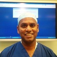
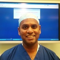
Mr Amir Sandiford - Orthopaedic Surgeon
Private Service, Orthopaedics
Today
8:30 AM to 3:30 PM.
Description
Mr Sandiford is a consultant Orthopaedic and Trauma surgeon based at the Joint Reconstruction Unit, Southland Hospital and a senior Clinical Lecturer at the University of Otago, New Zealand.
He specialises in primary and revision hip and knee reconstruction and prosthetic joint infection(PJI). He has published internationally in these fields.
He qualified from the University of the West Indies, Cave Hill Campus, Barbados in 2000. His basic training in Orthopaedics and Hip and Knee reconstruction was obtained in London, UK. This started under the mentorship of Ms Sarah Muirhead- Allwood. He then undertook specialist training at Guys and St Thomas’ Hospital , London followed by fellowship training in Vancouver, Exeter, Nijmegen and London.
He was a consultant surgeon at Guys and St Thomas’ Hospital and St Georges Hospitals in London, UK prior to moving to New Zealand.
He was awarded a Current Concepts in Joint Replacement Fellowship in 2014, a British Hip Society Travelling Fellowship in 2016 and the prestigious American British Canadian (ABC) Travelling Fellowship in 2018.
He was also awarded a Masters degree with Merit in Sports and Exercise Medicine from the University of Wales Institute, Cardiff and was recently awarded a Fellowship of the Royal College of Sport and Exercise Physicians of Ireland.
Mr Sandiford’s clinical and research interests have focused around hip and knee prosthesis design, Prosthetic Joint Infection and medical education. He has published widely on implant design, surgical techniques, clinical outcomes and return to function and sporting activity after joint replacement surgery.
Clinical Interests:
- Primary hip and knee reconstruction
- Complex hip and knee reconstruction
- Revision hip and knee reconstruction
- Periprosthetic fracture management
- Prosthetic joint infection
- Sports injuries
- Carpal Tunnel Surgery
Consultants
-

Mr Amir Sandiford
Orthopaedic Surgeon
How do I access this service?
Referral
Fees and Charges Categorisation
Fees apply
Fees and Charges Description
Amir is a Southern Cross Affiliated Provider and NIB first choice member.
Hours
8:30 AM to 3:30 PM.
| Mon | 8:30 AM – 3:30 PM |
|---|---|
| Tue | 8:00 AM – 5:00 PM |
| Wed – Thu | 8:30 AM – 3:30 PM |
Languages Spoken
English
Procedures / Treatments
For elderly patients joint replacement surgery is commonly required to treat damaged joints from wearing out, arthritis or other forms of joint disease including rheumatoid arthritis. In these procedures the damaged joint surface is removed and replaced with artificial surfaces normally made from metal (chromium cobalt alloy, titanium), plastic (high density polyethelene) or ceramic which act as alternate bearing surfaces for the damaged joint. These operations are major procedures which require the patient to be in hospital for several days and followed by a significant period of rehabilitation. The hospital has several ways of approaching the procedure for replacement and the specifics for the procedure will be covered at the time of assessment and booking of surgery. Occasionally blood transfusions are required; if you have some concerns raise this with your surgeon during consultation.
For elderly patients joint replacement surgery is commonly required to treat damaged joints from wearing out, arthritis or other forms of joint disease including rheumatoid arthritis. In these procedures the damaged joint surface is removed and replaced with artificial surfaces normally made from metal (chromium cobalt alloy, titanium), plastic (high density polyethelene) or ceramic which act as alternate bearing surfaces for the damaged joint. These operations are major procedures which require the patient to be in hospital for several days and followed by a significant period of rehabilitation. The hospital has several ways of approaching the procedure for replacement and the specifics for the procedure will be covered at the time of assessment and booking of surgery. Occasionally blood transfusions are required; if you have some concerns raise this with your surgeon during consultation.
For elderly patients joint replacement surgery is commonly required to treat damaged joints from wearing out, arthritis or other forms of joint disease including rheumatoid arthritis. In these procedures the damaged joint surface is removed and replaced with artificial surfaces normally made from metal (chromium cobalt alloy, titanium), plastic (high density polyethelene) or ceramic which act as alternate bearing surfaces for the damaged joint.
These operations are major procedures which require the patient to be in hospital for several days and followed by a significant period of rehabilitation. The hospital has several ways of approaching the procedure for replacement and the specifics for the procedure will be covered at the time of assessment and booking of surgery.
Occasionally blood transfusions are required; if you have some concerns raise this with your surgeon during consultation.
An incision (cut) is made on the side of the thigh to allow the surgeon access to the hip joint. The diseased and damaged parts of the hip joint are removed and replaced with smooth, artificial metal ‘ball’ and plastic ‘socket’ parts.
An incision (cut) is made on the side of the thigh to allow the surgeon access to the hip joint. The diseased and damaged parts of the hip joint are removed and replaced with smooth, artificial metal ‘ball’ and plastic ‘socket’ parts.
An incision (cut) is made on the side of the thigh to allow the surgeon access to the hip joint. The diseased and damaged parts of the hip joint are removed and replaced with smooth, artificial metal ‘ball’ and plastic ‘socket’ parts.
Knee surgery is an operation that helps fix problems in your knee, like an injury, arthritis, a torn ligament, or damaged cartilage. Surgery might be: Arthroscopic: less invasive surgery. The surgeon makes small cuts and uses a tiny camera to see inside the knee. They can then fix or take out the damaged parts. Open: for more complicated problems, the surgeon makes a larger cut to get a better look and fix the damaged parts directly. Joint Replacement: when the knee is badly damaged, the surgeon might remove the damaged areas and replace them with parts made of special materials.
Knee surgery is an operation that helps fix problems in your knee, like an injury, arthritis, a torn ligament, or damaged cartilage. Surgery might be: Arthroscopic: less invasive surgery. The surgeon makes small cuts and uses a tiny camera to see inside the knee. They can then fix or take out the damaged parts. Open: for more complicated problems, the surgeon makes a larger cut to get a better look and fix the damaged parts directly. Joint Replacement: when the knee is badly damaged, the surgeon might remove the damaged areas and replace them with parts made of special materials.
Knee surgery is an operation that helps fix problems in your knee, like an injury, arthritis, a torn ligament, or damaged cartilage. Surgery might be:
- Arthroscopic: less invasive surgery. The surgeon makes small cuts and uses a tiny camera to see inside the knee. They can then fix or take out the damaged parts.
- Open: for more complicated problems, the surgeon makes a larger cut to get a better look and fix the damaged parts directly.
- Joint Replacement: when the knee is badly damaged, the surgeon might remove the damaged areas and replace them with parts made of special materials.
This is a surgical procedure performed on a knee joint that has become painful and/or impaired because of disease, injury or wear and tear. In total knee replacement, artificial materials (metal and plastic) are used to replace the following damaged surfaces within the knee joint: the end of the thigh bone (femur) the end of the shin bone (tibia) the back of the kneecap (patella) This operation is a major procedure which requires you to be in hospital for several days and will be followed by a significant period of rehabilitation. Occasionally blood transfusions are required; if you have some concerns raise this with your surgeon during consultation. For more information about total knee replacement please click here.
This is a surgical procedure performed on a knee joint that has become painful and/or impaired because of disease, injury or wear and tear. In total knee replacement, artificial materials (metal and plastic) are used to replace the following damaged surfaces within the knee joint: the end of the thigh bone (femur) the end of the shin bone (tibia) the back of the kneecap (patella) This operation is a major procedure which requires you to be in hospital for several days and will be followed by a significant period of rehabilitation. Occasionally blood transfusions are required; if you have some concerns raise this with your surgeon during consultation. For more information about total knee replacement please click here.
This is a surgical procedure performed on a knee joint that has become painful and/or impaired because of disease, injury or wear and tear.
In total knee replacement, artificial materials (metal and plastic) are used to replace the following damaged surfaces within the knee joint:
- the end of the thigh bone (femur)
- the end of the shin bone (tibia)
- the back of the kneecap (patella)
This operation is a major procedure which requires you to be in hospital for several days and will be followed by a significant period of rehabilitation.
Occasionally blood transfusions are required; if you have some concerns raise this with your surgeon during consultation.
For more information about total knee replacement please click here.
The anterior cruciate ligament (ACL) is a strong, stabilising ligament running through the centre of the knee between the femur (thigh bone) and tibia (shin bone). When the ACL is torn, frequently as the result of a sporting injury, arthroscopic surgery known as ACL Reconstruction is performed. The procedure involves replacement of the damaged ligament with tissue grafted from elsewhere, usually the patellar or hamstring tendon. The ends of the grafted tendon are attached to the femur at one end and the tibia at the other using screws or staples. For more information about ACL reconstruction please click here.
The anterior cruciate ligament (ACL) is a strong, stabilising ligament running through the centre of the knee between the femur (thigh bone) and tibia (shin bone). When the ACL is torn, frequently as the result of a sporting injury, arthroscopic surgery known as ACL Reconstruction is performed. The procedure involves replacement of the damaged ligament with tissue grafted from elsewhere, usually the patellar or hamstring tendon. The ends of the grafted tendon are attached to the femur at one end and the tibia at the other using screws or staples. For more information about ACL reconstruction please click here.
The anterior cruciate ligament (ACL) is a strong, stabilising ligament running through the centre of the knee between the femur (thigh bone) and tibia (shin bone).
When the ACL is torn, frequently as the result of a sporting injury, arthroscopic surgery known as ACL Reconstruction is performed. The procedure involves replacement of the damaged ligament with tissue grafted from elsewhere, usually the patellar or hamstring tendon. The ends of the grafted tendon are attached to the femur at one end and the tibia at the other using screws or staples.
For more information about ACL reconstruction please click here.
A large number of orthopaedic procedures on joints are performed using an arthroscope, where a fibre optic telescope is used to look inside the joint. Through this type of keyhole surgery, fine instruments can be introduced through small incisions (portals) to allow surgery to be performed without the need for large cuts. This allows many procedures to be performed as a day stay and allows quicker return to normal function of the joint. Arthroscopic surgery is less painful than open surgery and decreases the risk of healing problems. Arthroscopy allows access to parts of the joints which can not be accessed by other types of surgery.
A large number of orthopaedic procedures on joints are performed using an arthroscope, where a fibre optic telescope is used to look inside the joint. Through this type of keyhole surgery, fine instruments can be introduced through small incisions (portals) to allow surgery to be performed without the need for large cuts. This allows many procedures to be performed as a day stay and allows quicker return to normal function of the joint. Arthroscopic surgery is less painful than open surgery and decreases the risk of healing problems. Arthroscopy allows access to parts of the joints which can not be accessed by other types of surgery.
A large number of orthopaedic procedures on joints are performed using an arthroscope, where a fibre optic telescope is used to look inside the joint. Through this type of keyhole surgery, fine instruments can be introduced through small incisions (portals) to allow surgery to be performed without the need for large cuts. This allows many procedures to be performed as a day stay and allows quicker return to normal function of the joint.
Arthroscopic surgery is less painful than open surgery and decreases the risk of healing problems. Arthroscopy allows access to parts of the joints which can not be accessed by other types of surgery.
The menisci are two circular strips of cartilage that form a cushioning layer between the ends of the femur (thigh bone) and tibia (shin bone) in the knee joint. Together the medial and lateral menisci, on the inside and outside of the knee, respectively, act as shock absorbers and distribute the weight of the body across the knee joint. The menisci can become torn through injury or damaged from age-related wear and tear and may require surgery. The most common meniscal surgery is partial meniscectomy in which the torn portion of the meniscus is cut away so that the cartilage surface is smooth again. In some cases meniscal repair is carried out, in this case the torn edges of the meniscus are sutured together. Both procedures are performed arthroscopically. For more information please click on the following link for meniscal tears and for meniscal transplant surgery.
The menisci are two circular strips of cartilage that form a cushioning layer between the ends of the femur (thigh bone) and tibia (shin bone) in the knee joint. Together the medial and lateral menisci, on the inside and outside of the knee, respectively, act as shock absorbers and distribute the weight of the body across the knee joint. The menisci can become torn through injury or damaged from age-related wear and tear and may require surgery. The most common meniscal surgery is partial meniscectomy in which the torn portion of the meniscus is cut away so that the cartilage surface is smooth again. In some cases meniscal repair is carried out, in this case the torn edges of the meniscus are sutured together. Both procedures are performed arthroscopically. For more information please click on the following link for meniscal tears and for meniscal transplant surgery.
The menisci are two circular strips of cartilage that form a cushioning layer between the ends of the femur (thigh bone) and tibia (shin bone) in the knee joint. Together the medial and lateral menisci, on the inside and outside of the knee, respectively, act as shock absorbers and distribute the weight of the body across the knee joint.
The menisci can become torn through injury or damaged from age-related wear and tear and may require surgery.
The most common meniscal surgery is partial meniscectomy in which the torn portion of the meniscus is cut away so that the cartilage surface is smooth again.
In some cases meniscal repair is carried out, in this case the torn edges of the meniscus are sutured together.
Both procedures are performed arthroscopically.
For more information please click on the following link for meniscal tears and for meniscal transplant surgery.
Carpal Tunnel Syndrome is caused by a pinched nerve in the wrist that causes tingling, numbness and pain in your hand. Surgery to relieve carpal tunnel syndrome involves making an incision (cut) from the middle of the palm of your hand to your wrist. Tissue that is pressing on the nerve is then cut to release the pressure. This operation is usually performed under local anaesthetic (the area being treated is numb but you are awake).
Carpal Tunnel Syndrome is caused by a pinched nerve in the wrist that causes tingling, numbness and pain in your hand. Surgery to relieve carpal tunnel syndrome involves making an incision (cut) from the middle of the palm of your hand to your wrist. Tissue that is pressing on the nerve is then cut to release the pressure. This operation is usually performed under local anaesthetic (the area being treated is numb but you are awake).
Carpal Tunnel Syndrome is caused by a pinched nerve in the wrist that causes tingling, numbness and pain in your hand.
Surgery to relieve carpal tunnel syndrome involves making an incision (cut) from the middle of the palm of your hand to your wrist. Tissue that is pressing on the nerve is then cut to release the pressure.
This operation is usually performed under local anaesthetic (the area being treated is numb but you are awake).
In many cases tendons will be lengthened to improve the muscle balance around a joint or tendons will be transferred to give overall better joint function. This occurs in children with neuromuscular conditions but also applies to a number of other conditions. Most of these procedures involve some sort of splintage after the surgery followed by a period of rehabilitation, normally supervised by a physiotherapist.
In many cases tendons will be lengthened to improve the muscle balance around a joint or tendons will be transferred to give overall better joint function. This occurs in children with neuromuscular conditions but also applies to a number of other conditions. Most of these procedures involve some sort of splintage after the surgery followed by a period of rehabilitation, normally supervised by a physiotherapist.
In many cases tendons will be lengthened to improve the muscle balance around a joint or tendons will be transferred to give overall better joint function. This occurs in children with neuromuscular conditions but also applies to a number of other conditions.
Most of these procedures involve some sort of splintage after the surgery followed by a period of rehabilitation, normally supervised by a physiotherapist.
This procedure is used when osteoarthritic damage to the cartilage on one side of the knee has caused the angle of the knee joint to change so that most of the body's weight is borne by the affected side, adding to the wear on that side. High Tibial Osteotomy involves reshaping and realignment of the bone so that weight becomes more evenly distributed between the inside and outside of the knee, thereby reducing the workload on the damaged side. You will probably have to stay in hospital for several days after surgery followed by up to 6 months rehabilitation. For more information about osteotomy please click here.
This procedure is used when osteoarthritic damage to the cartilage on one side of the knee has caused the angle of the knee joint to change so that most of the body's weight is borne by the affected side, adding to the wear on that side. High Tibial Osteotomy involves reshaping and realignment of the bone so that weight becomes more evenly distributed between the inside and outside of the knee, thereby reducing the workload on the damaged side. You will probably have to stay in hospital for several days after surgery followed by up to 6 months rehabilitation. For more information about osteotomy please click here.
This procedure is used when osteoarthritic damage to the cartilage on one side of the knee has caused the angle of the knee joint to change so that most of the body's weight is borne by the affected side, adding to the wear on that side.
High Tibial Osteotomy involves reshaping and realignment of the bone so that weight becomes more evenly distributed between the inside and outside of the knee, thereby reducing the workload on the damaged side.
You will probably have to stay in hospital for several days after surgery followed by up to 6 months rehabilitation.
For more information about osteotomy please click here.
Osteotomy is the division of a crooked or bent bone to improve alignment of the limb. These procedures normally involve some form of internal fixation, such as rods or plates, or external fixation which involves external wires and pins to hold the bone. The type of procedure for fixation will be explained when the surgery is planned.
Osteotomy is the division of a crooked or bent bone to improve alignment of the limb. These procedures normally involve some form of internal fixation, such as rods or plates, or external fixation which involves external wires and pins to hold the bone. The type of procedure for fixation will be explained when the surgery is planned.
Osteotomy is the division of a crooked or bent bone to improve alignment of the limb.
These procedures normally involve some form of internal fixation, such as rods or plates, or external fixation which involves external wires and pins to hold the bone. The type of procedure for fixation will be explained when the surgery is planned.
Disability Assistance
Wheelchair access, Wheelchair accessible toilet, Mobility parking space
Additional Details
Face to face / Kanohi ki te Kanohi, Phone
Parking
Free patient parking is provided
Pharmacy
Find your nearest pharmacy here
Contact Details
223 Spey St, Invercargill
Southland
8:30 AM to 3:30 PM.
-
Phone
(03) 218 4077
Healthlink EDI
sandifor
Email
Website
223 Spey Street
Invercargill
Southland 9810
Street Address
223 Spey Street
Invercargill
Southland 9810
Was this page helpful?
This page was last updated at 10:25AM on October 8, 2025. This information is reviewed and edited by Mr Amir Sandiford - Orthopaedic Surgeon.

