Central Auckland, East Auckland, North Auckland, South Auckland, West Auckland > Private Hospitals & Specialists >
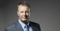
Mr Michael Hanlon - Knee & Musculoskeletal Tumour Surgeon
Private Service, Orthopaedics
Description
- Sports related injury and associated surgery
- Arthroscopic meniscal surgery
- Arthroscopic cartilage surgery
- Ligament reconstructive surgery
- Surgical management of degenerative disorders of the knee
- Re-alignment osteotomy surgery
- Partial knee replacement
- Primary total knee replacement
- Complex knee revision surgery encompassing major bone loss, and deformity surgery
- Surgical management of musculoskeletal tumours
- Benign and malignant bone tumours
- Benign and malignant soft tissue tumours
Please visit the Domain Orthopaedics website to read more.
Consultants
-

Mr Michael Hanlon
Knee & Musculoskeletal Tumour Surgeon
Referral Expectations
You need to bring with you to your appointment:
Hours
Please contact Marlene at my reception to arrange a convenient appointment.
I hold my clinics at the following times:
- 20 Titoki Street, Parnell: all day on every other Monday; every Thursday and Friday afternoon.
- Silverdale Medical Centre: Fridays fortnightly
Procedures / Treatments
Knee surgery is an operation that helps fix problems in your knee, like an injury, arthritis, a torn ligament, or damaged cartilage. Surgery might be: Arthroscopic: less invasive surgery. The surgeon makes small cuts and uses a tiny camera to see inside the knee. They can then fix or take out the damaged parts. Open: for more complicated problems, the surgeon makes a larger cut to get a better look and fix the damaged parts directly. Joint Replacement: when the knee is badly damaged, the surgeon might remove the damaged areas and replace them with parts made of special materials.
Knee surgery is an operation that helps fix problems in your knee, like an injury, arthritis, a torn ligament, or damaged cartilage. Surgery might be: Arthroscopic: less invasive surgery. The surgeon makes small cuts and uses a tiny camera to see inside the knee. They can then fix or take out the damaged parts. Open: for more complicated problems, the surgeon makes a larger cut to get a better look and fix the damaged parts directly. Joint Replacement: when the knee is badly damaged, the surgeon might remove the damaged areas and replace them with parts made of special materials.
Knee surgery is an operation that helps fix problems in your knee, like an injury, arthritis, a torn ligament, or damaged cartilage. Surgery might be:
- Arthroscopic: less invasive surgery. The surgeon makes small cuts and uses a tiny camera to see inside the knee. They can then fix or take out the damaged parts.
- Open: for more complicated problems, the surgeon makes a larger cut to get a better look and fix the damaged parts directly.
- Joint Replacement: when the knee is badly damaged, the surgeon might remove the damaged areas and replace them with parts made of special materials.
This is a surgical procedure performed on a knee joint that has become painful and/or impaired because of disease, injury or wear and tear. In total knee replacement, artificial materials (metal and plastic) are used to replace the following damaged surfaces within the knee joint: the end of the thigh bone (femur) the end of the shin bone (tibia) the back of the kneecap (patella) This operation is a major procedure which requires you to be in hospital for several days and will be followed by a significant period of rehabilitation. Occasionally blood transfusions are required; if you have some concerns raise this with your surgeon during consultation. For more we reccommend the AAOS total knee replacement website information
This is a surgical procedure performed on a knee joint that has become painful and/or impaired because of disease, injury or wear and tear. In total knee replacement, artificial materials (metal and plastic) are used to replace the following damaged surfaces within the knee joint: the end of the thigh bone (femur) the end of the shin bone (tibia) the back of the kneecap (patella) This operation is a major procedure which requires you to be in hospital for several days and will be followed by a significant period of rehabilitation. Occasionally blood transfusions are required; if you have some concerns raise this with your surgeon during consultation. For more we reccommend the AAOS total knee replacement website information
This is a surgical procedure performed on a knee joint that has become painful and/or impaired because of disease, injury or wear and tear.
In total knee replacement, artificial materials (metal and plastic) are used to replace the following damaged surfaces within the knee joint:
- the end of the thigh bone (femur)
- the end of the shin bone (tibia)
- the back of the kneecap (patella)
This operation is a major procedure which requires you to be in hospital for several days and will be followed by a significant period of rehabilitation.
Occasionally blood transfusions are required; if you have some concerns raise this with your surgeon during consultation.
For more we reccommend the AAOS total knee replacement website information
The anterior cruciate ligament (ACL) is a strong, stabilising ligament running through the centre of the knee between the femur (thigh bone) and tibia (shin bone). When the ACL is torn, frequently as the result of a sporting injury, arthroscopic surgery known as ACL Reconstruction is performed. The procedure involves replacement of the damaged ligament with tissue grafted from elsewhere, usually the patellar or hamstring tendon. The ends of the grafted tendon are attached to the femur at one end and the tibia at the other using screws or staples. For more we reccommend the AAOS ACL reconstruction website information.
The anterior cruciate ligament (ACL) is a strong, stabilising ligament running through the centre of the knee between the femur (thigh bone) and tibia (shin bone). When the ACL is torn, frequently as the result of a sporting injury, arthroscopic surgery known as ACL Reconstruction is performed. The procedure involves replacement of the damaged ligament with tissue grafted from elsewhere, usually the patellar or hamstring tendon. The ends of the grafted tendon are attached to the femur at one end and the tibia at the other using screws or staples. For more we reccommend the AAOS ACL reconstruction website information.
The anterior cruciate ligament (ACL) is a strong, stabilising ligament running through the centre of the knee between the femur (thigh bone) and tibia (shin bone).
When the ACL is torn, frequently as the result of a sporting injury, arthroscopic surgery known as ACL Reconstruction is performed. The procedure involves replacement of the damaged ligament with tissue grafted from elsewhere, usually the patellar or hamstring tendon. The ends of the grafted tendon are attached to the femur at one end and the tibia at the other using screws or staples.
For more we reccommend the AAOS ACL reconstruction website information.
This procedure is used when osteoarthritic damage to the cartilage on one side of the knee has caused the angle of the knee joint to change so that most of the body's weight is borne by the affected side, adding to the wear on that side. High Tibial Osteotomy involves reshaping and realignment of the bone so that weight becomes more evenly distributed between the inside and outside of the knee, thereby reducing the workload on the damaged side. You will probably have to stay in hospital for several days after surgery followed by up to 6 months rehabilitation. For more we reccommend the AAOS osteotomy website information
This procedure is used when osteoarthritic damage to the cartilage on one side of the knee has caused the angle of the knee joint to change so that most of the body's weight is borne by the affected side, adding to the wear on that side. High Tibial Osteotomy involves reshaping and realignment of the bone so that weight becomes more evenly distributed between the inside and outside of the knee, thereby reducing the workload on the damaged side. You will probably have to stay in hospital for several days after surgery followed by up to 6 months rehabilitation. For more we reccommend the AAOS osteotomy website information
This procedure is used when osteoarthritic damage to the cartilage on one side of the knee has caused the angle of the knee joint to change so that most of the body's weight is borne by the affected side, adding to the wear on that side.
High Tibial Osteotomy involves reshaping and realignment of the bone so that weight becomes more evenly distributed between the inside and outside of the knee, thereby reducing the workload on the damaged side.
You will probably have to stay in hospital for several days after surgery followed by up to 6 months rehabilitation.
For more we reccommend the AAOS osteotomy website information
A large number of orthopaedic procedures on joints are performed using an arthroscope, where a fibre optic telescope is used to look inside the joint. Through this type of keyhole surgery, fine instruments can be introduced through small incisions (portals) to allow surgery to be performed without the need for large cuts. This allows many procedures to be performed as a day stay and allows quicker return to normal function of the joint. Arthroscopic surgery is less painful than open surgery and decreases the risk of healing problems. Arthroscopy allows access to parts of the joints which can not be accessed by other types of surgery. For more we reccommend the AAOS arthroscopy website information
A large number of orthopaedic procedures on joints are performed using an arthroscope, where a fibre optic telescope is used to look inside the joint. Through this type of keyhole surgery, fine instruments can be introduced through small incisions (portals) to allow surgery to be performed without the need for large cuts. This allows many procedures to be performed as a day stay and allows quicker return to normal function of the joint. Arthroscopic surgery is less painful than open surgery and decreases the risk of healing problems. Arthroscopy allows access to parts of the joints which can not be accessed by other types of surgery. For more we reccommend the AAOS arthroscopy website information
A large number of orthopaedic procedures on joints are performed using an arthroscope, where a fibre optic telescope is used to look inside the joint. Through this type of keyhole surgery, fine instruments can be introduced through small incisions (portals) to allow surgery to be performed without the need for large cuts. This allows many procedures to be performed as a day stay and allows quicker return to normal function of the joint.
Arthroscopic surgery is less painful than open surgery and decreases the risk of healing problems. Arthroscopy allows access to parts of the joints which can not be accessed by other types of surgery.
For more we reccommend the AAOS arthroscopy website information
The menisci are two circular strips of cartilage that form a cushioning layer between the ends of the femur (thigh bone) and tibia (shin bone) in the knee joint. Together the medial and lateral menisci, on the inside and outside of the knee, respectively, act as shock absorbers and distribute the weight of the body across the knee joint. The menisci can become torn through injury or damaged from age-related wear and tear and may require surgery. The most common meniscal surgery is partial meniscectomy in which the torn portion of the meniscus is cut away so that the cartilage surface is smooth again. In some cases meniscal repair is carried out, in this case the torn edges of the meniscus are sutured together. Both procedures are performed arthroscopically. For more information please click on the following link for meniscal tears and for meniscal transplant surgery.
The menisci are two circular strips of cartilage that form a cushioning layer between the ends of the femur (thigh bone) and tibia (shin bone) in the knee joint. Together the medial and lateral menisci, on the inside and outside of the knee, respectively, act as shock absorbers and distribute the weight of the body across the knee joint. The menisci can become torn through injury or damaged from age-related wear and tear and may require surgery. The most common meniscal surgery is partial meniscectomy in which the torn portion of the meniscus is cut away so that the cartilage surface is smooth again. In some cases meniscal repair is carried out, in this case the torn edges of the meniscus are sutured together. Both procedures are performed arthroscopically. For more information please click on the following link for meniscal tears and for meniscal transplant surgery.
The menisci are two circular strips of cartilage that form a cushioning layer between the ends of the femur (thigh bone) and tibia (shin bone) in the knee joint. Together the medial and lateral menisci, on the inside and outside of the knee, respectively, act as shock absorbers and distribute the weight of the body across the knee joint.
The menisci can become torn through injury or damaged from age-related wear and tear and may require surgery.
The most common meniscal surgery is partial meniscectomy in which the torn portion of the meniscus is cut away so that the cartilage surface is smooth again.
In some cases meniscal repair is carried out, in this case the torn edges of the meniscus are sutured together.
Both procedures are performed arthroscopically.
For more information please click on the following link for meniscal tears and for meniscal transplant surgery.
Management of both benign and malignant bone and soft tissue tumours consisting of local and systemic staging, biopsy and definitive surgical management of these conditions in conjunction with limb salvage techniques.
Management of both benign and malignant bone and soft tissue tumours consisting of local and systemic staging, biopsy and definitive surgical management of these conditions in conjunction with limb salvage techniques.
Service types: Musculoskeletal tumours.
Management of both benign and malignant bone and soft tissue tumours consisting of local and systemic staging, biopsy and definitive surgical management of these conditions in conjunction with limb salvage techniques.
Public Transport
The Auckland Transport Journey Planner will help you to plan your journey.
Parking
There is free dedicated off street underground parking at the clinic for my patients. Entrance to the right hand side of the building.
Website
Contact Details
20 Titoki Street, Parnell, Auckland
Central Auckland
-
Phone
(09) 307 4283
-
Fax
(09) 307 4280
Healthlink EDI
domainor
Email
Website
Domain Orthopaedics
20 Titoki Street
Parnell
Auckland
Street Address
Domain Orthopaedics
20 Titoki Street
Parnell
Auckland
Postal Address
Domain Orthopaedics
20 Titoki Street
Parnell
Auckland
Silverdale Medical Centre, 7 Polarity Rise, Silverdale, Auckland
North Auckland
-
Phone
(09) 307 4283
-
Fax
(09) 307 4280
Healthlink EDI
domainor
Email
Website
Was this page helpful?
This page was last updated at 2:16PM on November 19, 2025. This information is reviewed and edited by Mr Michael Hanlon - Knee & Musculoskeletal Tumour Surgeon.

