Central Auckland, East Auckland, South Auckland > Private Hospitals & Specialists >
Ormiston Hospital Orthopaedic Surgery
Private Surgical Service, Orthopaedics
Description
Orthopaedic Surgery is the branch of surgery which concerns conditions involving the musculoskeletal system.
Consultants
-

Mr Jarome Bentley
Orthopaedic Surgeon
-
Mr Shaneel Deo
Orthopaedic Surgeon
-

Mr Shihab Faraj
Orthopaedic Surgeon
-
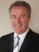
Mr Gary French
Orthopaedic Surgeon
-

Mr Nick Gormack
Orthopaedic Surgeon
-

Mr Wolfgang Heiss-Dunlop
Orthopaedic Surgeon
-

Mr Phillip Insull
Orthopaedic Surgeon
-

Mr Simon Manners
Orthopaedic Surgeon
-

Mr John Mutu-Grigg
Orthopaedic Surgeon
-

Mr Chris Ngar
Orthopaedic Surgeon
-

Dr Melissa Rossaak
Orthopaedic Surgeon
-
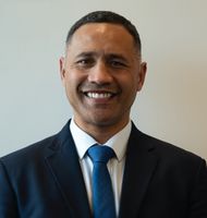
Mr Jeremy Stanley
Orthopaedic Surgeon
-

Mr Chris Taylor
Orthopaedic Surgeon
Procedures / Treatments
Two or three small incisions (cuts) are made in the ankle and a small telescopic instrument with a tiny camera attached (arthroscope) is inserted. This allows the surgeon to look inside the joint, identify problems and, in some cases, operate. Tiny instruments can be passed through the arthroscope to remove bony spurs, damaged cartilage or inflamed tissue.
Two or three small incisions (cuts) are made in the ankle and a small telescopic instrument with a tiny camera attached (arthroscope) is inserted. This allows the surgeon to look inside the joint, identify problems and, in some cases, operate. Tiny instruments can be passed through the arthroscope to remove bony spurs, damaged cartilage or inflamed tissue.
An incision (cut) is made in the front of, and several smaller cuts on the outside of, the ankle. The damaged ankle joint is replaced with a metal and plastic implant.
An incision (cut) is made in the front of, and several smaller cuts on the outside of, the ankle. The damaged ankle joint is replaced with a metal and plastic implant.
Surgery to relieve carpal tunnel syndrome involves making an incision (cut) from the middle of the palm of your hand to your wrist. Tissue that is pressing on the nerve is then cut to release the pressure.
Surgery to relieve carpal tunnel syndrome involves making an incision (cut) from the middle of the palm of your hand to your wrist. Tissue that is pressing on the nerve is then cut to release the pressure.
An incision (cut) is made over the relevant part of the spine and the bulging part of the painful disc is cut off and removed.
An incision (cut) is made over the relevant part of the spine and the bulging part of the painful disc is cut off and removed.
Small incisions (cuts) are made in the hip area and a small telescopic instrument with a tiny camera attached (arthroscope) is inserted. This allows the surgeon to look inside the joint, identify problems and, in some cases, operate. Tiny instruments can be passed through the arthroscope to remove loose, damaged or inflamed tissue.
Small incisions (cuts) are made in the hip area and a small telescopic instrument with a tiny camera attached (arthroscope) is inserted. This allows the surgeon to look inside the joint, identify problems and, in some cases, operate. Tiny instruments can be passed through the arthroscope to remove loose, damaged or inflamed tissue.
An incision (cut) is made on the side of the thigh to allow the surgeon access to the hip joint. The diseased and damaged parts of the hip joint are removed and replaced with smooth, artificial metal ‘ball’ and plastic ‘socket’ parts.
An incision (cut) is made on the side of the thigh to allow the surgeon access to the hip joint. The diseased and damaged parts of the hip joint are removed and replaced with smooth, artificial metal ‘ball’ and plastic ‘socket’ parts.
Several small incisions (cuts) are made on the knee through which is inserted a small telescopic instrument with a tiny camera attached (arthroscope). This allows the surgeon to look inside the joint, identify problems and, in some cases, make repairs to damaged tissue.
Several small incisions (cuts) are made on the knee through which is inserted a small telescopic instrument with a tiny camera attached (arthroscope). This allows the surgeon to look inside the joint, identify problems and, in some cases, make repairs to damaged tissue.
An incision (cut) is made on the front of the knee to allow the surgeon access to the knee joint. The damaged and painful areas of the thigh bone (femur) and lower leg bone (tibia), including the knee joint, are removed and replaced with metal and plastic parts.
An incision (cut) is made on the front of the knee to allow the surgeon access to the knee joint. The damaged and painful areas of the thigh bone (femur) and lower leg bone (tibia), including the knee joint, are removed and replaced with metal and plastic parts.
Several small incisions (cuts) are made in the shoulder through which is inserted a small telescopic instrument with a tiny camera attached (arthroscope). The surgeon is then able to remove any bony spurs or inflamed tissue and mend torn tendons of the rotator cuff group.
Several small incisions (cuts) are made in the shoulder through which is inserted a small telescopic instrument with a tiny camera attached (arthroscope). The surgeon is then able to remove any bony spurs or inflamed tissue and mend torn tendons of the rotator cuff group.
This surgery involves making several small incisions (cuts) on the shoulder through which is inserted a small telescopic instrument with a tiny camera attached (arthroscope). This allows the surgeon to look inside the shoulder, identify problems and, in some cases, make repairs to damaged tissue.
This surgery involves making several small incisions (cuts) on the shoulder through which is inserted a small telescopic instrument with a tiny camera attached (arthroscope). This allows the surgeon to look inside the shoulder, identify problems and, in some cases, make repairs to damaged tissue.
An incision (cut) is made over the relevant part of the spine. Two or more vertebrae (the small bones that make up the spinal column) are fused together with bone grafts and/or metal rods to form a single bone.
An incision (cut) is made over the relevant part of the spine. Two or more vertebrae (the small bones that make up the spinal column) are fused together with bone grafts and/or metal rods to form a single bone.
An incision (cut) is made over the damaged tendon. The damaged ends of the tendon are sewn together and, if necessary, reattached to surrounding tissue.
An incision (cut) is made over the damaged tendon. The damaged ends of the tendon are sewn together and, if necessary, reattached to surrounding tissue.
Other
Website
Contact Details
Ormiston Hospital, 125 Ormiston Road, Flat Bush, Auckland
South Auckland
-
Phone
(09) 250 1157
Email
Website
125 Ormiston Road
Flat Bush
Auckland 2016
Street Address
125 Ormiston Road
Flat Bush
Auckland 2016
Postal Address
PO Box 38921
Howick
Auckland 2145
Was this page helpful?
This page was last updated at 12:09PM on August 1, 2023. This information is reviewed and edited by Ormiston Hospital Orthopaedic Surgery.

