Central Auckland > Private Hospitals & Specialists > Southern Cross Hospitals >
Southern Cross Brightside Hospital - Orthopaedic Surgery
Private Surgical Service, Orthopaedics
Description
Southern Cross' Brightside Hospital is a modern private surgical hospital located in central Auckland.
Brightside Hospital was originally established on its Auckland site, in Epsom, in the 1940s and was completely rebuilt to modern, high tech specifications and re-opened in 2000.
Brightside offers a wide range of surgical services and postoperative care to around 4,500 patients each year. This four-theatre hospital provides 43 inpatient beds and 6 day stay chairs and has a staff of nearly 100.
With an established reputation for gynaecological, orthopaedic and urological services, Brightside was also the first private hospital in New Zealand to offer prostate brachytherapy, a new type of treatment for prostate cancer. This specialist service employs advanced techniques and technologies for prostate implants, and the medical specialists operating at the hospital have helped Brightside to be involved in leading the way in urological treatment in New Zealand in some respects.
Plastic surgery, oral surgery and general surgery are also amongst the surgical services offered at Brightside Hospital.
Recent developments include the introduction of an Intermediate Care Facility (ICF), opened in 2012 and providing a specialised postoperative ward for patients with medical conditions or surgical issues that require an enhanced level of monitoring and care. The four-bed ward caters for patients who unexpectedly require some extra post-surgical care, or those with medical issues - such as heart or blood pressure conditions - that may have the potential to cause complications during or after surgery.
Consultants
-

Dr Charlotte Allen
Orthopaedic Surgeon
-

Mr Craig Ball
Orthopaedic Surgeon
-
Mr Michael Caughey
Orthopaedic Surgeon
-

Mr Clayton Chan
Orthopaedic Surgeon
-

Mr Brendan Coleman
Orthopaedic Surgeon
-
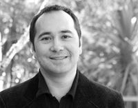
Mr Adam Dalgleish
Orthopaedic Surgeon
-

Mr Tony Danesh-Clough
Orthopaedic Surgeon
-
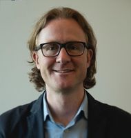
Mr Matt Debenham
Orthopaedic Surgeon
-
Mr Shaneel Deo
Orthopaedic Surgeon
-
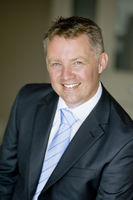
Mr Adam Durrant
Orthopaedic Surgeon
-

Mr John English
Orthopaedic Surgeon
-

Mr Shihab Faraj
Orthopaedic Surgeon
-

Mr Michael Flint
Orthopaedic Surgeon
-
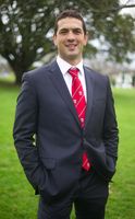
Mr Chris Fougere
Orthopaedic Surgeon
-
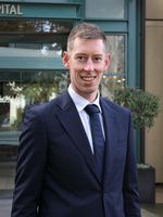
Mr Nick Gormack
Orthopaedic Surgeon
-
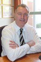
Mr Alastair Hadlow
Orthopaedic Surgeon
-
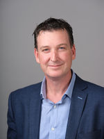
Mr Peter Hucker
Orthopaedic Surgeon
-

Mr Kevin Karpik
Orthopaedic Surgeon
-

Mr Simon Manners
Orthopaedic Surgeon
-
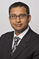
Mr Dean Mistry
Orthopaedic Surgeon
-
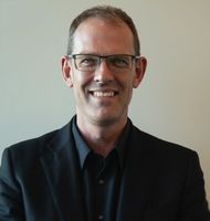
Mr Michael Rosenfeldt
Orthopaedic Surgeon
-
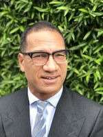
Mr Janus Schaumkel
Orthopaedic Surgeon
Procedures / Treatments
Two or three small incisions (cuts) are made in the ankle and a small telescopic instrument with a tiny camera attached (arthroscope) is inserted. This allows the surgeon to look inside the joint, identify problems and, in some cases, operate. Tiny instruments can be passed through the arthroscope to remove bony spurs, damaged cartilage or inflamed tissue.
Two or three small incisions (cuts) are made in the ankle and a small telescopic instrument with a tiny camera attached (arthroscope) is inserted. This allows the surgeon to look inside the joint, identify problems and, in some cases, operate. Tiny instruments can be passed through the arthroscope to remove bony spurs, damaged cartilage or inflamed tissue.
Two or three small incisions (cuts) are made in the ankle and a small telescopic instrument with a tiny camera attached (arthroscope) is inserted. This allows the surgeon to look inside the joint, identify problems and, in some cases, operate. Tiny instruments can be passed through the arthroscope to remove bony spurs, damaged cartilage or inflamed tissue.
An incision (cut) is made in the front of, and several smaller cuts on the outside of, the ankle. The damaged ankle joint is replaced with a metal and plastic implant.
An incision (cut) is made in the front of, and several smaller cuts on the outside of, the ankle. The damaged ankle joint is replaced with a metal and plastic implant.
An incision (cut) is made in the front of, and several smaller cuts on the outside of, the ankle. The damaged ankle joint is replaced with a metal and plastic implant.
Carpal Tunnel Syndrome is caused by a pinched nerve in the wrist that causes tingling, numbness and pain in your hand. Surgery to relieve carpal tunnel syndrome involves making an incision (cut) from the middle of the palm of your hand to your wrist. Tissue that is pressing on the nerve is then cut to release the pressure.
Carpal Tunnel Syndrome is caused by a pinched nerve in the wrist that causes tingling, numbness and pain in your hand. Surgery to relieve carpal tunnel syndrome involves making an incision (cut) from the middle of the palm of your hand to your wrist. Tissue that is pressing on the nerve is then cut to release the pressure.
Carpal Tunnel Syndrome is caused by a pinched nerve in the wrist that causes tingling, numbness and pain in your hand.
Surgery to relieve carpal tunnel syndrome involves making an incision (cut) from the middle of the palm of your hand to your wrist. Tissue that is pressing on the nerve is then cut to release the pressure.
Discectomy is an operation to remove part or all of a damaged spinal disc that is pressing on nerves, helping to relieve pain and improve movement. Microdiscectomy:a microscope is used by the surgeon to guide tiny instruments to remove the disc or disc fragments.
Discectomy is an operation to remove part or all of a damaged spinal disc that is pressing on nerves, helping to relieve pain and improve movement. Microdiscectomy:a microscope is used by the surgeon to guide tiny instruments to remove the disc or disc fragments.
Discectomy is an operation to remove part or all of a damaged spinal disc that is pressing on nerves, helping to relieve pain and improve movement.
Microdiscectomy:a microscope is used by the surgeon to guide tiny instruments to remove the disc or disc fragments.
Small incisions (cuts) are made in the hip area and a small telescopic instrument with a tiny camera attached (arthroscope) is inserted. This allows the surgeon to look inside the joint, identify problems and, in some cases, operate. Tiny instruments can be passed through the arthroscope to remove loose, damaged or inflamed tissue.
Small incisions (cuts) are made in the hip area and a small telescopic instrument with a tiny camera attached (arthroscope) is inserted. This allows the surgeon to look inside the joint, identify problems and, in some cases, operate. Tiny instruments can be passed through the arthroscope to remove loose, damaged or inflamed tissue.
Small incisions (cuts) are made in the hip area and a small telescopic instrument with a tiny camera attached (arthroscope) is inserted. This allows the surgeon to look inside the joint, identify problems and, in some cases, operate. Tiny instruments can be passed through the arthroscope to remove loose, damaged or inflamed tissue.
An incision (cut) is made on the side of the thigh to allow the surgeon access to the hip joint. The diseased and damaged parts of the hip joint are removed and replaced with smooth, artificial metal ‘ball’ and plastic ‘socket’ parts.
An incision (cut) is made on the side of the thigh to allow the surgeon access to the hip joint. The diseased and damaged parts of the hip joint are removed and replaced with smooth, artificial metal ‘ball’ and plastic ‘socket’ parts.
An incision (cut) is made on the side of the thigh to allow the surgeon access to the hip joint. The diseased and damaged parts of the hip joint are removed and replaced with smooth, artificial metal ‘ball’ and plastic ‘socket’ parts.
Several small incisions (cuts) are made on the knee through which is inserted a small telescopic instrument with a tiny camera attached (arthroscope). This allows the surgeon to look inside the joint, identify problems and, in some cases, make repairs to damaged tissue.
Several small incisions (cuts) are made on the knee through which is inserted a small telescopic instrument with a tiny camera attached (arthroscope). This allows the surgeon to look inside the joint, identify problems and, in some cases, make repairs to damaged tissue.
Several small incisions (cuts) are made on the knee through which is inserted a small telescopic instrument with a tiny camera attached (arthroscope). This allows the surgeon to look inside the joint, identify problems and, in some cases, make repairs to damaged tissue.
An incision (cut) is made on the front of the knee to allow the surgeon access to the knee joint. The damaged and painful areas of the thigh bone (femur) and lower leg bone (tibia), including the knee joint, are removed and replaced with metal and plastic parts.
An incision (cut) is made on the front of the knee to allow the surgeon access to the knee joint. The damaged and painful areas of the thigh bone (femur) and lower leg bone (tibia), including the knee joint, are removed and replaced with metal and plastic parts.
An incision (cut) is made on the front of the knee to allow the surgeon access to the knee joint. The damaged and painful areas of the thigh bone (femur) and lower leg bone (tibia), including the knee joint, are removed and replaced with metal and plastic parts.
Several small incisions (cuts) are made in the shoulder through which is inserted a small telescopic instrument with a tiny camera attached (arthroscope). The surgeon is then able to remove any bony spurs or inflamed tissue and mend torn tendons of the rotator cuff group.
Several small incisions (cuts) are made in the shoulder through which is inserted a small telescopic instrument with a tiny camera attached (arthroscope). The surgeon is then able to remove any bony spurs or inflamed tissue and mend torn tendons of the rotator cuff group.
Several small incisions (cuts) are made in the shoulder through which is inserted a small telescopic instrument with a tiny camera attached (arthroscope). The surgeon is then able to remove any bony spurs or inflamed tissue and mend torn tendons of the rotator cuff group.
This surgery involves making several small incisions (cuts) on the shoulder through which is inserted a small telescopic instrument with a tiny camera attached (arthroscope). This allows the surgeon to look inside the shoulder, identify problems and, in some cases, make repairs to damaged tissue.
This surgery involves making several small incisions (cuts) on the shoulder through which is inserted a small telescopic instrument with a tiny camera attached (arthroscope). This allows the surgeon to look inside the shoulder, identify problems and, in some cases, make repairs to damaged tissue.
This surgery involves making several small incisions (cuts) on the shoulder through which is inserted a small telescopic instrument with a tiny camera attached (arthroscope). This allows the surgeon to look inside the shoulder, identify problems and, in some cases, make repairs to damaged tissue.
An incision (cut) is made over the relevant part of the spine. Two or more vertebrae (the small bones that make up the spinal column) are fused together with bone grafts and/or metal rods to form a single bone.
An incision (cut) is made over the relevant part of the spine. Two or more vertebrae (the small bones that make up the spinal column) are fused together with bone grafts and/or metal rods to form a single bone.
An incision (cut) is made over the relevant part of the spine. Two or more vertebrae (the small bones that make up the spinal column) are fused together with bone grafts and/or metal rods to form a single bone.
An incision (cut) is made over the damaged tendon. The damaged ends of the tendon are sewn together and, if necessary, reattached to surrounding tissue.
An incision (cut) is made over the damaged tendon. The damaged ends of the tendon are sewn together and, if necessary, reattached to surrounding tissue.
An incision (cut) is made over the damaged tendon. The damaged ends of the tendon are sewn together and, if necessary, reattached to surrounding tissue.
Visiting Hours
- Weekdays: 11:00 to 20:00
- Weekends: 11:00 to 20:00
Parking
Free visitor parking is provided at the hospital.
Contact Details
Southern Cross Brightside Hospital
Central Auckland
-
Phone
(09) 925 4200
Email
Website
Surgeons can be contacted at their private consultation rooms.
Street Address
3 Brightside Road
Epsom
Auckland 1023
Postal Address
PO Box 26064
Epsom
Auckland 1344
Was this page helpful?
This page was last updated at 11:41AM on April 23, 2025. This information is reviewed and edited by Southern Cross Brightside Hospital - Orthopaedic Surgery.


