Waikato > Private Hospitals & Specialists > Southern Cross Hospitals >
Southern Cross Hamilton Hospital - Orthopaedic Surgery
Private Surgical Service, Orthopaedics, Spinal
Description
Situated in a quiet, central part of Hamilton, Southern Cross Hospital has been frequently and extensively modernised. Our hospital currently has 8 fully equipped operating theatres, offering the latest technology, as well as modern day-stay facilities and 60 ensuited patient rooms for those staying overnight. Also on site, we have a purpose-built six bed intensive care unit (ICU), high dependency care and access to radiology services.
Consultants
-

Mr Joe Baker
Orthopaedic Spine Surgeon
-
Mr Peter Black
Orthopaedic Surgeon
-
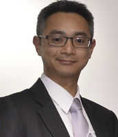
Mr Godwin Choy
Orthopaedic Surgeon
-
Mr Hamish Deverall
Orthopaedic Surgeon
-

Dr Reinhold Gregor
Orthopaedic Surgeon
-

Mr Satyen Jesani
Orthopaedic Surgeon
-

Dr Jillian Lee
Orthopaedic Surgeon
-

Mr Jonathan Lescheid
Orthopaedic Surgeon
-

Mr Anthony Maher
Orthopaedic Surgeon
-

Dr May Mak
Orthopaedic Surgeon
-
Mr Stephen McChesney
Orthopaedic Surgeon
-
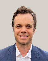
Mr Stephen McGrath
Orthopaedic Surgeon
-

Mr Sandeep Patel
Orthopaedic Surgeon
-

Mr Earle Savage
Orthopaedic Surgeon
-

Mr Richard Somerville
Orthopaedic Surgeon
-

Mr Richard Willoughby
Orthopaedic Surgeon
Procedures / Treatments
The anterior cruciate ligament (ACL) is a strong, stabilising ligament running through the centre of the knee between the femur (thigh bone) and tibia (shin bone). When the ACL is torn, frequently as the result of a sporting injury, arthroscopic surgery known as ACL Reconstruction is performed. The procedure involves replacement of the damaged ligament with tissue grafted from elsewhere, usually the patellar or hamstring tendon. The ends of the grafted tendon are attached to the femur at one end and the tibia at the other using screws or staples. For more information about ACL reconstruction please click here.
The anterior cruciate ligament (ACL) is a strong, stabilising ligament running through the centre of the knee between the femur (thigh bone) and tibia (shin bone). When the ACL is torn, frequently as the result of a sporting injury, arthroscopic surgery known as ACL Reconstruction is performed. The procedure involves replacement of the damaged ligament with tissue grafted from elsewhere, usually the patellar or hamstring tendon. The ends of the grafted tendon are attached to the femur at one end and the tibia at the other using screws or staples. For more information about ACL reconstruction please click here.
The anterior cruciate ligament (ACL) is a strong, stabilising ligament running through the centre of the knee between the femur (thigh bone) and tibia (shin bone).
When the ACL is torn, frequently as the result of a sporting injury, arthroscopic surgery known as ACL Reconstruction is performed. The procedure involves replacement of the damaged ligament with tissue grafted from elsewhere, usually the patellar or hamstring tendon. The ends of the grafted tendon are attached to the femur at one end and the tibia at the other using screws or staples.
For more information about ACL reconstruction please click here.
Two or three small incisions (cuts) are made in the ankle and a small telescopic instrument with a tiny camera attached (arthroscope) is inserted. This allows the surgeon to look inside the joint, identify problems and, in some cases, operate. Tiny instruments can be passed through the arthroscope to remove bony spurs, damaged cartilage or inflamed tissue.
Two or three small incisions (cuts) are made in the ankle and a small telescopic instrument with a tiny camera attached (arthroscope) is inserted. This allows the surgeon to look inside the joint, identify problems and, in some cases, operate. Tiny instruments can be passed through the arthroscope to remove bony spurs, damaged cartilage or inflamed tissue.
Two or three small incisions (cuts) are made in the ankle and a small telescopic instrument with a tiny camera attached (arthroscope) is inserted. This allows the surgeon to look inside the joint, identify problems and, in some cases, operate. Tiny instruments can be passed through the arthroscope to remove bony spurs, damaged cartilage or inflamed tissue.
An incision (cut) is made in the front of, and several smaller cuts on the outside of, the ankle. The damaged ankle joint is replaced with a metal and plastic implant.
An incision (cut) is made in the front of, and several smaller cuts on the outside of, the ankle. The damaged ankle joint is replaced with a metal and plastic implant.
An incision (cut) is made in the front of, and several smaller cuts on the outside of, the ankle. The damaged ankle joint is replaced with a metal and plastic implant.
A large number of orthopaedic procedures on joints are performed using an arthroscope, where a fibre optic telescope is used to look inside the joint. Through this type of keyhole surgery, fine instruments can be introduced through small incisions (portals) to allow surgery to be performed without the need for large cuts. This allows many procedures to be performed as a day stay and allows quicker return to normal function of the joint. Arthroscopic surgery is less painful than open surgery and decreases the risk of healing problems. Arthroscopy allows access to parts of the joints which can not be accessed by other types of surgery.
A large number of orthopaedic procedures on joints are performed using an arthroscope, where a fibre optic telescope is used to look inside the joint. Through this type of keyhole surgery, fine instruments can be introduced through small incisions (portals) to allow surgery to be performed without the need for large cuts. This allows many procedures to be performed as a day stay and allows quicker return to normal function of the joint. Arthroscopic surgery is less painful than open surgery and decreases the risk of healing problems. Arthroscopy allows access to parts of the joints which can not be accessed by other types of surgery.
A large number of orthopaedic procedures on joints are performed using an arthroscope, where a fibre optic telescope is used to look inside the joint. Through this type of keyhole surgery, fine instruments can be introduced through small incisions (portals) to allow surgery to be performed without the need for large cuts. This allows many procedures to be performed as a day stay and allows quicker return to normal function of the joint.
Arthroscopic surgery is less painful than open surgery and decreases the risk of healing problems. Arthroscopy allows access to parts of the joints which can not be accessed by other types of surgery.
A bunion is a lump of bone and soft tissue that forms where the big toe joins the foot. Typically caused by ill-fitting shoes, bunions may require surgery to relieve pain and allow a return to normal activities. Click here for more information.
A bunion is a lump of bone and soft tissue that forms where the big toe joins the foot. Typically caused by ill-fitting shoes, bunions may require surgery to relieve pain and allow a return to normal activities. Click here for more information.
A bunion is a lump of bone and soft tissue that forms where the big toe joins the foot. Typically caused by ill-fitting shoes, bunions may require surgery to relieve pain and allow a return to normal activities.
Click here for more information.
Carpal Tunnel Syndrome is caused by a pinched nerve in the wrist that causes tingling, numbness and pain in your hand. Surgery to relieve carpal tunnel syndrome involves making an incision (cut) from the middle of the palm of your hand to your wrist. Tissue that is pressing on the nerve is then cut to release the pressure.
Carpal Tunnel Syndrome is caused by a pinched nerve in the wrist that causes tingling, numbness and pain in your hand. Surgery to relieve carpal tunnel syndrome involves making an incision (cut) from the middle of the palm of your hand to your wrist. Tissue that is pressing on the nerve is then cut to release the pressure.
Carpal Tunnel Syndrome is caused by a pinched nerve in the wrist that causes tingling, numbness and pain in your hand.
Surgery to relieve carpal tunnel syndrome involves making an incision (cut) from the middle of the palm of your hand to your wrist. Tissue that is pressing on the nerve is then cut to release the pressure.
Discectomy is an operation to remove part or all of a damaged spinal disc that is pressing on nerves, helping to relieve pain and improve movement. Microdiscectomy:a microscope is used by the surgeon to guide tiny instruments to remove the disc or disc fragments.
Discectomy is an operation to remove part or all of a damaged spinal disc that is pressing on nerves, helping to relieve pain and improve movement. Microdiscectomy:a microscope is used by the surgeon to guide tiny instruments to remove the disc or disc fragments.
Discectomy is an operation to remove part or all of a damaged spinal disc that is pressing on nerves, helping to relieve pain and improve movement.
Microdiscectomy:a microscope is used by the surgeon to guide tiny instruments to remove the disc or disc fragments.
This condition occurs when there is abnormal thickening of the deep tissue between the palm of your hand and your fingers. This thickening occurs very gradually and will start to make your fingers curl toward your palm. If this condition gets to the stage where it significantly limits your hand function, surgery may be recommended. This usually involves removal of the thickened tissue, allowing you to straighten your fingers again.
This condition occurs when there is abnormal thickening of the deep tissue between the palm of your hand and your fingers. This thickening occurs very gradually and will start to make your fingers curl toward your palm. If this condition gets to the stage where it significantly limits your hand function, surgery may be recommended. This usually involves removal of the thickened tissue, allowing you to straighten your fingers again.
This condition occurs when there is abnormal thickening of the deep tissue between the palm of your hand and your fingers. This thickening occurs very gradually and will start to make your fingers curl toward your palm.
If this condition gets to the stage where it significantly limits your hand function, surgery may be recommended. This usually involves removal of the thickened tissue, allowing you to straighten your fingers again.
Elbow surgery is an operation that helps fix problems in the elbow, like injuries or wear and tear from arthritis. Surgery might be: Arthroscopic: less invasive surgery. The surgeon makes small cuts and uses a tiny camera to see inside the elbow. They can then fix or take out the damaged parts. Open: for more complicated problems, the surgeon makes a larger cut to get a better look and fix the damaged parts directly. Joint Replacement: when the elbow joint is badly damaged, the surgeon might remove the damaged areas and replace them with parts made of special materials.
Elbow surgery is an operation that helps fix problems in the elbow, like injuries or wear and tear from arthritis. Surgery might be: Arthroscopic: less invasive surgery. The surgeon makes small cuts and uses a tiny camera to see inside the elbow. They can then fix or take out the damaged parts. Open: for more complicated problems, the surgeon makes a larger cut to get a better look and fix the damaged parts directly. Joint Replacement: when the elbow joint is badly damaged, the surgeon might remove the damaged areas and replace them with parts made of special materials.
Elbow surgery is an operation that helps fix problems in the elbow, like injuries or wear and tear from arthritis. Surgery might be:
- Arthroscopic: less invasive surgery. The surgeon makes small cuts and uses a tiny camera to see inside the elbow. They can then fix or take out the damaged parts.
- Open: for more complicated problems, the surgeon makes a larger cut to get a better look and fix the damaged parts directly.
- Joint Replacement: when the elbow joint is badly damaged, the surgeon might remove the damaged areas and replace them with parts made of special materials.
Foot and ankle surgeries are operations that help fix problems in your feet and ankles, like broken bones, arthritis, or injuries to the ligaments and tendons. Surgery might be: Arthroscopic: less invasive surgery. The surgeon makes small cuts and uses a tiny camera to see inside. They can then fix or take out the damaged parts. Open: for more complicated problems, the surgeon makes a larger cut to get a better look and fix the damaged parts directly. Joint Replacement (ankle): when the ankle is badly damaged, the surgeon might remove the damaged areas and replace them with parts made of special materials.
Foot and ankle surgeries are operations that help fix problems in your feet and ankles, like broken bones, arthritis, or injuries to the ligaments and tendons. Surgery might be: Arthroscopic: less invasive surgery. The surgeon makes small cuts and uses a tiny camera to see inside. They can then fix or take out the damaged parts. Open: for more complicated problems, the surgeon makes a larger cut to get a better look and fix the damaged parts directly. Joint Replacement (ankle): when the ankle is badly damaged, the surgeon might remove the damaged areas and replace them with parts made of special materials.
Foot and ankle surgeries are operations that help fix problems in your feet and ankles, like broken bones, arthritis, or injuries to the ligaments and tendons. Surgery might be:
- Arthroscopic: less invasive surgery. The surgeon makes small cuts and uses a tiny camera to see inside. They can then fix or take out the damaged parts.
- Open: for more complicated problems, the surgeon makes a larger cut to get a better look and fix the damaged parts directly.
- Joint Replacement (ankle): when the ankle is badly damaged, the surgeon might remove the damaged areas and replace them with parts made of special materials.
Orthopaedic surgeons have expertise in the treatment of fractured (broken) bones, particularly in the assessment of damage that may have occurred around the fracture. Follow-up of a fracture may involve monitoring the progress of the healing bone, checking the position of the bone in a cast and deciding when other steps in management such as re-manipulation of the fracture or removal of a cast is required. Click here for more information about fractures.
Orthopaedic surgeons have expertise in the treatment of fractured (broken) bones, particularly in the assessment of damage that may have occurred around the fracture. Follow-up of a fracture may involve monitoring the progress of the healing bone, checking the position of the bone in a cast and deciding when other steps in management such as re-manipulation of the fracture or removal of a cast is required. Click here for more information about fractures.
Orthopaedic surgeons have expertise in the treatment of fractured (broken) bones, particularly in the assessment of damage that may have occurred around the fracture.
Follow-up of a fracture may involve monitoring the progress of the healing bone, checking the position of the bone in a cast and deciding when other steps in management such as re-manipulation of the fracture or removal of a cast is required.
Click here for more information about fractures.
Hand and wrist surgeries are operations that help fix problems in your hands and wrists, like broken bones, arthritis, or injuries to the tendons, ligaments, or nerves. Surgery might be: Arthroscopic: less invasive surgery. The surgeon makes small cuts and uses a tiny camera to see inside. They can then fix or take out the damaged parts. Open: for more complicated problems, the surgeon makes a larger cut to get a better look and fix the damaged parts directly.
Hand and wrist surgeries are operations that help fix problems in your hands and wrists, like broken bones, arthritis, or injuries to the tendons, ligaments, or nerves. Surgery might be: Arthroscopic: less invasive surgery. The surgeon makes small cuts and uses a tiny camera to see inside. They can then fix or take out the damaged parts. Open: for more complicated problems, the surgeon makes a larger cut to get a better look and fix the damaged parts directly.
Hand and wrist surgeries are operations that help fix problems in your hands and wrists, like broken bones, arthritis, or injuries to the tendons, ligaments, or nerves. Surgery might be:
- Arthroscopic: less invasive surgery. The surgeon makes small cuts and uses a tiny camera to see inside. They can then fix or take out the damaged parts.
- Open: for more complicated problems, the surgeon makes a larger cut to get a better look and fix the damaged parts directly.
Small incisions (cuts) are made in the hip area and a small telescopic instrument with a tiny camera attached (arthroscope) is inserted. This allows the surgeon to look inside the joint, identify problems and, in some cases, operate. Tiny instruments can be passed through the arthroscope to remove loose, damaged or inflamed tissue.
Small incisions (cuts) are made in the hip area and a small telescopic instrument with a tiny camera attached (arthroscope) is inserted. This allows the surgeon to look inside the joint, identify problems and, in some cases, operate. Tiny instruments can be passed through the arthroscope to remove loose, damaged or inflamed tissue.
Small incisions (cuts) are made in the hip area and a small telescopic instrument with a tiny camera attached (arthroscope) is inserted. This allows the surgeon to look inside the joint, identify problems and, in some cases, operate. Tiny instruments can be passed through the arthroscope to remove loose, damaged or inflamed tissue.
An incision (cut) is made on the side of the thigh to allow the surgeon access to the hip joint. The diseased and damaged parts of the hip joint are removed and replaced with smooth, artificial metal ‘ball’ and plastic ‘socket’ parts.
An incision (cut) is made on the side of the thigh to allow the surgeon access to the hip joint. The diseased and damaged parts of the hip joint are removed and replaced with smooth, artificial metal ‘ball’ and plastic ‘socket’ parts.
An incision (cut) is made on the side of the thigh to allow the surgeon access to the hip joint. The diseased and damaged parts of the hip joint are removed and replaced with smooth, artificial metal ‘ball’ and plastic ‘socket’ parts.
Several small incisions (cuts) are made on the knee through which is inserted a small telescopic instrument with a tiny camera attached (arthroscope). This allows the surgeon to look inside the joint, identify problems and, in some cases, make repairs to damaged tissue.
Several small incisions (cuts) are made on the knee through which is inserted a small telescopic instrument with a tiny camera attached (arthroscope). This allows the surgeon to look inside the joint, identify problems and, in some cases, make repairs to damaged tissue.
Several small incisions (cuts) are made on the knee through which is inserted a small telescopic instrument with a tiny camera attached (arthroscope). This allows the surgeon to look inside the joint, identify problems and, in some cases, make repairs to damaged tissue.
An incision (cut) is made on the front of the knee to allow the surgeon access to the knee joint. The damaged and painful areas of the thigh bone (femur) and lower leg bone (tibia), including the knee joint, are removed and replaced with metal and plastic parts.
An incision (cut) is made on the front of the knee to allow the surgeon access to the knee joint. The damaged and painful areas of the thigh bone (femur) and lower leg bone (tibia), including the knee joint, are removed and replaced with metal and plastic parts.
An incision (cut) is made on the front of the knee to allow the surgeon access to the knee joint. The damaged and painful areas of the thigh bone (femur) and lower leg bone (tibia), including the knee joint, are removed and replaced with metal and plastic parts.
Knee surgery is an operation that helps fix problems in your knee, like an injury, arthritis, a torn ligament, or damaged cartilage. Surgery might be: Arthroscopic: less invasive surgery. The surgeon makes small cuts and uses a tiny camera to see inside the knee. They can then fix or take out the damaged parts. Open: for more complicated problems, the surgeon makes a larger cut to get a better look and fix the damaged parts directly. Joint Replacement: when the knee is badly damaged, the surgeon might remove the damaged areas and replace them with parts made of special materials.
Knee surgery is an operation that helps fix problems in your knee, like an injury, arthritis, a torn ligament, or damaged cartilage. Surgery might be: Arthroscopic: less invasive surgery. The surgeon makes small cuts and uses a tiny camera to see inside the knee. They can then fix or take out the damaged parts. Open: for more complicated problems, the surgeon makes a larger cut to get a better look and fix the damaged parts directly. Joint Replacement: when the knee is badly damaged, the surgeon might remove the damaged areas and replace them with parts made of special materials.
Knee surgery is an operation that helps fix problems in your knee, like an injury, arthritis, a torn ligament, or damaged cartilage. Surgery might be:
- Arthroscopic: less invasive surgery. The surgeon makes small cuts and uses a tiny camera to see inside the knee. They can then fix or take out the damaged parts.
- Open: for more complicated problems, the surgeon makes a larger cut to get a better look and fix the damaged parts directly.
- Joint Replacement: when the knee is badly damaged, the surgeon might remove the damaged areas and replace them with parts made of special materials.
Several small incisions (cuts) are made in the shoulder through which is inserted a small telescopic instrument with a tiny camera attached (arthroscope). The surgeon is then able to remove any bony spurs or inflamed tissue and mend torn tendons of the rotator cuff group.
Several small incisions (cuts) are made in the shoulder through which is inserted a small telescopic instrument with a tiny camera attached (arthroscope). The surgeon is then able to remove any bony spurs or inflamed tissue and mend torn tendons of the rotator cuff group.
Several small incisions (cuts) are made in the shoulder through which is inserted a small telescopic instrument with a tiny camera attached (arthroscope). The surgeon is then able to remove any bony spurs or inflamed tissue and mend torn tendons of the rotator cuff group.
This surgery involves making several small incisions (cuts) on the shoulder through which is inserted a small telescopic instrument with a tiny camera attached (arthroscope). This allows the surgeon to look inside the shoulder, identify problems and, in some cases, make repairs to damaged tissue.
This surgery involves making several small incisions (cuts) on the shoulder through which is inserted a small telescopic instrument with a tiny camera attached (arthroscope). This allows the surgeon to look inside the shoulder, identify problems and, in some cases, make repairs to damaged tissue.
This surgery involves making several small incisions (cuts) on the shoulder through which is inserted a small telescopic instrument with a tiny camera attached (arthroscope). This allows the surgeon to look inside the shoulder, identify problems and, in some cases, make repairs to damaged tissue.
Shoulder surgery is an operation that helps fix problems in your shoulder, like torn muscles or tendons or wear and tear from arthritis. Surgery might be: Arthroscopic: less invasive surgery. The surgeon makes small cuts and uses a tiny camera to see inside the shoulder. They can then fix or take out the damaged parts. Open: for more complicated problems, the surgeon makes a larger cut to get a better look and fix the damaged parts directly. Joint Replacement: when the shoulder is badly damaged, the surgeon might remove the damaged areas and replace them with parts made of special materials.
Shoulder surgery is an operation that helps fix problems in your shoulder, like torn muscles or tendons or wear and tear from arthritis. Surgery might be: Arthroscopic: less invasive surgery. The surgeon makes small cuts and uses a tiny camera to see inside the shoulder. They can then fix or take out the damaged parts. Open: for more complicated problems, the surgeon makes a larger cut to get a better look and fix the damaged parts directly. Joint Replacement: when the shoulder is badly damaged, the surgeon might remove the damaged areas and replace them with parts made of special materials.
Shoulder surgery is an operation that helps fix problems in your shoulder, like torn muscles or tendons or wear and tear from arthritis. Surgery might be:
- Arthroscopic: less invasive surgery. The surgeon makes small cuts and uses a tiny camera to see inside the shoulder. They can then fix or take out the damaged parts.
- Open: for more complicated problems, the surgeon makes a larger cut to get a better look and fix the damaged parts directly.
- Joint Replacement: when the shoulder is badly damaged, the surgeon might remove the damaged areas and replace them with parts made of special materials.
An incision (cut) is made over the relevant part of the spine. Two or more vertebrae (the small bones that make up the spinal column) are fused together with bone grafts and/or metal rods to form a single bone.
An incision (cut) is made over the relevant part of the spine. Two or more vertebrae (the small bones that make up the spinal column) are fused together with bone grafts and/or metal rods to form a single bone.
An incision (cut) is made over the relevant part of the spine. Two or more vertebrae (the small bones that make up the spinal column) are fused together with bone grafts and/or metal rods to form a single bone.
An incision (cut) is made over the damaged tendon. The damaged ends of the tendon are sewn together and, if necessary, reattached to surrounding tissue.
An incision (cut) is made over the damaged tendon. The damaged ends of the tendon are sewn together and, if necessary, reattached to surrounding tissue.
An incision (cut) is made over the damaged tendon. The damaged ends of the tendon are sewn together and, if necessary, reattached to surrounding tissue.
Between the vertebrae in your spine are flat, round discs that act as shock absorbers for the spinal bones. Sometimes some of the gel-like substance in the center of the disc (nucleus) bulges out through the tough outer ring (annulus) and into the spinal canal. This is known as a herniated or ruptured disc and the pressure it puts on the spinal nerves often causes symptoms such as pain, numbness and tingling. Initial treatment for a herniated disc may involve low level activity, nonsteroidal anti-inflammatory medication and physiotherapy. If these approaches fail to reduce or remove the pain, surgical treatment may be considered. Discectomy This surgery is performed to remove part or all of a herniated intervertebral disc. Open discectomy – involves making an incision (cut) over the vertebra and stripping back the muscles to expose the herniated disc. The entire disc, or parts of it are removed, thus relieving pressure on the spinal nerves. Microdiscectomy – this is a ‘minimally invasive’ surgical technique, meaning it requires smaller incisions and no muscle stripping is required. Tiny, specialised instruments are used to remove the disc or disc fragments. Laminectomy or Laminotomy These procedures involve making an incision down the centre of the back and removing some or all of the bony arch (lamina) of a vertebra. In a laminectomy, all or most of the lamina is surgically removed while a laminotomy involves partial removal of the lamina. By making more room in the spinal canal, these procedures reduce pressure on the spinal nerves. They also give the surgeon better access to the disc and other parts of the spine if further procedures e.g. discectomy, spinal fusion, are required. Spinal fusion In this procedure, individual vertebrae are fused together so that no movement can occur between the vertebrae and hence pain is reduced. Spinal fusion may be required for disc herniation in the cervical region of the spine as well as for some cases of vertebral fracture and to prevent pain-inducing movements.
Between the vertebrae in your spine are flat, round discs that act as shock absorbers for the spinal bones. Sometimes some of the gel-like substance in the center of the disc (nucleus) bulges out through the tough outer ring (annulus) and into the spinal canal. This is known as a herniated or ruptured disc and the pressure it puts on the spinal nerves often causes symptoms such as pain, numbness and tingling. Initial treatment for a herniated disc may involve low level activity, nonsteroidal anti-inflammatory medication and physiotherapy. If these approaches fail to reduce or remove the pain, surgical treatment may be considered. Discectomy This surgery is performed to remove part or all of a herniated intervertebral disc. Open discectomy – involves making an incision (cut) over the vertebra and stripping back the muscles to expose the herniated disc. The entire disc, or parts of it are removed, thus relieving pressure on the spinal nerves. Microdiscectomy – this is a ‘minimally invasive’ surgical technique, meaning it requires smaller incisions and no muscle stripping is required. Tiny, specialised instruments are used to remove the disc or disc fragments. Laminectomy or Laminotomy These procedures involve making an incision down the centre of the back and removing some or all of the bony arch (lamina) of a vertebra. In a laminectomy, all or most of the lamina is surgically removed while a laminotomy involves partial removal of the lamina. By making more room in the spinal canal, these procedures reduce pressure on the spinal nerves. They also give the surgeon better access to the disc and other parts of the spine if further procedures e.g. discectomy, spinal fusion, are required. Spinal fusion In this procedure, individual vertebrae are fused together so that no movement can occur between the vertebrae and hence pain is reduced. Spinal fusion may be required for disc herniation in the cervical region of the spine as well as for some cases of vertebral fracture and to prevent pain-inducing movements.
Between the vertebrae in your spine are flat, round discs that act as shock absorbers for the spinal bones. Sometimes some of the gel-like substance in the center of the disc (nucleus) bulges out through the tough outer ring (annulus) and into the spinal canal. This is known as a herniated or ruptured disc and the pressure it puts on the spinal nerves often causes symptoms such as pain, numbness and tingling.
Initial treatment for a herniated disc may involve low level activity, nonsteroidal anti-inflammatory medication and physiotherapy. If these approaches fail to reduce or remove the pain, surgical treatment may be considered.
Discectomy
This surgery is performed to remove part or all of a herniated intervertebral disc.
Open discectomy – involves making an incision (cut) over the vertebra and stripping back the muscles to expose the herniated disc. The entire disc, or parts of it are removed, thus relieving pressure on the spinal nerves.
Microdiscectomy – this is a ‘minimally invasive’ surgical technique, meaning it requires smaller incisions and no muscle stripping is required. Tiny, specialised instruments are used to remove the disc or disc fragments.
Laminectomy or Laminotomy
These procedures involve making an incision down the centre of the back and removing some or all of the bony arch (lamina) of a vertebra.
In a laminectomy, all or most of the lamina is surgically removed while a laminotomy involves partial removal of the lamina.
By making more room in the spinal canal, these procedures reduce pressure on the spinal nerves. They also give the surgeon better access to the disc and other parts of the spine if further procedures e.g. discectomy, spinal fusion, are required.
Spinal fusion
In this procedure, individual vertebrae are fused together so that no movement can occur between the vertebrae and hence pain is reduced. Spinal fusion may be required for disc herniation in the cervical region of the spine as well as for some cases of vertebral fracture and to prevent pain-inducing movements.
This procedure is used when osteoarthritic damage to the cartilage on one side of the knee has caused the angle of the knee joint to change so that most of the body's weight is borne by the affected side, adding to the wear on that side. High Tibial Osteotomy involves reshaping and realignment of the bone so that weight becomes more evenly distributed between the inside and outside of the knee, thereby reducing the workload on the damaged side. You will probably have to stay in hospital for several days after surgery followed by up to 6 months rehabilitation. For more information about osteotomy please click here.
This procedure is used when osteoarthritic damage to the cartilage on one side of the knee has caused the angle of the knee joint to change so that most of the body's weight is borne by the affected side, adding to the wear on that side. High Tibial Osteotomy involves reshaping and realignment of the bone so that weight becomes more evenly distributed between the inside and outside of the knee, thereby reducing the workload on the damaged side. You will probably have to stay in hospital for several days after surgery followed by up to 6 months rehabilitation. For more information about osteotomy please click here.
This procedure is used when osteoarthritic damage to the cartilage on one side of the knee has caused the angle of the knee joint to change so that most of the body's weight is borne by the affected side, adding to the wear on that side.
High Tibial Osteotomy involves reshaping and realignment of the bone so that weight becomes more evenly distributed between the inside and outside of the knee, thereby reducing the workload on the damaged side.
You will probably have to stay in hospital for several days after surgery followed by up to 6 months rehabilitation.
For more information about osteotomy please click here.
For elderly patients joint replacement surgery is commonly required to treat damaged joints from wearing out, arthritis or other forms of joint disease including rheumatoid arthritis. In these procedures the damaged joint surface is removed and replaced with artificial surfaces normally made from metal (chromium cobalt alloy, titanium), plastic (high density polyethelene) or ceramic which act as alternate bearing surfaces for the damaged joint. These operations are major procedures which require the patient to be in hospital for several days and followed by a significant period of rehabilitation. The hospital has several ways of approaching the procedure for replacement and the specifics for the procedure will be covered at the time of assessment and booking of surgery. Occasionally blood transfusions are required; if you have some concerns raise this with your surgeon during consultation.
For elderly patients joint replacement surgery is commonly required to treat damaged joints from wearing out, arthritis or other forms of joint disease including rheumatoid arthritis. In these procedures the damaged joint surface is removed and replaced with artificial surfaces normally made from metal (chromium cobalt alloy, titanium), plastic (high density polyethelene) or ceramic which act as alternate bearing surfaces for the damaged joint. These operations are major procedures which require the patient to be in hospital for several days and followed by a significant period of rehabilitation. The hospital has several ways of approaching the procedure for replacement and the specifics for the procedure will be covered at the time of assessment and booking of surgery. Occasionally blood transfusions are required; if you have some concerns raise this with your surgeon during consultation.
For elderly patients joint replacement surgery is commonly required to treat damaged joints from wearing out, arthritis or other forms of joint disease including rheumatoid arthritis. In these procedures the damaged joint surface is removed and replaced with artificial surfaces normally made from metal (chromium cobalt alloy, titanium), plastic (high density polyethelene) or ceramic which act as alternate bearing surfaces for the damaged joint.
These operations are major procedures which require the patient to be in hospital for several days and followed by a significant period of rehabilitation. The hospital has several ways of approaching the procedure for replacement and the specifics for the procedure will be covered at the time of assessment and booking of surgery.
Occasionally blood transfusions are required; if you have some concerns raise this with your surgeon during consultation.
The menisci are two circular strips of cartilage that form a cushioning layer between the ends of the femur (thigh bone) and tibia (shin bone) in the knee joint. Together the medial and lateral menisci, on the inside and outside of the knee, respectively, act as shock absorbers and distribute the weight of the body across the knee joint. The menisci can become torn through injury or damaged from age-related wear and tear and may require surgery. The most common meniscal surgery is partial meniscectomy in which the torn portion of the meniscus is cut away so that the cartilage surface is smooth again. In some cases meniscal repair is carried out, in this case the torn edges of the meniscus are sutured together. Both procedures are performed arthroscopically. For more information please click on the following link for meniscal tears and for meniscal transplant surgery.
The menisci are two circular strips of cartilage that form a cushioning layer between the ends of the femur (thigh bone) and tibia (shin bone) in the knee joint. Together the medial and lateral menisci, on the inside and outside of the knee, respectively, act as shock absorbers and distribute the weight of the body across the knee joint. The menisci can become torn through injury or damaged from age-related wear and tear and may require surgery. The most common meniscal surgery is partial meniscectomy in which the torn portion of the meniscus is cut away so that the cartilage surface is smooth again. In some cases meniscal repair is carried out, in this case the torn edges of the meniscus are sutured together. Both procedures are performed arthroscopically. For more information please click on the following link for meniscal tears and for meniscal transplant surgery.
The menisci are two circular strips of cartilage that form a cushioning layer between the ends of the femur (thigh bone) and tibia (shin bone) in the knee joint. Together the medial and lateral menisci, on the inside and outside of the knee, respectively, act as shock absorbers and distribute the weight of the body across the knee joint.
The menisci can become torn through injury or damaged from age-related wear and tear and may require surgery.
The most common meniscal surgery is partial meniscectomy in which the torn portion of the meniscus is cut away so that the cartilage surface is smooth again.
In some cases meniscal repair is carried out, in this case the torn edges of the meniscus are sutured together.
Both procedures are performed arthroscopically.
For more information please click on the following link for meniscal tears and for meniscal transplant surgery.
These procedures involve making an incision down the centre of the back and removing some or all of the bony arch (lamina) of a vertebra. In a laminectomy, all or most of the lamina is surgically removed while a laminotomy involves partial removal of the lamina. By making more room in the spinal canal, these procedures reduce pressure on the spinal nerves. They also give the surgeon better access to the disc and other parts of the spine if further procedures e.g. discectomy, spinal fusion, are required.
These procedures involve making an incision down the centre of the back and removing some or all of the bony arch (lamina) of a vertebra. In a laminectomy, all or most of the lamina is surgically removed while a laminotomy involves partial removal of the lamina. By making more room in the spinal canal, these procedures reduce pressure on the spinal nerves. They also give the surgeon better access to the disc and other parts of the spine if further procedures e.g. discectomy, spinal fusion, are required.
These procedures involve making an incision down the centre of the back and removing some or all of the bony arch (lamina) of a vertebra.
In a laminectomy, all or most of the lamina is surgically removed while a laminotomy involves partial removal of the lamina.
By making more room in the spinal canal, these procedures reduce pressure on the spinal nerves. They also give the surgeon better access to the disc and other parts of the spine if further procedures e.g. discectomy, spinal fusion, are required.
These are growths or masses that develop in bone or soft tissue such as muscles or nerves. They may be benign (noncancerous) or malignant (cancerous and spreading to surrounding tissue and to other parts of the body). Treatment of musculoskeletal tumours ranges from just monitoring for benign tumours to various combinations of radiotherapy, chemotherapy and surgery for malignant tumours.
These are growths or masses that develop in bone or soft tissue such as muscles or nerves. They may be benign (noncancerous) or malignant (cancerous and spreading to surrounding tissue and to other parts of the body). Treatment of musculoskeletal tumours ranges from just monitoring for benign tumours to various combinations of radiotherapy, chemotherapy and surgery for malignant tumours.
These are growths or masses that develop in bone or soft tissue such as muscles or nerves. They may be benign (noncancerous) or malignant (cancerous and spreading to surrounding tissue and to other parts of the body).
Treatment of musculoskeletal tumours ranges from just monitoring for benign tumours to various combinations of radiotherapy, chemotherapy and surgery for malignant tumours.
Orthopaedic deformities can be congenital or acquired as the result of injury, infection or tumour. Resulting in crooked limbs or discrepancies in limb length, such deformities can affect appearance and function and can often cause significant pain. Osteotomy is the division of a crooked or bent bone to improve alignment of the limb. These procedures normally involve some form of internal fixation, such as rods or plates, or external fixation which involves external wires and pins to hold the bone. The type of procedure for fixation will be explained when the surgery is planned. Some of the more common orthopaedic deformities are: Intoeing Bow legs (genu varum) Club foot (talipes) Developmental dislocation of the hip Bunions Limb length discrepancy
Orthopaedic deformities can be congenital or acquired as the result of injury, infection or tumour. Resulting in crooked limbs or discrepancies in limb length, such deformities can affect appearance and function and can often cause significant pain. Osteotomy is the division of a crooked or bent bone to improve alignment of the limb. These procedures normally involve some form of internal fixation, such as rods or plates, or external fixation which involves external wires and pins to hold the bone. The type of procedure for fixation will be explained when the surgery is planned. Some of the more common orthopaedic deformities are: Intoeing Bow legs (genu varum) Club foot (talipes) Developmental dislocation of the hip Bunions Limb length discrepancy
Orthopaedic deformities can be congenital or acquired as the result of injury, infection or tumour. Resulting in crooked limbs or discrepancies in limb length, such deformities can affect appearance and function and can often cause significant pain.
Osteotomy is the division of a crooked or bent bone to improve alignment of the limb. These procedures normally involve some form of internal fixation, such as rods or plates, or external fixation which involves external wires and pins to hold the bone. The type of procedure for fixation will be explained when the surgery is planned.
Some of the more common orthopaedic deformities are:
Otherwise known as degenerative arthritis. OA occurs when there is a breakdown of the cartilage, leaving the bones unprotected. It is very common and usually affects people as they get older. You can get it at any age and are more likely to if you have previously injured a joint, or are overweight. The symptoms can be very mild with just occasional pain with activity. Worsening symptoms include pain with activity and stiffness with rest. Joints can become swollen and restricted in movement. Joints can change shape as the bone changes in response to loss of protection. You otherwise feel well. The diagnosis is made on the basis of the history, examination findings and sometimes x-rays. The severity of joint damage seen on x-ray does not always correlate with the degree of pain you experience. Treatment includes guided exercises, weight reduction if needed, pain relief and sometimes surgery. For more information see www.arthritis.org.nz
Otherwise known as degenerative arthritis. OA occurs when there is a breakdown of the cartilage, leaving the bones unprotected. It is very common and usually affects people as they get older. You can get it at any age and are more likely to if you have previously injured a joint, or are overweight. The symptoms can be very mild with just occasional pain with activity. Worsening symptoms include pain with activity and stiffness with rest. Joints can become swollen and restricted in movement. Joints can change shape as the bone changes in response to loss of protection. You otherwise feel well. The diagnosis is made on the basis of the history, examination findings and sometimes x-rays. The severity of joint damage seen on x-ray does not always correlate with the degree of pain you experience. Treatment includes guided exercises, weight reduction if needed, pain relief and sometimes surgery. For more information see www.arthritis.org.nz
Otherwise known as degenerative arthritis. OA occurs when there is a breakdown of the cartilage, leaving the bones unprotected. It is very common and usually affects people as they get older.
You can get it at any age and are more likely to if you have previously injured a joint, or are overweight.
The symptoms can be very mild with just occasional pain with activity. Worsening symptoms include pain with activity and stiffness with rest. Joints can become swollen and restricted in movement. Joints can change shape as the bone changes in response to loss of protection. You otherwise feel well.
The diagnosis is made on the basis of the history, examination findings and sometimes x-rays. The severity of joint damage seen on x-ray does not always correlate with the degree of pain you experience.
Treatment includes guided exercises, weight reduction if needed, pain relief and sometimes surgery. For more information see www.arthritis.org.nz
Osteotomy is the division of a crooked or bent bone to improve alignment of the limb. These procedures normally involve some form of internal fixation, such as rods or plates, or external fixation which involves external wires and pins to hold the bone. The type of procedure for fixation will be explained when the surgery is planned.
Osteotomy is the division of a crooked or bent bone to improve alignment of the limb. These procedures normally involve some form of internal fixation, such as rods or plates, or external fixation which involves external wires and pins to hold the bone. The type of procedure for fixation will be explained when the surgery is planned.
Osteotomy is the division of a crooked or bent bone to improve alignment of the limb.
These procedures normally involve some form of internal fixation, such as rods or plates, or external fixation which involves external wires and pins to hold the bone. The type of procedure for fixation will be explained when the surgery is planned.
Revision joint replacement is the repair or replacement of an existing joint replacement.
Revision joint replacement is the repair or replacement of an existing joint replacement.
Revision joint replacement is the repair or replacement of an existing joint replacement.
Scoliosis is where there is a sideways curve in the spine. In most cases the scoliosis is mild and no treatment is required. Treatment, if necessary, may include use of a brace or surgery in severe cases.
Scoliosis is where there is a sideways curve in the spine. In most cases the scoliosis is mild and no treatment is required. Treatment, if necessary, may include use of a brace or surgery in severe cases.
Scoliosis is where there is a sideways curve in the spine. In most cases the scoliosis is mild and no treatment is required. Treatment, if necessary, may include use of a brace or surgery in severe cases.
An incision (cut) is made in the shoulder to allow the surgeon access to the shoulder joint. The diseased and damaged parts of the shoulder joint are removed and replaced with smooth, artificial metal ‘ball’ and plastic ‘socket’ parts.
An incision (cut) is made in the shoulder to allow the surgeon access to the shoulder joint. The diseased and damaged parts of the shoulder joint are removed and replaced with smooth, artificial metal ‘ball’ and plastic ‘socket’ parts.
An incision (cut) is made in the shoulder to allow the surgeon access to the shoulder joint. The diseased and damaged parts of the shoulder joint are removed and replaced with smooth, artificial metal ‘ball’ and plastic ‘socket’ parts.
In many cases tendons will be lengthened to improve the muscle balance around a joint or tendons will be transferred to give overall better joint function. This occurs in children with neuromuscular conditions but also applies to a number of other conditions. Most of these procedures involve some sort of splintage after the surgery followed by a period of rehabilitation, normally supervised by a physiotherapist.
In many cases tendons will be lengthened to improve the muscle balance around a joint or tendons will be transferred to give overall better joint function. This occurs in children with neuromuscular conditions but also applies to a number of other conditions. Most of these procedures involve some sort of splintage after the surgery followed by a period of rehabilitation, normally supervised by a physiotherapist.
In many cases tendons will be lengthened to improve the muscle balance around a joint or tendons will be transferred to give overall better joint function. This occurs in children with neuromuscular conditions but also applies to a number of other conditions.
Most of these procedures involve some sort of splintage after the surgery followed by a period of rehabilitation, normally supervised by a physiotherapist.
Tumours may be found within the spinal cord itself, between the spinal cord and its tough outer covering, the dura, or outside the dura. They may be primary (they arise in the in the spine or nearby tissue) or metastatic (they have originated in another part of the body and traveled to the spine, usually via the bloodstream). Spinal tumours may be treated by any combination of surgery, radiotherapy and chemotherapy. Surgery may be performed to take a small sample of tissue to examine under the microscope (biopsy) or to remove the tumour. Typically, the patient will be lying face downwards and a procedure known as a laminectomy is performed (the bone overlying the spinal cord is removed). This gives the surgeon access to the spinal cord and allows removal of the tumour.
Tumours may be found within the spinal cord itself, between the spinal cord and its tough outer covering, the dura, or outside the dura. They may be primary (they arise in the in the spine or nearby tissue) or metastatic (they have originated in another part of the body and traveled to the spine, usually via the bloodstream). Spinal tumours may be treated by any combination of surgery, radiotherapy and chemotherapy. Surgery may be performed to take a small sample of tissue to examine under the microscope (biopsy) or to remove the tumour. Typically, the patient will be lying face downwards and a procedure known as a laminectomy is performed (the bone overlying the spinal cord is removed). This gives the surgeon access to the spinal cord and allows removal of the tumour.
Tumours may be found within the spinal cord itself, between the spinal cord and its tough outer covering, the dura, or outside the dura. They may be primary (they arise in the in the spine or nearby tissue) or metastatic (they have originated in another part of the body and traveled to the spine, usually via the bloodstream).
Spinal tumours may be treated by any combination of surgery, radiotherapy and chemotherapy. Surgery may be performed to take a small sample of tissue to examine under the microscope (biopsy) or to remove the tumour. Typically, the patient will be lying face downwards and a procedure known as a laminectomy is performed (the bone overlying the spinal cord is removed). This gives the surgeon access to the spinal cord and allows removal of the tumour.
This is a surgical procedure performed on a knee joint that has become painful and/or impaired because of disease, injury or wear and tear. In total knee replacement, artificial materials (metal and plastic) are used to replace the following damaged surfaces within the knee joint: the end of the thigh bone (femur) the end of the shin bone (tibia) the back of the kneecap (patella) This operation is a major procedure which requires you to be in hospital for several days and will be followed by a significant period of rehabilitation. Occasionally blood transfusions are required; if you have some concerns raise this with your surgeon during consultation. For more information about total knee replacement please click here.
This is a surgical procedure performed on a knee joint that has become painful and/or impaired because of disease, injury or wear and tear. In total knee replacement, artificial materials (metal and plastic) are used to replace the following damaged surfaces within the knee joint: the end of the thigh bone (femur) the end of the shin bone (tibia) the back of the kneecap (patella) This operation is a major procedure which requires you to be in hospital for several days and will be followed by a significant period of rehabilitation. Occasionally blood transfusions are required; if you have some concerns raise this with your surgeon during consultation. For more information about total knee replacement please click here.
This is a surgical procedure performed on a knee joint that has become painful and/or impaired because of disease, injury or wear and tear.
In total knee replacement, artificial materials (metal and plastic) are used to replace the following damaged surfaces within the knee joint:
- the end of the thigh bone (femur)
- the end of the shin bone (tibia)
- the back of the kneecap (patella)
This operation is a major procedure which requires you to be in hospital for several days and will be followed by a significant period of rehabilitation.
Occasionally blood transfusions are required; if you have some concerns raise this with your surgeon during consultation.
For more information about total knee replacement please click here.
Visiting Hours
Weekdays 09:00 to 13:00 & 14:30 to 20:00
Weekends 09:00 to 13:00 & 14:30 to 20:00
Public Transport
The BUSIT website provides good public transport information. See here
Parking
Over 25 parking spaces are provided for patients and visitors.
Pharmacy
Contact Details
Southern Cross Hamilton Hospital
Waikato
-
Phone
(07) 959 5700
-
Fax
(07) 959 5702
Email
Website
21 Puutikitiki Street
Hamilton East
Hamilton
Waikato 3216
Street Address
21 Puutikitiki Street
Hamilton East
Hamilton
Waikato 3216
Postal Address
PO Box 4173
Hamilton East
Hamilton 3247
Was this page helpful?
This page was last updated at 11:43AM on February 5, 2026. This information is reviewed and edited by Southern Cross Hamilton Hospital - Orthopaedic Surgery.

