Wellington > Private Hospitals & Specialists > Southern Cross Hospitals >
Southern Cross Wellington Hospital - Orthopaedic Surgery
Private Surgical Service, Orthopaedics
Description
At Southern Cross Hospital in Wellington we want you to feel well cared for and that you leave our hospital satisfied with the care you have received. A hospital visit can be an anxious time and we will do all we can to make your experience a comfortable and positive one. A very capable team will take care of you.
Investing in quality
At our Wellington hospital, our promise is a quality-driven service. We deliver quality care, not only to Southern Cross members, but also to patients with other private medical insurers.
Increasingly, we are providing services to those who pay for surgery themselves, and to patients funded by ACC and through other specific public funding arrangements.
Whilst in our Wellington hospital, patients can expect:
- a professional, caring nursing team, whose focus is on the patients’ wellbeing and comfort
- comfortable private rooms for overnight stay, with a large ensuite, television and radio facilities
- nourishing, quality meals for overnight patients (with attention to your special dietary needs on request)
- a modern, well-equipped day-stay facility for those not requiring overnight stay
- support from our team to ensure that from admission to discharge, the administrative procedures run as smoothly as possible
- flexible visiting hours and visiting arrangements to ensure a restful environment for all of our patients
- free parking facilities.
As demand for new services grows within the Wellington region, we continue to invest in our facilities to ensure that we respond to the needs of specialists and patients in the greater Wellington region and beyond.
The specialists who currently consult at our facilities in Wellington are:
Orthopaedic Surgery is provided by surgeons listed below
Consultants
-

Mr Justin Chou
Orthopaedic Surgeon
-
Dr Koen De Ridder
Orthopaedic Surgeon
-
Mr Ilia Elkinson
Orthopaedic Surgeon
-

Mr Giles Foley
Orthopaedic Surgeon
-
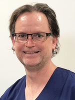
Dr Tim Gregg
Orthopaedic Surgeon
-

Mr Chris Hoffman
Orthopaedic Surgeon
-
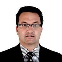
Mr Ryan Johnstone
Orthopaedic Surgeon
-

Mr Grant Kiddle
Orthopaedic Surgeon
-
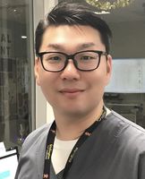
Mr Woo-Sung Kim
Orthopaedic Surgeon
-
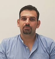
Dr Shay Mandler
Orthopaedic Surgeon
-

Dr Andrew Matthews
Orthopaedic Surgeon
-

Mr John Mortimer
Orthopaedic Surgeon
-
Mr Jonathon Richards
Orthopaedic Surgeon
-

Dr Robert Rowan
Orthopaedic Surgeon
-
Mr Mark Sherwood
Orthopaedic Surgeon
-

Dr Saesol Stallard
Orthopaedic Surgeon
Procedures / Treatments
Two or three small incisions (cuts) are made in the ankle and a small telescopic instrument with a tiny camera attached (arthroscope) is inserted. This allows the surgeon to look inside the joint, identify problems and, in some cases, operate. Tiny instruments can be passed through the arthroscope to remove bony spurs, damaged cartilage or inflamed tissue.
Two or three small incisions (cuts) are made in the ankle and a small telescopic instrument with a tiny camera attached (arthroscope) is inserted. This allows the surgeon to look inside the joint, identify problems and, in some cases, operate. Tiny instruments can be passed through the arthroscope to remove bony spurs, damaged cartilage or inflamed tissue.
Two or three small incisions (cuts) are made in the ankle and a small telescopic instrument with a tiny camera attached (arthroscope) is inserted. This allows the surgeon to look inside the joint, identify problems and, in some cases, operate. Tiny instruments can be passed through the arthroscope to remove bony spurs, damaged cartilage or inflamed tissue.
An incision (cut) is made in the front of, and several smaller cuts on the outside of, the ankle. The damaged ankle joint is replaced with a metal and plastic implant.
An incision (cut) is made in the front of, and several smaller cuts on the outside of, the ankle. The damaged ankle joint is replaced with a metal and plastic implant.
An incision (cut) is made in the front of, and several smaller cuts on the outside of, the ankle. The damaged ankle joint is replaced with a metal and plastic implant.
Carpal Tunnel Syndrome is caused by a pinched nerve in the wrist that causes tingling, numbness and pain in your hand. Surgery to relieve carpal tunnel syndrome involves making an incision (cut) from the middle of the palm of your hand to your wrist. Tissue that is pressing on the nerve is then cut to release the pressure.
Carpal Tunnel Syndrome is caused by a pinched nerve in the wrist that causes tingling, numbness and pain in your hand. Surgery to relieve carpal tunnel syndrome involves making an incision (cut) from the middle of the palm of your hand to your wrist. Tissue that is pressing on the nerve is then cut to release the pressure.
Carpal Tunnel Syndrome is caused by a pinched nerve in the wrist that causes tingling, numbness and pain in your hand.
Surgery to relieve carpal tunnel syndrome involves making an incision (cut) from the middle of the palm of your hand to your wrist. Tissue that is pressing on the nerve is then cut to release the pressure.
Discectomy is an operation to remove part or all of a damaged spinal disc that is pressing on nerves, helping to relieve pain and improve movement. Microdiscectomy:a microscope is used by the surgeon to guide tiny instruments to remove the disc or disc fragments.
Discectomy is an operation to remove part or all of a damaged spinal disc that is pressing on nerves, helping to relieve pain and improve movement. Microdiscectomy:a microscope is used by the surgeon to guide tiny instruments to remove the disc or disc fragments.
Discectomy is an operation to remove part or all of a damaged spinal disc that is pressing on nerves, helping to relieve pain and improve movement.
Microdiscectomy:a microscope is used by the surgeon to guide tiny instruments to remove the disc or disc fragments.
Foot and ankle surgeries are operations that help fix problems in your feet and ankles, like broken bones, arthritis, or injuries to the ligaments and tendons. Surgery might be: Arthroscopic: less invasive surgery. The surgeon makes small cuts and uses a tiny camera to see inside. They can then fix or take out the damaged parts. Open: for more complicated problems, the surgeon makes a larger cut to get a better look and fix the damaged parts directly. Joint Replacement (ankle): when the ankle is badly damaged, the surgeon might remove the damaged areas and replace them with parts made of special materials.
Foot and ankle surgeries are operations that help fix problems in your feet and ankles, like broken bones, arthritis, or injuries to the ligaments and tendons. Surgery might be: Arthroscopic: less invasive surgery. The surgeon makes small cuts and uses a tiny camera to see inside. They can then fix or take out the damaged parts. Open: for more complicated problems, the surgeon makes a larger cut to get a better look and fix the damaged parts directly. Joint Replacement (ankle): when the ankle is badly damaged, the surgeon might remove the damaged areas and replace them with parts made of special materials.
Foot and ankle surgeries are operations that help fix problems in your feet and ankles, like broken bones, arthritis, or injuries to the ligaments and tendons. Surgery might be:
- Arthroscopic: less invasive surgery. The surgeon makes small cuts and uses a tiny camera to see inside. They can then fix or take out the damaged parts.
- Open: for more complicated problems, the surgeon makes a larger cut to get a better look and fix the damaged parts directly.
- Joint Replacement (ankle): when the ankle is badly damaged, the surgeon might remove the damaged areas and replace them with parts made of special materials.
Small incisions (cuts) are made in the hip area and a small telescopic instrument with a tiny camera attached (arthroscope) is inserted. This allows the surgeon to look inside the joint, identify problems and, in some cases, operate. Tiny instruments can be passed through the arthroscope to remove loose, damaged or inflamed tissue.
Small incisions (cuts) are made in the hip area and a small telescopic instrument with a tiny camera attached (arthroscope) is inserted. This allows the surgeon to look inside the joint, identify problems and, in some cases, operate. Tiny instruments can be passed through the arthroscope to remove loose, damaged or inflamed tissue.
Small incisions (cuts) are made in the hip area and a small telescopic instrument with a tiny camera attached (arthroscope) is inserted. This allows the surgeon to look inside the joint, identify problems and, in some cases, operate. Tiny instruments can be passed through the arthroscope to remove loose, damaged or inflamed tissue.
An incision (cut) is made on the side of the thigh to allow the surgeon access to the hip joint. The diseased and damaged parts of the hip joint are removed and replaced with smooth, artificial metal ‘ball’ and plastic ‘socket’ parts.
An incision (cut) is made on the side of the thigh to allow the surgeon access to the hip joint. The diseased and damaged parts of the hip joint are removed and replaced with smooth, artificial metal ‘ball’ and plastic ‘socket’ parts.
An incision (cut) is made on the side of the thigh to allow the surgeon access to the hip joint. The diseased and damaged parts of the hip joint are removed and replaced with smooth, artificial metal ‘ball’ and plastic ‘socket’ parts.
Several small incisions (cuts) are made on the knee through which is inserted a small telescopic instrument with a tiny camera attached (arthroscope). This allows the surgeon to look inside the joint, identify problems and, in some cases, make repairs to damaged tissue.
Several small incisions (cuts) are made on the knee through which is inserted a small telescopic instrument with a tiny camera attached (arthroscope). This allows the surgeon to look inside the joint, identify problems and, in some cases, make repairs to damaged tissue.
Several small incisions (cuts) are made on the knee through which is inserted a small telescopic instrument with a tiny camera attached (arthroscope). This allows the surgeon to look inside the joint, identify problems and, in some cases, make repairs to damaged tissue.
An incision (cut) is made on the front of the knee to allow the surgeon access to the knee joint. The damaged and painful areas of the thigh bone (femur) and lower leg bone (tibia), including the knee joint, are removed and replaced with metal and plastic parts.
An incision (cut) is made on the front of the knee to allow the surgeon access to the knee joint. The damaged and painful areas of the thigh bone (femur) and lower leg bone (tibia), including the knee joint, are removed and replaced with metal and plastic parts.
An incision (cut) is made on the front of the knee to allow the surgeon access to the knee joint. The damaged and painful areas of the thigh bone (femur) and lower leg bone (tibia), including the knee joint, are removed and replaced with metal and plastic parts.
Knee surgery is an operation that helps fix problems in your knee, like an injury, arthritis, a torn ligament, or damaged cartilage. Surgery might be: Arthroscopic: less invasive surgery. The surgeon makes small cuts and uses a tiny camera to see inside the knee. They can then fix or take out the damaged parts. Open: for more complicated problems, the surgeon makes a larger cut to get a better look and fix the damaged parts directly. Joint Replacement: when the knee is badly damaged, the surgeon might remove the damaged areas and replace them with parts made of special materials.
Knee surgery is an operation that helps fix problems in your knee, like an injury, arthritis, a torn ligament, or damaged cartilage. Surgery might be: Arthroscopic: less invasive surgery. The surgeon makes small cuts and uses a tiny camera to see inside the knee. They can then fix or take out the damaged parts. Open: for more complicated problems, the surgeon makes a larger cut to get a better look and fix the damaged parts directly. Joint Replacement: when the knee is badly damaged, the surgeon might remove the damaged areas and replace them with parts made of special materials.
Knee surgery is an operation that helps fix problems in your knee, like an injury, arthritis, a torn ligament, or damaged cartilage. Surgery might be:
- Arthroscopic: less invasive surgery. The surgeon makes small cuts and uses a tiny camera to see inside the knee. They can then fix or take out the damaged parts.
- Open: for more complicated problems, the surgeon makes a larger cut to get a better look and fix the damaged parts directly.
- Joint Replacement: when the knee is badly damaged, the surgeon might remove the damaged areas and replace them with parts made of special materials.
Several small incisions (cuts) are made in the shoulder through which is inserted a small telescopic instrument with a tiny camera attached (arthroscope). The surgeon is then able to remove any bony spurs or inflamed tissue and mend torn tendons of the rotator cuff group.
Several small incisions (cuts) are made in the shoulder through which is inserted a small telescopic instrument with a tiny camera attached (arthroscope). The surgeon is then able to remove any bony spurs or inflamed tissue and mend torn tendons of the rotator cuff group.
Several small incisions (cuts) are made in the shoulder through which is inserted a small telescopic instrument with a tiny camera attached (arthroscope). The surgeon is then able to remove any bony spurs or inflamed tissue and mend torn tendons of the rotator cuff group.
This surgery involves making several small incisions (cuts) on the shoulder through which is inserted a small telescopic instrument with a tiny camera attached (arthroscope). This allows the surgeon to look inside the shoulder, identify problems and, in some cases, make repairs to damaged tissue.
This surgery involves making several small incisions (cuts) on the shoulder through which is inserted a small telescopic instrument with a tiny camera attached (arthroscope). This allows the surgeon to look inside the shoulder, identify problems and, in some cases, make repairs to damaged tissue.
This surgery involves making several small incisions (cuts) on the shoulder through which is inserted a small telescopic instrument with a tiny camera attached (arthroscope). This allows the surgeon to look inside the shoulder, identify problems and, in some cases, make repairs to damaged tissue.
Shoulder surgery is an operation that helps fix problems in your shoulder, like torn muscles or tendons or wear and tear from arthritis. Surgery might be: Arthroscopic: less invasive surgery. The surgeon makes small cuts and uses a tiny camera to see inside the shoulder. They can then fix or take out the damaged parts. Open: for more complicated problems, the surgeon makes a larger cut to get a better look and fix the damaged parts directly. Joint Replacement: when the shoulder is badly damaged, the surgeon might remove the damaged areas and replace them with parts made of special materials.
Shoulder surgery is an operation that helps fix problems in your shoulder, like torn muscles or tendons or wear and tear from arthritis. Surgery might be: Arthroscopic: less invasive surgery. The surgeon makes small cuts and uses a tiny camera to see inside the shoulder. They can then fix or take out the damaged parts. Open: for more complicated problems, the surgeon makes a larger cut to get a better look and fix the damaged parts directly. Joint Replacement: when the shoulder is badly damaged, the surgeon might remove the damaged areas and replace them with parts made of special materials.
Shoulder surgery is an operation that helps fix problems in your shoulder, like torn muscles or tendons or wear and tear from arthritis. Surgery might be:
- Arthroscopic: less invasive surgery. The surgeon makes small cuts and uses a tiny camera to see inside the shoulder. They can then fix or take out the damaged parts.
- Open: for more complicated problems, the surgeon makes a larger cut to get a better look and fix the damaged parts directly.
- Joint Replacement: when the shoulder is badly damaged, the surgeon might remove the damaged areas and replace them with parts made of special materials.
An incision (cut) is made over the relevant part of the spine. Two or more vertebrae (the small bones that make up the spinal column) are fused together with bone grafts and/or metal rods to form a single bone.
An incision (cut) is made over the relevant part of the spine. Two or more vertebrae (the small bones that make up the spinal column) are fused together with bone grafts and/or metal rods to form a single bone.
An incision (cut) is made over the relevant part of the spine. Two or more vertebrae (the small bones that make up the spinal column) are fused together with bone grafts and/or metal rods to form a single bone.
An incision (cut) is made over the damaged tendon. The damaged ends of the tendon are sewn together and, if necessary, reattached to surrounding tissue.
An incision (cut) is made over the damaged tendon. The damaged ends of the tendon are sewn together and, if necessary, reattached to surrounding tissue.
An incision (cut) is made over the damaged tendon. The damaged ends of the tendon are sewn together and, if necessary, reattached to surrounding tissue.
Visiting Hours
- Weekdays: 8.00am to 8.00pm
- Weekends: 8.00am to 8.00pm
Parking
Free patient parking is provided.
Contact Details
Southern Cross Wellington Hospital
Wellington
-
Phone
04 910 2160
Healthlink EDI
wgtnmspc
Email
Website
90 Hanson Street
Newtown
Wellington 6021
Street Address
90 Hanson Street
Newtown
Wellington 6021
Postal Address
PO Box 7233
Newtown
Wellington 6242
Was this page helpful?
This page was last updated at 1:32PM on June 10, 2025. This information is reviewed and edited by Southern Cross Wellington Hospital - Orthopaedic Surgery.

