
MidCentral, Central Lakes > Private Hospitals & Specialists >
Tim Love – Hip, Knee and Spine Orthopaedic Surgeon
Private Service, Orthopaedics
Description
Tim works as a visiting Orthopaedic Surgeon to Te Whatu Ora MidCentral and consults privately at Aorangi Orthopaedic Centre in Palmerston North and also holds clinics at the Centre of Medical Excellence in Queenstown.
What is Orthopaedics?
Consultants
-
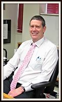
Mr Tim Love
Hip, Knee and Spine Orthopaedic Surgeon
Ages
Youth / Rangatahi, Adult / Pakeke, Older adult / Kaumātua
How do I access this service?
Referral, Contact us
Referral Expectations
You need to bring with you:
Fees and Charges Categorisation
Fees apply
Fees and Charges Description
- Southern Cross Health Insurance Affiliated Provider for Consultations
- nib First Choice Partner
Languages Spoken
English
Procedures / Treatments
For elderly patients joint replacement surgery is commonly required to treat damaged joints from wearing out, arthritis or other forms of joint disease including rheumatoid arthritis. In these procedures the damaged joint surface is removed and replaced with artificial surfaces normally made from metal (chromium cobalt alloy, titanium), plastic (high density polyethelene) or ceramic which act as alternate bearing surfaces for the damaged joint. These operations are major procedures which require the patient to be in hospital for several days and followed by a significant period of rehabilitation. The hospital has several ways of approaching the procedure for replacement and the specifics for the procedure will be covered at the time of assessment and booking of surgery. Occasionally blood transfusions are required; if you have some concerns raise this with your surgeon during consultation.
For elderly patients joint replacement surgery is commonly required to treat damaged joints from wearing out, arthritis or other forms of joint disease including rheumatoid arthritis. In these procedures the damaged joint surface is removed and replaced with artificial surfaces normally made from metal (chromium cobalt alloy, titanium), plastic (high density polyethelene) or ceramic which act as alternate bearing surfaces for the damaged joint. These operations are major procedures which require the patient to be in hospital for several days and followed by a significant period of rehabilitation. The hospital has several ways of approaching the procedure for replacement and the specifics for the procedure will be covered at the time of assessment and booking of surgery. Occasionally blood transfusions are required; if you have some concerns raise this with your surgeon during consultation.
For elderly patients joint replacement surgery is commonly required to treat damaged joints from wearing out, arthritis or other forms of joint disease including rheumatoid arthritis. In these procedures the damaged joint surface is removed and replaced with artificial surfaces normally made from metal (chromium cobalt alloy, titanium), plastic (high density polyethelene) or ceramic which act as alternate bearing surfaces for the damaged joint.
These operations are major procedures which require the patient to be in hospital for several days and followed by a significant period of rehabilitation. The hospital has several ways of approaching the procedure for replacement and the specifics for the procedure will be covered at the time of assessment and booking of surgery.
Occasionally blood transfusions are required; if you have some concerns raise this with your surgeon during consultation.
Knee surgery is an operation that helps fix problems in your knee, like an injury, arthritis, a torn ligament, or damaged cartilage. Surgery might be: Arthroscopic: less invasive surgery. The surgeon makes small cuts and uses a tiny camera to see inside the knee. They can then fix or take out the damaged parts. Open: for more complicated problems, the surgeon makes a larger cut to get a better look and fix the damaged parts directly. Joint Replacement: when the knee is badly damaged, the surgeon might remove the damaged areas and replace them with parts made of special materials.
Knee surgery is an operation that helps fix problems in your knee, like an injury, arthritis, a torn ligament, or damaged cartilage. Surgery might be: Arthroscopic: less invasive surgery. The surgeon makes small cuts and uses a tiny camera to see inside the knee. They can then fix or take out the damaged parts. Open: for more complicated problems, the surgeon makes a larger cut to get a better look and fix the damaged parts directly. Joint Replacement: when the knee is badly damaged, the surgeon might remove the damaged areas and replace them with parts made of special materials.
Knee surgery is an operation that helps fix problems in your knee, like an injury, arthritis, a torn ligament, or damaged cartilage. Surgery might be:
- Arthroscopic: less invasive surgery. The surgeon makes small cuts and uses a tiny camera to see inside the knee. They can then fix or take out the damaged parts.
- Open: for more complicated problems, the surgeon makes a larger cut to get a better look and fix the damaged parts directly.
- Joint Replacement: when the knee is badly damaged, the surgeon might remove the damaged areas and replace them with parts made of special materials.
This is a surgical procedure performed on a knee joint that has become painful and/or impaired because of disease, injury or wear and tear. In total knee replacement, artificial materials (metal and plastic) are used to replace the following damaged surfaces within the knee joint: the end of the thigh bone (femur) the end of the shin bone (tibia) the back of the kneecap (patella) This operation is a major procedure which requires you to be in hospital for several days and will be followed by a significant period of rehabilitation. Occasionally blood transfusions are required; if you have some concerns raise this with your surgeon during consultation. For more information about total knee replacement please click here.
This is a surgical procedure performed on a knee joint that has become painful and/or impaired because of disease, injury or wear and tear. In total knee replacement, artificial materials (metal and plastic) are used to replace the following damaged surfaces within the knee joint: the end of the thigh bone (femur) the end of the shin bone (tibia) the back of the kneecap (patella) This operation is a major procedure which requires you to be in hospital for several days and will be followed by a significant period of rehabilitation. Occasionally blood transfusions are required; if you have some concerns raise this with your surgeon during consultation. For more information about total knee replacement please click here.
This is a surgical procedure performed on a knee joint that has become painful and/or impaired because of disease, injury or wear and tear.
In total knee replacement, artificial materials (metal and plastic) are used to replace the following damaged surfaces within the knee joint:
- the end of the thigh bone (femur)
- the end of the shin bone (tibia)
- the back of the kneecap (patella)
This operation is a major procedure which requires you to be in hospital for several days and will be followed by a significant period of rehabilitation.
Occasionally blood transfusions are required; if you have some concerns raise this with your surgeon during consultation.
For more information about total knee replacement please click here.
An incision (cut) is made on the front of the knee to allow the surgeon access to the knee joint. The damaged and painful areas of the thigh bone (femur) and lower leg bone (tibia), including the knee joint, are removed and replaced with metal and plastic parts.
An incision (cut) is made on the front of the knee to allow the surgeon access to the knee joint. The damaged and painful areas of the thigh bone (femur) and lower leg bone (tibia), including the knee joint, are removed and replaced with metal and plastic parts.
An incision (cut) is made on the front of the knee to allow the surgeon access to the knee joint. The damaged and painful areas of the thigh bone (femur) and lower leg bone (tibia), including the knee joint, are removed and replaced with metal and plastic parts.
An incision (cut) is made on the side of the thigh to allow the surgeon access to the hip joint. The diseased and damaged parts of the hip joint are removed and replaced with smooth, artificial metal ‘ball’ and plastic ‘socket’ parts.
An incision (cut) is made on the side of the thigh to allow the surgeon access to the hip joint. The diseased and damaged parts of the hip joint are removed and replaced with smooth, artificial metal ‘ball’ and plastic ‘socket’ parts.
An incision (cut) is made on the side of the thigh to allow the surgeon access to the hip joint. The diseased and damaged parts of the hip joint are removed and replaced with smooth, artificial metal ‘ball’ and plastic ‘socket’ parts.
Between the vertebrae in your spine are flat, round discs that act as shock absorbers for the spinal bones. Sometimes some of the gel-like substance in the center of the disc (nucleus) bulges out through the tough outer ring (annulus) and into the spinal canal. This is known as a herniated or ruptured disc and the pressure it puts on the spinal nerves often causes symptoms such as pain, numbness and tingling. Initial treatment for a herniated disc may involve low level activity, nonsteroidal anti-inflammatory medication and physiotherapy. If these approaches fail to reduce or remove the pain, surgical treatment may be considered. Discectomy This surgery is performed to remove part or all of a herniated intervertebral disc. Open discectomy – involves making an incision (cut) over the vertebra and stripping back the muscles to expose the herniated disc. The entire disc, or parts of it are removed, thus relieving pressure on the spinal nerves. Microdiscectomy – this is a ‘minimally invasive’ surgical technique, meaning it requires smaller incisions and no muscle stripping is required. Tiny, specialised instruments are used to remove the disc or disc fragments. Laminectomy or Laminotomy These procedures involve making an incision down the centre of the back and removing some or all of the bony arch (lamina) of a vertebra. In a laminectomy, all or most of the lamina is surgically removed while a laminotomy involves partial removal of the lamina. By making more room in the spinal canal, these procedures reduce pressure on the spinal nerves. They also give the surgeon better access to the disc and other parts of the spine if further procedures e.g. discectomy, spinal fusion, are required. Spinal fusion In this procedure, individual vertebrae are fused together so that no movement can occur between the vertebrae and hence pain is reduced. Spinal fusion may be required for disc herniation in the cervical region of the spine as well as for some cases of vertebral fracture and to prevent pain-inducing movements.
Between the vertebrae in your spine are flat, round discs that act as shock absorbers for the spinal bones. Sometimes some of the gel-like substance in the center of the disc (nucleus) bulges out through the tough outer ring (annulus) and into the spinal canal. This is known as a herniated or ruptured disc and the pressure it puts on the spinal nerves often causes symptoms such as pain, numbness and tingling. Initial treatment for a herniated disc may involve low level activity, nonsteroidal anti-inflammatory medication and physiotherapy. If these approaches fail to reduce or remove the pain, surgical treatment may be considered. Discectomy This surgery is performed to remove part or all of a herniated intervertebral disc. Open discectomy – involves making an incision (cut) over the vertebra and stripping back the muscles to expose the herniated disc. The entire disc, or parts of it are removed, thus relieving pressure on the spinal nerves. Microdiscectomy – this is a ‘minimally invasive’ surgical technique, meaning it requires smaller incisions and no muscle stripping is required. Tiny, specialised instruments are used to remove the disc or disc fragments. Laminectomy or Laminotomy These procedures involve making an incision down the centre of the back and removing some or all of the bony arch (lamina) of a vertebra. In a laminectomy, all or most of the lamina is surgically removed while a laminotomy involves partial removal of the lamina. By making more room in the spinal canal, these procedures reduce pressure on the spinal nerves. They also give the surgeon better access to the disc and other parts of the spine if further procedures e.g. discectomy, spinal fusion, are required. Spinal fusion In this procedure, individual vertebrae are fused together so that no movement can occur between the vertebrae and hence pain is reduced. Spinal fusion may be required for disc herniation in the cervical region of the spine as well as for some cases of vertebral fracture and to prevent pain-inducing movements.
Between the vertebrae in your spine are flat, round discs that act as shock absorbers for the spinal bones. Sometimes some of the gel-like substance in the center of the disc (nucleus) bulges out through the tough outer ring (annulus) and into the spinal canal. This is known as a herniated or ruptured disc and the pressure it puts on the spinal nerves often causes symptoms such as pain, numbness and tingling.
Initial treatment for a herniated disc may involve low level activity, nonsteroidal anti-inflammatory medication and physiotherapy. If these approaches fail to reduce or remove the pain, surgical treatment may be considered.
Discectomy
This surgery is performed to remove part or all of a herniated intervertebral disc.
Open discectomy – involves making an incision (cut) over the vertebra and stripping back the muscles to expose the herniated disc. The entire disc, or parts of it are removed, thus relieving pressure on the spinal nerves.
Microdiscectomy – this is a ‘minimally invasive’ surgical technique, meaning it requires smaller incisions and no muscle stripping is required. Tiny, specialised instruments are used to remove the disc or disc fragments.
Laminectomy or Laminotomy
These procedures involve making an incision down the centre of the back and removing some or all of the bony arch (lamina) of a vertebra.
In a laminectomy, all or most of the lamina is surgically removed while a laminotomy involves partial removal of the lamina.
By making more room in the spinal canal, these procedures reduce pressure on the spinal nerves. They also give the surgeon better access to the disc and other parts of the spine if further procedures e.g. discectomy, spinal fusion, are required.
Spinal fusion
In this procedure, individual vertebrae are fused together so that no movement can occur between the vertebrae and hence pain is reduced. Spinal fusion may be required for disc herniation in the cervical region of the spine as well as for some cases of vertebral fracture and to prevent pain-inducing movements.
Tumours may be found within the spinal cord itself, between the spinal cord and its tough outer covering, the dura, or outside the dura. They may be primary (they arise in the in the spine or nearby tissue) or metastatic (they have originated in another part of the body and traveled to the spine, usually via the bloodstream). Spinal tumours may be treated by any combination of surgery, radiotherapy and chemotherapy. Surgery may be performed to take a small sample of tissue to examine under the microscope (biopsy) or to remove the tumour. Typically, the patient will be lying face downwards and a procedure known as a laminectomy is performed (the bone overlying the spinal cord is removed). This gives the surgeon access to the spinal cord and allows removal of the tumour.
Tumours may be found within the spinal cord itself, between the spinal cord and its tough outer covering, the dura, or outside the dura. They may be primary (they arise in the in the spine or nearby tissue) or metastatic (they have originated in another part of the body and traveled to the spine, usually via the bloodstream). Spinal tumours may be treated by any combination of surgery, radiotherapy and chemotherapy. Surgery may be performed to take a small sample of tissue to examine under the microscope (biopsy) or to remove the tumour. Typically, the patient will be lying face downwards and a procedure known as a laminectomy is performed (the bone overlying the spinal cord is removed). This gives the surgeon access to the spinal cord and allows removal of the tumour.
Tumours may be found within the spinal cord itself, between the spinal cord and its tough outer covering, the dura, or outside the dura. They may be primary (they arise in the in the spine or nearby tissue) or metastatic (they have originated in another part of the body and traveled to the spine, usually via the bloodstream).
Spinal tumours may be treated by any combination of surgery, radiotherapy and chemotherapy. Surgery may be performed to take a small sample of tissue to examine under the microscope (biopsy) or to remove the tumour. Typically, the patient will be lying face downwards and a procedure known as a laminectomy is performed (the bone overlying the spinal cord is removed). This gives the surgeon access to the spinal cord and allows removal of the tumour.
A large number of orthopaedic procedures on joints are performed using an arthroscope, where a fibre optic telescope is used to look inside the joint. Through this type of keyhole surgery, fine instruments can be introduced through small incisions (portals) to allow surgery to be performed without the need for large cuts. This allows many procedures to be performed as a day stay and allows quicker return to normal function of the joint. Arthroscopic surgery is less painful than open surgery and decreases the risk of healing problems. Arthroscopy allows access to parts of the joints which can not be accessed by other types of surgery. For more information about arthroscopy please click here.
A large number of orthopaedic procedures on joints are performed using an arthroscope, where a fibre optic telescope is used to look inside the joint. Through this type of keyhole surgery, fine instruments can be introduced through small incisions (portals) to allow surgery to be performed without the need for large cuts. This allows many procedures to be performed as a day stay and allows quicker return to normal function of the joint. Arthroscopic surgery is less painful than open surgery and decreases the risk of healing problems. Arthroscopy allows access to parts of the joints which can not be accessed by other types of surgery. For more information about arthroscopy please click here.
A large number of orthopaedic procedures on joints are performed using an arthroscope, where a fibre optic telescope is used to look inside the joint. Through this type of keyhole surgery, fine instruments can be introduced through small incisions (portals) to allow surgery to be performed without the need for large cuts. This allows many procedures to be performed as a day stay and allows quicker return to normal function of the joint.
Arthroscopic surgery is less painful than open surgery and decreases the risk of healing problems. Arthroscopy allows access to parts of the joints which can not be accessed by other types of surgery.
For more information about arthroscopy please click here.
Osteotomy is the division of a crooked or bent bone to improve alignment of the limb. These procedures normally involve some form of internal fixation, such as rods or plates, or external fixation which involves external wires and pins to hold the bone. The type of procedure for fixation will be explained when the surgery is planned.
Osteotomy is the division of a crooked or bent bone to improve alignment of the limb. These procedures normally involve some form of internal fixation, such as rods or plates, or external fixation which involves external wires and pins to hold the bone. The type of procedure for fixation will be explained when the surgery is planned.
Osteotomy is the division of a crooked or bent bone to improve alignment of the limb.
These procedures normally involve some form of internal fixation, such as rods or plates, or external fixation which involves external wires and pins to hold the bone. The type of procedure for fixation will be explained when the surgery is planned.
In many cases tendons will be lengthened to improve the muscle balance around a joint or tendons will be transferred to give overall better joint function. This occurs in children with neuromuscular conditions but also applies to a number of other conditions. Most of these procedures involve some sort of splintage after the surgery followed by a period of rehabilitation, normally supervised by a physiotherapist.
In many cases tendons will be lengthened to improve the muscle balance around a joint or tendons will be transferred to give overall better joint function. This occurs in children with neuromuscular conditions but also applies to a number of other conditions. Most of these procedures involve some sort of splintage after the surgery followed by a period of rehabilitation, normally supervised by a physiotherapist.
In many cases tendons will be lengthened to improve the muscle balance around a joint or tendons will be transferred to give overall better joint function. This occurs in children with neuromuscular conditions but also applies to a number of other conditions.
Most of these procedures involve some sort of splintage after the surgery followed by a period of rehabilitation, normally supervised by a physiotherapist.
Bunions are a bony lump that occurs on the inner side of the big toe joint. Smaller bunionettes can occur on the side of the little toe. Bunions usually occur in conjunction with an excessive angulation of the big toe (varus deformity). Occasionally a different condition where arthritis develops in the joint (Hallux Rigidus) can be mistaken for a bunion. Generally bunion surgery requires correction of the angulation AND removal of the bunion. The surgery is straightforward but it can take several months to recover fully and be able to wear shoes comfortably. Severe bunions or Hallux Rigidus may be treated by fusing the joint permanently.
Bunions are a bony lump that occurs on the inner side of the big toe joint. Smaller bunionettes can occur on the side of the little toe. Bunions usually occur in conjunction with an excessive angulation of the big toe (varus deformity). Occasionally a different condition where arthritis develops in the joint (Hallux Rigidus) can be mistaken for a bunion. Generally bunion surgery requires correction of the angulation AND removal of the bunion. The surgery is straightforward but it can take several months to recover fully and be able to wear shoes comfortably. Severe bunions or Hallux Rigidus may be treated by fusing the joint permanently.
Bunions are a bony lump that occurs on the inner side of the big toe joint. Smaller bunionettes can occur on the side of the little toe. Bunions usually occur in conjunction with an excessive angulation of the big toe (varus deformity). Occasionally a different condition where arthritis develops in the joint (Hallux Rigidus) can be mistaken for a bunion.
Generally bunion surgery requires correction of the angulation AND removal of the bunion. The surgery is straightforward but it can take several months to recover fully and be able to wear shoes comfortably. Severe bunions or Hallux Rigidus may be treated by fusing the joint permanently.
Dupuytren's contracture is a condition where the normally thin sheet of palmar fascia (gristle which toughens the palm and supports the skin on the palm and fingers) becomes abnormally thickened. As it does so it contracts and causes the fingers to curl up. It is a genetic condition and therefore runs in families but can "skip" generations. Males are more affected than females. There is a relationship to descending from Scandinavian countries. Surgery is a treatment and not a cure. The bands of thickened tissue can be removed to help the mobility of the hand and fingers. However, full correction is unlikely and the condition can slowly recur. Surgery is usually only done when the contractures of the fingers are significant as operating too soon can paradoxically worsen the condition.
Dupuytren's contracture is a condition where the normally thin sheet of palmar fascia (gristle which toughens the palm and supports the skin on the palm and fingers) becomes abnormally thickened. As it does so it contracts and causes the fingers to curl up. It is a genetic condition and therefore runs in families but can "skip" generations. Males are more affected than females. There is a relationship to descending from Scandinavian countries. Surgery is a treatment and not a cure. The bands of thickened tissue can be removed to help the mobility of the hand and fingers. However, full correction is unlikely and the condition can slowly recur. Surgery is usually only done when the contractures of the fingers are significant as operating too soon can paradoxically worsen the condition.
Dupuytren's contracture is a condition where the normally thin sheet of palmar fascia (gristle which toughens the palm and supports the skin on the palm and fingers) becomes abnormally thickened. As it does so it contracts and causes the fingers to curl up. It is a genetic condition and therefore runs in families but can "skip" generations. Males are more affected than females. There is a relationship to descending from Scandinavian countries. Surgery is a treatment and not a cure. The bands of thickened tissue can be removed to help the mobility of the hand and fingers. However, full correction is unlikely and the condition can slowly recur. Surgery is usually only done when the contractures of the fingers are significant as operating too soon can paradoxically worsen the condition.
Disability Assistance
Wheelchair access, Wheelchair accessible toilet, Mobility parking space
Additional Details
Face to face / Kanohi ki te Kanohi, Phone
Parking
Free patient parking is provided at both locations.
Pharmacy
Find your nearest pharmacy here
Contact Details
175 Grey Street, Palmerston North
MidCentral
-
Phone
(06) 357 6024
-
Fax
(06) 356 5663
Email
Website
Was this page helpful?
This page was last updated at 11:43AM on May 14, 2025. This information is reviewed and edited by Tim Love – Hip, Knee and Spine Orthopaedic Surgeon.