Central Auckland, East Auckland, North Auckland, South Auckland, West Auckland > Private Hospitals & Specialists >
UniSports Orthopaedics
Private Service, Orthopaedics, Sports Medicine
Today
8:00 AM to 5:00 PM.
Description
Welcome to UniSports Orthopaedics.
The UniSports Orthopaedics team are leaders in their field and have undertaken extensive postgraduate training at international centres of excellence.
We are focused on the management and treatment of sports and musculoskeletal injuries. UniSports Orthopaedics is based at UniSports Sports Medicine along with Axis Sports Medicine Specialists and UniSports Physiotherapy.
As well as our general patient care, we currently provide orthopaedic support for leading sports teams including the All Blacks, Team NZ, New Zealand Warriors, Auckland Blues, NZ Breakers and High Performance Sport NZ.
Working alongside our consultant surgeons is a team of experienced nurses as well as recently-qualified international orthopaedic surgeons furthering their education in sports medicine:
UniSports Orthopaedic Fellows. A Fellow is a recently-qualified Orthopaedic Surgeon who is furthering their education in sports medicine surgery.
Consultants
Note: Please note below that some people are not available at all locations.
-
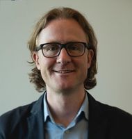
Mr Matt Debenham
Orthopaedic Surgeon
Available at 261 Morrin Road, Saint Johns, Auckland
-
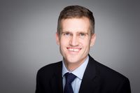
Mr Paul Monk
Orthopaedic Surgeon
Available at 261 Morrin Road, Saint Johns, Auckland, 2 Wagener Place, Mt Albert, Auckland
-
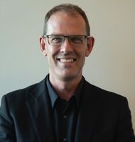
Mr Michael Rosenfeldt
Orthopaedic Surgeon
Available at 261 Morrin Road, Saint Johns, Auckland, 4 St Marks Road, Remuera, Auckland
-
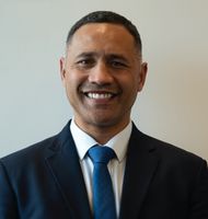
Mr Jeremy Stanley
Orthopaedic Surgeon
Available at 261 Morrin Road, Saint Johns, Auckland, Cavendish Clinic, 175 Cavendish Drive, Manukau, Auckland, 53 Lincoln Road, Henderson, Auckland, Franklin Hospital, 2 Wagener Place, Mt Albert, Auckland
-
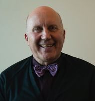
Mr Bruce Twaddle
Orthopaedic Surgeon
Available at 261 Morrin Road, Saint Johns, Auckland, Forté 2, 132 Peterborough Street, Christchurch Central, Queenstown Centre of Medical Excellence, 12 Twelfth Avenue, Kawarau Park, Queenstown
-
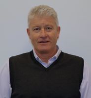
Mr Stewart Walsh
Orthopaedic Surgeon
Available at 261 Morrin Road, Saint Johns, Auckland
Referral Expectations
A referral from a Physiotherapist, Sports Physician or General Practitioner is required before an appointment with an orthopaedic consultant can be scheduled.
Fees and Charges Description
UniSports Orthopaedics is a Southern Cross affiliated provider for certain procedures and treatments. We can assist you with your referral and insurance claim.
Hours
8:00 AM to 5:00 PM.
| Mon – Fri | 8:00 AM – 5:00 PM |
|---|
Please contact us during these hours to discuss your condition and arrange appointments.
Services Provided
Foot and ankle surgeries are operations that help fix problems in your feet and ankles, like broken bones, arthritis, or injuries to the ligaments and tendons. Surgery might be: Arthroscopic: less invasive surgery. The surgeon makes small cuts and uses a tiny camera to see inside. They can then fix or take out the damaged parts. Open: for more complicated problems, the surgeon makes a larger cut to get a better look and fix the damaged parts directly. Joint Replacement (ankle): when the ankle is badly damaged, the surgeon might remove the damaged areas and replace them with parts made of special materials.
Foot and ankle surgeries are operations that help fix problems in your feet and ankles, like broken bones, arthritis, or injuries to the ligaments and tendons. Surgery might be: Arthroscopic: less invasive surgery. The surgeon makes small cuts and uses a tiny camera to see inside. They can then fix or take out the damaged parts. Open: for more complicated problems, the surgeon makes a larger cut to get a better look and fix the damaged parts directly. Joint Replacement (ankle): when the ankle is badly damaged, the surgeon might remove the damaged areas and replace them with parts made of special materials.
Foot and ankle surgeries are operations that help fix problems in your feet and ankles, like broken bones, arthritis, or injuries to the ligaments and tendons. Surgery might be:
- Arthroscopic: less invasive surgery. The surgeon makes small cuts and uses a tiny camera to see inside. They can then fix or take out the damaged parts.
- Open: for more complicated problems, the surgeon makes a larger cut to get a better look and fix the damaged parts directly.
- Joint Replacement (ankle): when the ankle is badly damaged, the surgeon might remove the damaged areas and replace them with parts made of special materials.
Knee surgery is an operation that helps fix problems in your knee, like an injury, arthritis, a torn ligament, or damaged cartilage. Surgery might be: Arthroscopic: less invasive surgery. The surgeon makes small cuts and uses a tiny camera to see inside the knee. They can then fix or take out the damaged parts. Open: for more complicated problems, the surgeon makes a larger cut to get a better look and fix the damaged parts directly. Joint Replacement: when the knee is badly damaged, the surgeon might remove the damaged areas and replace them with parts made of special materials.
Knee surgery is an operation that helps fix problems in your knee, like an injury, arthritis, a torn ligament, or damaged cartilage. Surgery might be: Arthroscopic: less invasive surgery. The surgeon makes small cuts and uses a tiny camera to see inside the knee. They can then fix or take out the damaged parts. Open: for more complicated problems, the surgeon makes a larger cut to get a better look and fix the damaged parts directly. Joint Replacement: when the knee is badly damaged, the surgeon might remove the damaged areas and replace them with parts made of special materials.
Knee surgery is an operation that helps fix problems in your knee, like an injury, arthritis, a torn ligament, or damaged cartilage. Surgery might be:
- Arthroscopic: less invasive surgery. The surgeon makes small cuts and uses a tiny camera to see inside the knee. They can then fix or take out the damaged parts.
- Open: for more complicated problems, the surgeon makes a larger cut to get a better look and fix the damaged parts directly.
- Joint Replacement: when the knee is badly damaged, the surgeon might remove the damaged areas and replace them with parts made of special materials.
Orthopaedics is an area that deals with conditions of the musculoskeletal system (bones and joints of the limbs and spine). The specialty covers a range of different types of conditions starting with congenital (conditions which children are born with) through to degenerative (conditions relating to the wearing out of joints). The field of orthopaedics includes trauma, where bones are broken or injuries are sustained to limbs. Other conditions that are covered by orthopaedics are metabolic conditions, neurological and inflammatory conditions. Read more about orthopaedics at Unisports Orthopaedics here.
Orthopaedics is an area that deals with conditions of the musculoskeletal system (bones and joints of the limbs and spine). The specialty covers a range of different types of conditions starting with congenital (conditions which children are born with) through to degenerative (conditions relating to the wearing out of joints). The field of orthopaedics includes trauma, where bones are broken or injuries are sustained to limbs. Other conditions that are covered by orthopaedics are metabolic conditions, neurological and inflammatory conditions. Read more about orthopaedics at Unisports Orthopaedics here.
Orthopaedics is an area that deals with conditions of the musculoskeletal system (bones and joints of the limbs and spine). The specialty covers a range of different types of conditions starting with congenital (conditions which children are born with) through to degenerative (conditions relating to the wearing out of joints). The field of orthopaedics includes trauma, where bones are broken or injuries are sustained to limbs.
Other conditions that are covered by orthopaedics are metabolic conditions, neurological and inflammatory conditions.
Read more about orthopaedics at Unisports Orthopaedics here.
A large number of orthopaedic procedures on joints are performed using an arthroscope, where a fibre optic telescope is used to look inside the joint. Through this type of keyhole surgery, fine instruments can be introduced through small incisions (portals) to allow surgery to be performed without the need for large cuts. This allows many procedures to be performed as a day stay and allows quicker return to normal function of the joint. Arthroscopic surgery is less painful than open surgery and decreases the risk of healing problems. Arthroscopy allows access to parts of the joints which can not be accessed by other types of surgery.
A large number of orthopaedic procedures on joints are performed using an arthroscope, where a fibre optic telescope is used to look inside the joint. Through this type of keyhole surgery, fine instruments can be introduced through small incisions (portals) to allow surgery to be performed without the need for large cuts. This allows many procedures to be performed as a day stay and allows quicker return to normal function of the joint. Arthroscopic surgery is less painful than open surgery and decreases the risk of healing problems. Arthroscopy allows access to parts of the joints which can not be accessed by other types of surgery.
A large number of orthopaedic procedures on joints are performed using an arthroscope, where a fibre optic telescope is used to look inside the joint. Through this type of keyhole surgery, fine instruments can be introduced through small incisions (portals) to allow surgery to be performed without the need for large cuts. This allows many procedures to be performed as a day stay and allows quicker return to normal function of the joint.
Arthroscopic surgery is less painful than open surgery and decreases the risk of healing problems. Arthroscopy allows access to parts of the joints which can not be accessed by other types of surgery.
Shoulder surgery is an operation that helps fix problems in your shoulder, like torn muscles or tendons or wear and tear from arthritis. Surgery might be: Arthroscopic: less invasive surgery. The surgeon makes small cuts and uses a tiny camera to see inside the shoulder. They can then fix or take out the damaged parts. Open: for more complicated problems, the surgeon makes a larger cut to get a better look and fix the damaged parts directly. Joint Replacement: when the shoulder is badly damaged, the surgeon might remove the damaged areas and replace them with parts made of special materials.
Shoulder surgery is an operation that helps fix problems in your shoulder, like torn muscles or tendons or wear and tear from arthritis. Surgery might be: Arthroscopic: less invasive surgery. The surgeon makes small cuts and uses a tiny camera to see inside the shoulder. They can then fix or take out the damaged parts. Open: for more complicated problems, the surgeon makes a larger cut to get a better look and fix the damaged parts directly. Joint Replacement: when the shoulder is badly damaged, the surgeon might remove the damaged areas and replace them with parts made of special materials.
Shoulder surgery is an operation that helps fix problems in your shoulder, like torn muscles or tendons or wear and tear from arthritis. Surgery might be:
- Arthroscopic: less invasive surgery. The surgeon makes small cuts and uses a tiny camera to see inside the shoulder. They can then fix or take out the damaged parts.
- Open: for more complicated problems, the surgeon makes a larger cut to get a better look and fix the damaged parts directly.
- Joint Replacement: when the shoulder is badly damaged, the surgeon might remove the damaged areas and replace them with parts made of special materials.
In many cases tendons will be lengthened to improve the muscle balance around a joint or tendons will be transferred to give overall better joint function. This occurs in children with neuromuscular conditions but also applies to a number of other conditions. Most of these procedures involve some sort of splintage after the surgery followed by a period of rehabilitation, normally supervised by a physiotherapist.
In many cases tendons will be lengthened to improve the muscle balance around a joint or tendons will be transferred to give overall better joint function. This occurs in children with neuromuscular conditions but also applies to a number of other conditions. Most of these procedures involve some sort of splintage after the surgery followed by a period of rehabilitation, normally supervised by a physiotherapist.
In many cases tendons will be lengthened to improve the muscle balance around a joint or tendons will be transferred to give overall better joint function. This occurs in children with neuromuscular conditions but also applies to a number of other conditions.
Most of these procedures involve some sort of splintage after the surgery followed by a period of rehabilitation, normally supervised by a physiotherapist.
For elderly patients joint replacement surgery is commonly required to treat damaged joints from wearing out, arthritis or other forms of joint disease including rheumatoid arthritis. In these procedures the damaged joint surface is removed and replaced with artificial surfaces normally made from metal (chromium cobalt alloy, titanium), plastic (high density polyethelene) or ceramic which act as alternate bearing surfaces for the damaged joint. These operations are major procedures which require the patient to be in hospital for several days and followed by a significant period of rehabilitation. The hospital has several ways of approaching the procedure for replacement and the specifics for the procedure will be covered at the time of assessment and booking of surgery. Occasionally blood transfusions are required; if you have some concerns raise this with your surgeon during consultation.
For elderly patients joint replacement surgery is commonly required to treat damaged joints from wearing out, arthritis or other forms of joint disease including rheumatoid arthritis. In these procedures the damaged joint surface is removed and replaced with artificial surfaces normally made from metal (chromium cobalt alloy, titanium), plastic (high density polyethelene) or ceramic which act as alternate bearing surfaces for the damaged joint. These operations are major procedures which require the patient to be in hospital for several days and followed by a significant period of rehabilitation. The hospital has several ways of approaching the procedure for replacement and the specifics for the procedure will be covered at the time of assessment and booking of surgery. Occasionally blood transfusions are required; if you have some concerns raise this with your surgeon during consultation.
For elderly patients joint replacement surgery is commonly required to treat damaged joints from wearing out, arthritis or other forms of joint disease including rheumatoid arthritis. In these procedures the damaged joint surface is removed and replaced with artificial surfaces normally made from metal (chromium cobalt alloy, titanium), plastic (high density polyethelene) or ceramic which act as alternate bearing surfaces for the damaged joint.
These operations are major procedures which require the patient to be in hospital for several days and followed by a significant period of rehabilitation. The hospital has several ways of approaching the procedure for replacement and the specifics for the procedure will be covered at the time of assessment and booking of surgery.
Occasionally blood transfusions are required; if you have some concerns raise this with your surgeon during consultation.
Several small incisions (cuts) are made on the knee through which is inserted a small telescopic instrument with a tiny camera attached (arthroscope). This allows the surgeon to look inside the joint, identify problems and, in some cases, make repairs to damaged tissue.
Several small incisions (cuts) are made on the knee through which is inserted a small telescopic instrument with a tiny camera attached (arthroscope). This allows the surgeon to look inside the joint, identify problems and, in some cases, make repairs to damaged tissue.
Several small incisions (cuts) are made on the knee through which is inserted a small telescopic instrument with a tiny camera attached (arthroscope). This allows the surgeon to look inside the joint, identify problems and, in some cases, make repairs to damaged tissue.
The anterior cruciate ligament (ACL) is a strong, stabilising ligament running through the centre of the knee between the femur (thigh bone) and tibia (shin bone). When the ACL is torn, frequently as the result of a sporting injury, arthroscopic surgery known as ACL Reconstruction is performed. The procedure involves replacement of the damaged ligament with tissue grafted from elsewhere, usually the patellar or hamstring tendon. The ends of the grafted tendon are attached to the femur at one end and the tibia at the other using screws or staples. For more information about ACL reconstruction please click here.
The anterior cruciate ligament (ACL) is a strong, stabilising ligament running through the centre of the knee between the femur (thigh bone) and tibia (shin bone). When the ACL is torn, frequently as the result of a sporting injury, arthroscopic surgery known as ACL Reconstruction is performed. The procedure involves replacement of the damaged ligament with tissue grafted from elsewhere, usually the patellar or hamstring tendon. The ends of the grafted tendon are attached to the femur at one end and the tibia at the other using screws or staples. For more information about ACL reconstruction please click here.
The anterior cruciate ligament (ACL) is a strong, stabilising ligament running through the centre of the knee between the femur (thigh bone) and tibia (shin bone).
When the ACL is torn, frequently as the result of a sporting injury, arthroscopic surgery known as ACL Reconstruction is performed. The procedure involves replacement of the damaged ligament with tissue grafted from elsewhere, usually the patellar or hamstring tendon. The ends of the grafted tendon are attached to the femur at one end and the tibia at the other using screws or staples.
For more information about ACL reconstruction please click here.
The menisci are two circular strips of cartilage that form a cushioning layer between the ends of the femur (thigh bone) and tibia (shin bone) in the knee joint. Together the medial and lateral menisci, on the inside and outside of the knee, respectively, act as shock absorbers and distribute the weight of the body across the knee joint. The menisci can become torn through injury or damaged from age-related wear and tear and may require surgery. The most common meniscal surgery is partial meniscectomy in which the torn portion of the meniscus is cut away so that the cartilage surface is smooth again. In some cases meniscal repair is carried out, in this case the torn edges of the meniscus are sutured together. Both procedures are performed arthroscopically. For more information please click on the following link for meniscal tears and for meniscal transplant surgery.
The menisci are two circular strips of cartilage that form a cushioning layer between the ends of the femur (thigh bone) and tibia (shin bone) in the knee joint. Together the medial and lateral menisci, on the inside and outside of the knee, respectively, act as shock absorbers and distribute the weight of the body across the knee joint. The menisci can become torn through injury or damaged from age-related wear and tear and may require surgery. The most common meniscal surgery is partial meniscectomy in which the torn portion of the meniscus is cut away so that the cartilage surface is smooth again. In some cases meniscal repair is carried out, in this case the torn edges of the meniscus are sutured together. Both procedures are performed arthroscopically. For more information please click on the following link for meniscal tears and for meniscal transplant surgery.
The menisci are two circular strips of cartilage that form a cushioning layer between the ends of the femur (thigh bone) and tibia (shin bone) in the knee joint. Together the medial and lateral menisci, on the inside and outside of the knee, respectively, act as shock absorbers and distribute the weight of the body across the knee joint.
The menisci can become torn through injury or damaged from age-related wear and tear and may require surgery.
The most common meniscal surgery is partial meniscectomy in which the torn portion of the meniscus is cut away so that the cartilage surface is smooth again.
In some cases meniscal repair is carried out, in this case the torn edges of the meniscus are sutured together.
Both procedures are performed arthroscopically.
For more information please click on the following link for meniscal tears and for meniscal transplant surgery.
This is a surgical procedure performed on a knee joint that has become painful and/or impaired because of disease, injury or wear and tear. In total knee replacement, artificial materials (metal and plastic) are used to replace the following damaged surfaces within the knee joint: the end of the thigh bone (femur) the end of the shin bone (tibia) the back of the kneecap (patella) This operation is a major procedure which requires you to be in hospital for several days and will be followed by a significant period of rehabilitation. Occasionally blood transfusions are required; if you have some concerns raise this with your surgeon during consultation. For more information about total knee replacement please click here.
This is a surgical procedure performed on a knee joint that has become painful and/or impaired because of disease, injury or wear and tear. In total knee replacement, artificial materials (metal and plastic) are used to replace the following damaged surfaces within the knee joint: the end of the thigh bone (femur) the end of the shin bone (tibia) the back of the kneecap (patella) This operation is a major procedure which requires you to be in hospital for several days and will be followed by a significant period of rehabilitation. Occasionally blood transfusions are required; if you have some concerns raise this with your surgeon during consultation. For more information about total knee replacement please click here.
This is a surgical procedure performed on a knee joint that has become painful and/or impaired because of disease, injury or wear and tear.
In total knee replacement, artificial materials (metal and plastic) are used to replace the following damaged surfaces within the knee joint:
- the end of the thigh bone (femur)
- the end of the shin bone (tibia)
- the back of the kneecap (patella)
This operation is a major procedure which requires you to be in hospital for several days and will be followed by a significant period of rehabilitation.
Occasionally blood transfusions are required; if you have some concerns raise this with your surgeon during consultation.
For more information about total knee replacement please click here.
Two or three small incisions (cuts) are made in the ankle and a small telescopic instrument with a tiny camera attached (arthroscope) is inserted. This allows the surgeon to look inside the joint, identify problems and, in some cases, operate. Tiny instruments can be passed through the arthroscope to remove bony spurs, damaged cartilage or inflamed tissue.
Two or three small incisions (cuts) are made in the ankle and a small telescopic instrument with a tiny camera attached (arthroscope) is inserted. This allows the surgeon to look inside the joint, identify problems and, in some cases, operate. Tiny instruments can be passed through the arthroscope to remove bony spurs, damaged cartilage or inflamed tissue.
Two or three small incisions (cuts) are made in the ankle and a small telescopic instrument with a tiny camera attached (arthroscope) is inserted. This allows the surgeon to look inside the joint, identify problems and, in some cases, operate. Tiny instruments can be passed through the arthroscope to remove bony spurs, damaged cartilage or inflamed tissue.
Several small incisions (cuts) are made in the shoulder through which is inserted a small telescopic instrument with a tiny camera attached (arthroscope). The surgeon is then able to remove any bony spurs or inflamed tissue and mend torn tendons of the rotator cuff group.
Several small incisions (cuts) are made in the shoulder through which is inserted a small telescopic instrument with a tiny camera attached (arthroscope). The surgeon is then able to remove any bony spurs or inflamed tissue and mend torn tendons of the rotator cuff group.
Several small incisions (cuts) are made in the shoulder through which is inserted a small telescopic instrument with a tiny camera attached (arthroscope). The surgeon is then able to remove any bony spurs or inflamed tissue and mend torn tendons of the rotator cuff group.
Public Transport
Use the Auckland Transport website for detailed public transport options.
Parking
There is plenty of free off-street parking available outside the clinic.
Pharmacy
Website
Contact Details
53 Lincoln Road, Henderson, Auckland
West Auckland
8:00 AM to 5:00 PM.
-
Phone
(09) 522 6300
-
Fax
(09) 521 9840
Healthlink EDI
uniortho
Email
Website
53 Lincoln Road
Henderson
Auckland
Auckland 0612
Street Address
53 Lincoln Road
Henderson
Auckland
Auckland 0612
Postal Address
261 Morrin Road, Level 1,
Building 730, St Johns,
Auckland 1072,
New Zealand
261 Morrin Road, Saint Johns, Auckland
Central Auckland
8:00 AM to 5:00 PM.
-
Phone
(09) 522 6300
-
Fax
(09) 521 9840
Healthlink EDI
uniortho
Email
Website
Cavendish Clinic, 175 Cavendish Drive, Manukau, Auckland
South Auckland
8:00 AM to 5:00 PM.
-
Phone
(09) 522 6300
-
Fax
(09) 521 9840
Healthlink EDI
uniortho
Email
Website
Forté 2, 132 Peterborough Street, Christchurch Central
Canterbury
8:00 AM to 5:00 PM.
-
Phone
(09) 522 6300
-
Fax
(09) 521 9840
Healthlink EDI
uniortho
Email
Website
Queenstown Centre of Medical Excellence, 12 Twelfth Avenue, Kawarau Park, Queenstown
Central Lakes
8:00 AM to 5:00 PM.
-
Phone
(09) 522 6300
-
Fax
(09) 521 9840
Healthlink EDI
uniortho
Email
Website
4 St Marks Road, Remuera, Auckland
Central Auckland
8:00 AM to 5:00 PM.
-
Phone
(09) 522 6300
-
Fax
(09) 521 9840
Healthlink EDI
uniortho
Email
Website
Franklin Hospital
South Auckland
8:00 AM to 5:00 PM.
-
Phone
(09) 522 6300
-
Fax
(09) 521 9840
Healthlink EDI
uniortho
Email
Website
2 Wagener Place, Mt Albert, Auckland
Central Auckland
8:00 AM to 5:00 PM.
-
Phone
(09) 522 6300
-
Fax
(09) 521 9840
Healthlink EDI
uniortho
Email
Website
Was this page helpful?
This page was last updated at 1:58PM on May 14, 2025. This information is reviewed and edited by UniSports Orthopaedics.

