Taranaki > Private Hospitals & Specialists > I-MED Radiology NZ >
I-MED Radiology - Phoenix (Taranaki Radiology)
Private Service, Radiology
Today
9:00 AM to 5:30 PM.
Description
Taranaki Radiology was established in January 2016 and is focused on technological excellence, timely access and professionalism throughout the patient imaging experience.
Our promise is to provide our patients with the right examination for the right reasons giving you and your referrer the best quality information about your health.
Services offered at our 95 on Vivian location:
What is Radiology?
- diagnose disease states, such as cancer or heart disease
- show the extent of injury to body structures
- to aid in interventional procedures, such as angiography.
The Team
- Medical Radiation Technologists (MRTs) or Radiographers perform your X-ray, and mammography examinations.
- Sonographers are MRTs who perform your ultrasound examinations.
- Radiologists are specialist doctors who read and understand your films. They will also be involved if you have an intravenous urogram (IVU), mammogram and a number of other ultrasound procedures. They interpret the results of the images and send them to your doctor.
Staff
We have an experienced and dynamic team of Radiologists and Medical Imaging Technologists supported by skilled Radiology Assistants.
We place significant emphasis on training our professional team of clinical and administrative staff. Every member of staff is carefully chosen to offer a comprehensive mix of skills, including total commitment to quality patient care.
Consultants
-
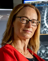
Dr Maren Krueger
Consultant Radiologist & Director
-

Dr Alina Leigh
Consultant Radiologist & Director
-

Dr Dana Tipene-Hook
Consultant Radiologist & Director
Ages
Child / Tamariki, Youth / Rangatahi, Adult / Pakeke, Older adult / Kaumātua
How do I access this service?
Walk in
Referral
GPs, Specialists, Midwives, Physiotherapists, Osteopaths, Chiropractors, Nurse Practitioners, Podiatrists and Dentists can all refer you to Taranaki Radiology.
Make an appointment
Referral Expectations
Referrers: find referral and PACS information here
Fees and Charges Description
We are an affiliated provider with Southern Cross Health Insurance and NIB Healthcare.
Hours
9:00 AM to 5:30 PM.
| Mon – Fri | 9:00 AM – 5:30 PM |
|---|
Closed 12.30pm - 1pm daily
Languages Spoken
English
Services Provided
An X-ray is a high frequency, high energy wave form. It cannot be seen with the naked eye, but can be picked up on photographic film. Although you may think of an X-ray as a picture of bones, a trained observer can also see air spaces, like the lungs (which look black) and fluid (which looks white, but not as white as bones). What to expect? You will have all metal objects removed from your body. You will be asked to remain still in a specific position and hold your breath on command. There are staff present, but they will not necessarily remain in the room, but will speak with you via an intercom system and will be viewing the procedure constantly through a windowed control room. The examination time will vary depending on the type of procedure required, but as a rule it will take around 30 minutes. Find out more about x-rays at Taranaki Radiology here
An X-ray is a high frequency, high energy wave form. It cannot be seen with the naked eye, but can be picked up on photographic film. Although you may think of an X-ray as a picture of bones, a trained observer can also see air spaces, like the lungs (which look black) and fluid (which looks white, but not as white as bones). What to expect? You will have all metal objects removed from your body. You will be asked to remain still in a specific position and hold your breath on command. There are staff present, but they will not necessarily remain in the room, but will speak with you via an intercom system and will be viewing the procedure constantly through a windowed control room. The examination time will vary depending on the type of procedure required, but as a rule it will take around 30 minutes. Find out more about x-rays at Taranaki Radiology here
An X-ray is a high frequency, high energy wave form. It cannot be seen with the naked eye, but can be picked up on photographic film. Although you may think of an X-ray as a picture of bones, a trained observer can also see air spaces, like the lungs (which look black) and fluid (which looks white, but not as white as bones).
What to expect?
You will have all metal objects removed from your body. You will be asked to remain still in a specific position and hold your breath on command. There are staff present, but they will not necessarily remain in the room, but will speak with you via an intercom system and will be viewing the procedure constantly through a windowed control room.
The examination time will vary depending on the type of procedure required, but as a rule it will take around 30 minutes.
Find out more about x-rays at Taranaki Radiology here
Find out about immigration x-rays at Taranaki Radiology here
Find out about immigration x-rays at Taranaki Radiology here
Service types: X-ray.
Find out about immigration x-rays at Taranaki Radiology here
Online Booking URL
Parking
Parking is available out the front and around the side of the building.
Pharmacy
Find your nearest pharmacy here
Website
Contact Details
95 Vivian Street, New Plymouth
Taranaki
9:00 AM to 5:30 PM.
-
Phone
(06) 759 4317 extension 709
-
Fax
(06) 758 4797
Email
Website
95 On Vivian Medical Hub, 95 Vivian Street
Lower Vogeltown
New Plymouth
Taranaki 4310
Street Address
95 On Vivian Medical Hub, 95 Vivian Street
Lower Vogeltown
New Plymouth
Taranaki 4310
Was this page helpful?
This page was last updated at 11:27AM on October 17, 2025. This information is reviewed and edited by I-MED Radiology - Phoenix (Taranaki Radiology).