North Auckland, West Auckland > Private Hospitals & Specialists > Southern Cross Hospitals >
Southern Cross North Harbour Hospital - Urology
Private Surgical Service, Urology
Description
Established in 1991, our North Harbour Hospital typically provides services to over 7,000 patients annually, and is widely known amongst patients and specialists in the area. It is their demand for our services that drives new investments, and that has delivered significant extensions, implementation of new technologies, and ongoing improvements to operating and nursing facilities. Recent upgrades have also seen improvements to recovery and day stay and pre-admission facilities and specialist consulting areas.
Within the grounds of the North Harbour Hospital campus is the Northern Clinic, which comprises Specialist consulting, Endoscopy services, Image Guided Healthcare, Radiology services and rehabilitation.
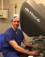
The hospital campus offers specialists and their patients access to new advanced operating theatres, including a robotically-assisted surgical system and other advanced technologies to support advanced imaging and a range of minimally invasive treatments. New recovery areas and other patient facilities have also been added, during 2017, along with new consulting rooms and a purpose-built endoscopy suite. These facilities offer patients a wide range of specialist services on one developing health campus which also includes a pharmacy, radiology unit, cardiology services, and a specially designed post-operative intermediate care facility - which helps to increase the scope and complexity of surgical services that can be provided within the private sector on Auckland's North Shore.
Consultants
-

Mr Tony Beaven
Urologist
-

Mr Jason Du
Urologist
-

Dr Eva Fong
Urologist
-

Mr Chris Hawke
Urologist
-

Mr Andrew Lienert
Urologist
-
Dr Sum Sum Lo
Urologist
-

Mr Michael Mackey
Urologist
-

Mr David Merrilees
Urologist
-

Mr Mischel Neill
Urologist
-

Mr Morgan Pokorny
Urologist
-

Mr Simon van Rij
Urologist
Procedures / Treatments
The foreskin is pulled away from the body of the penis and cut off, exposing the underlying head of the penis (glans). Stitches may be required to keep the remaining edges of the foreskin in place.
The foreskin is pulled away from the body of the penis and cut off, exposing the underlying head of the penis (glans). Stitches may be required to keep the remaining edges of the foreskin in place.
Incisions (cuts) are made in the abdomen (stomach) to allow access to the bladder. Tissue lying next to the bladder is attached to a solid structure within the pelvis, allowing the bladder neck to be supported, thus correcting urine leakage.
Incisions (cuts) are made in the abdomen (stomach) to allow access to the bladder. Tissue lying next to the bladder is attached to a solid structure within the pelvis, allowing the bladder neck to be supported, thus correcting urine leakage.
A long, thin tube with a tiny camera attached (cystoscope) is inserted into the urinary opening and through the urethra (the tube that carries urine from your bladder to the outside of your body) to your bladder. This allows the urologist to view any abnormalities in your lower urinary tract and, if necessary, take a small tissue sample to look at under the microscope (biopsy).
A long, thin tube with a tiny camera attached (cystoscope) is inserted into the urinary opening and through the urethra (the tube that carries urine from your bladder to the outside of your body) to your bladder. This allows the urologist to view any abnormalities in your lower urinary tract and, if necessary, take a small tissue sample to look at under the microscope (biopsy).
Incisions (cuts) are made in the side of the body, between the ribs and hip, to allow removal of one or both kidneys.
Incisions (cuts) are made in the side of the body, between the ribs and hip, to allow removal of one or both kidneys.
A tube is inserted into the kidney to allow urine to drain out. The tube may drain into a bag on the outside of your body (on your back) or may drain inside your body into the bladder.
A tube is inserted into the kidney to allow urine to drain out. The tube may drain into a bag on the outside of your body (on your back) or may drain inside your body into the bladder.
A small incision (cut) is made in the groin on the side of the undescended testicle and the testicle pulled down into the scrotum. Sometimes a small cut will need to be made in the scrotum as well.
A small incision (cut) is made in the groin on the side of the undescended testicle and the testicle pulled down into the scrotum. Sometimes a small cut will need to be made in the scrotum as well.
Scrotal: a small incision (cut) is made in the front of the scrotum and the testicles removed. This greatly reduces the amount of testosterone produced in the body. Inguinal: an incision is made in the groin to remove a testicle that: is undescended from childhood, has wasted away (atrophied), or has a tumour.
Scrotal: a small incision (cut) is made in the front of the scrotum and the testicles removed. This greatly reduces the amount of testosterone produced in the body. Inguinal: an incision is made in the groin to remove a testicle that: is undescended from childhood, has wasted away (atrophied), or has a tumour.
A thin wire is inserted into your lower back and guided using x-ray imaging to your kidney. A small incision (cut) is then made on your back and a narrow tube is inserted and follows the guide wire to the kidney. The kidney stone(s) is then removed or broken up.
A thin wire is inserted into your lower back and guided using x-ray imaging to your kidney. A small incision (cut) is then made on your back and a narrow tube is inserted and follows the guide wire to the kidney. The kidney stone(s) is then removed or broken up.
Incisions (cuts) are made in either the lower abdomen (stomach) or between the scrotum and the anus to allow removal of the enlarged parts of, or the entire, prostate gland.
Incisions (cuts) are made in either the lower abdomen (stomach) or between the scrotum and the anus to allow removal of the enlarged parts of, or the entire, prostate gland.
Small incisions (cuts) are made in the lower abdomen (stomach) and in the front wall of the vagina. Synthetic tissue is inserted to form a supportive sling under the urethra at the bladder neck to prevent urine leakage.
Small incisions (cuts) are made in the lower abdomen (stomach) and in the front wall of the vagina. Synthetic tissue is inserted to form a supportive sling under the urethra at the bladder neck to prevent urine leakage.
A long, thin tube with a tiny camera attached (resectoscope) is inserted into the urinary opening, through the urethra and into the bladder. Instruments are passed through the resectoscope and the tumour removed.
A long, thin tube with a tiny camera attached (resectoscope) is inserted into the urinary opening, through the urethra and into the bladder. Instruments are passed through the resectoscope and the tumour removed.
A long, thin tube with a tiny camera attached (resectoscope) is inserted into the urinary opening of the penis and through the urethra (the tube that carries urine from your bladder to the outside of your body) to your bladder. The urologist is then able to view the prostate gland and, by passing an instrument through the resectoscope, is able to remove the part of the gland that has become enlarged.
A long, thin tube with a tiny camera attached (resectoscope) is inserted into the urinary opening of the penis and through the urethra (the tube that carries urine from your bladder to the outside of your body) to your bladder. The urologist is then able to view the prostate gland and, by passing an instrument through the resectoscope, is able to remove the part of the gland that has become enlarged.
A long, thin tube with a tiny camera attached (ureteroscope) is inserted into the urinary opening, through the urethra (the tube that carries urine from your bladder to the outside of your body) and bladder to the ureters (the two tubes that drain urine from the kidneys to the bladder). This allows the urologist to view and, in some cases, treat any problems in the ureters.
A long, thin tube with a tiny camera attached (ureteroscope) is inserted into the urinary opening, through the urethra (the tube that carries urine from your bladder to the outside of your body) and bladder to the ureters (the two tubes that drain urine from the kidneys to the bladder). This allows the urologist to view and, in some cases, treat any problems in the ureters.
An incision (cut) is made in the penis and the narrowed part of the urethra (the tube that carries urine to the outside of your body) is removed and the urethra rejoined. In balloon urethroplasty, a thin tube with a balloon attached is inserted into the opening of the penis. When it reaches the narrowed part of the urethra, the balloon is inflated, thus widening the urethra.
An incision (cut) is made in the penis and the narrowed part of the urethra (the tube that carries urine to the outside of your body) is removed and the urethra rejoined. In balloon urethroplasty, a thin tube with a balloon attached is inserted into the opening of the penis. When it reaches the narrowed part of the urethra, the balloon is inflated, thus widening the urethra.
A tiny incision (cut) is made in the scrotum and a short length of the vas deferens (the tube carrying sperm away from the testicles where it is produced) is removed.
A tiny incision (cut) is made in the scrotum and a short length of the vas deferens (the tube carrying sperm away from the testicles where it is produced) is removed.
Visiting Hours
- Weekdays: 11:00 to 20:00
- Weekends: 11:00 to 20:00
Travel Directions
Please use entry B, 232 Wairau Road for Hospital entrance, car parking and patient drop off.
click here for street view location.
Parking
Patient and visitor parking provided.
Contact Details
Southern Cross North Harbour Hospital,
North Auckland
Phone: (09) 925 4400
Fax: (09) 925 4434
E-mail: northharbour@southerncrosshospitals.co.nz
Location details here
Surgeons can be contacted directly at their private consultation rooms.
232 Wairau Road
Totara Vale
Kaipātiki
Auckland 0629
Street Address
232 Wairau Road
Totara Vale
Kaipātiki
Auckland 0629
Postal Address
P.O. Box 101-488,
North Shore Mail Centre,
Auckland City, 0745
Was this page helpful?
This page was last updated at 2:37PM on October 3, 2024. This information is reviewed and edited by Southern Cross North Harbour Hospital - Urology.

