Central Auckland > Public Hospital Services > Starship Child Health >
Starship Paediatric Neurosurgery
Public Service, Neurosurgery, Paediatrics
Description
The Neurosurgical Department at Starship Children's Health treats children aged 0-15 yrs with diseases or trauma involving the central and peripheral nervous system. It covers the full range of neurosurgical problems, some of which include trauma (mild to severe head injuries), benign and malignant brain and spinal tumours, neurovascular conditions, congenital problems, craniofacial surgery, and management of hydrocephalus.
The Department currently covers an extensive area of the North Island from Northland, all of Auckland, Coromandel to Taranaki, Waikato, Lakeland and Tairāwhiti.
Consultants
-

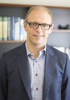
Mr Lawrence (Siu) Choi
Neurosurgeon
-

Mr Peter Heppner
Neurosurgeon
-

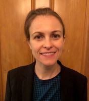
Ms Phoebe Matthews
Neurosurgeon
Referral Expectations
Referrals are triaged (sorted into groups according to urgency of need) by specialists and allocated a grading. If serious, i.e Grade A, the case is booked immediately for Outpatients.
Urgent acute cases go through the Children's Emergency Department via the Paediatric Neurosurgical registrar and after hours and in the weekend by the on call registrar.
Hours
Our service operates 24 hours a day, 7 days a week.
Procedures / Treatments
A cerebral (cranial) aneurysm is a weakened section in the wall of a blood vessel in the brain that bulges or balloons out. Possible causes of cerebral aneurysm include: a defect in the blood vessels present at birth, brain tumour, head trauma or atherosclerosis (fatty deposits start to block the arteries). A small aneurysm may produce no symptoms but as it grows it might cause vision problems, facial numbness or seizures. A ruptured or burst aneurysm can cause bleeding in and around the brain which may affect mental skills and bodily functions and may, in serious cases, lead to brain damage, stroke, coma or death. Surgical Clipping This is a treatment that can be used for both unruptured and ruptured aneurysms. The skull is opened surgically (craniotomy) and the aneurysm is isolated from the rest of the blood vessel using a small metal clip that seals off each end of the aneurysm. Endovascular Clipping This is a less invasive form of treatment that avoids the need for surgery. A catheter (a small, flexible tube) is inserted into an artery in the groin and gently pushed up to the brain. At the site of the aneurysm, the catheter releases soft wire coils that block the aneurysm from inside the blood vessel. Sometimes a tiny balloon is also released to help hold the coils in place.
A cerebral (cranial) aneurysm is a weakened section in the wall of a blood vessel in the brain that bulges or balloons out. Possible causes of cerebral aneurysm include: a defect in the blood vessels present at birth, brain tumour, head trauma or atherosclerosis (fatty deposits start to block the arteries). A small aneurysm may produce no symptoms but as it grows it might cause vision problems, facial numbness or seizures. A ruptured or burst aneurysm can cause bleeding in and around the brain which may affect mental skills and bodily functions and may, in serious cases, lead to brain damage, stroke, coma or death. Surgical Clipping This is a treatment that can be used for both unruptured and ruptured aneurysms. The skull is opened surgically (craniotomy) and the aneurysm is isolated from the rest of the blood vessel using a small metal clip that seals off each end of the aneurysm. Endovascular Clipping This is a less invasive form of treatment that avoids the need for surgery. A catheter (a small, flexible tube) is inserted into an artery in the groin and gently pushed up to the brain. At the site of the aneurysm, the catheter releases soft wire coils that block the aneurysm from inside the blood vessel. Sometimes a tiny balloon is also released to help hold the coils in place.
Brain tumours may be primary (they arise in the brain or spinal cord) or metastatic (they have originated in another part of the body and travelled to the brain). Primary tumours are the most common paediatric tumour and may either be benign (slow growing) or malignant (aggressive). The type of tumour is usually diagnosed after surgery or biopsy. Histology can take 7-10 working days. Surgery may be the only treatment approach for a brain tumour, or it may be used in combination with radiation therapy and/or chemotherapy. Typically, the skull is opened up (craniotomy) giving the surgeon access to the tumour and allowing removal of as much of the tumour as safely as possible. There are a small number of tumours that are inoperable, meaning that the tumour cannot be safely removed without significant neurological deficit and/or death. A biopsy is another surgical procedure often performed to aid in tumour diagnosis. A small hole is drilled into the skull and a sample of tissue removed for examination under the microscope. Radiation therapy uses high energy x-rays to kill abnormal cells, while chemotherapy uses chemicals (drugs) to destroy cancer cells. Prognosis depends on the type of tumour, location and if there is any tumour left after surgery.
Brain tumours may be primary (they arise in the brain or spinal cord) or metastatic (they have originated in another part of the body and travelled to the brain). Primary tumours are the most common paediatric tumour and may either be benign (slow growing) or malignant (aggressive). The type of tumour is usually diagnosed after surgery or biopsy. Histology can take 7-10 working days. Surgery may be the only treatment approach for a brain tumour, or it may be used in combination with radiation therapy and/or chemotherapy. Typically, the skull is opened up (craniotomy) giving the surgeon access to the tumour and allowing removal of as much of the tumour as safely as possible. There are a small number of tumours that are inoperable, meaning that the tumour cannot be safely removed without significant neurological deficit and/or death. A biopsy is another surgical procedure often performed to aid in tumour diagnosis. A small hole is drilled into the skull and a sample of tissue removed for examination under the microscope. Radiation therapy uses high energy x-rays to kill abnormal cells, while chemotherapy uses chemicals (drugs) to destroy cancer cells. Prognosis depends on the type of tumour, location and if there is any tumour left after surgery.
Tumours may be found within the spinal cord itself, between the spinal cord and its tough outer covering, the dura, or outside the dura. They may be primary (they arise in the spine or nearby tissue) or metastatic (they have originated in another part of the body and travelled to the spine). Most children's spinal tumours will be primary in nature. Spinal tumours may be treated by any combination of surgery, radiotherapy and chemotherapy. Surgery may be performed to take a small sample of tissue to examine under the microscope (biopsy) or to remove the tumour. Typically, the patient will be lying face downwards and a procedure known as a laminectomy is performed (the bone overlying the spinal cord is removed). This gives the surgeon access to the spinal cord and allows removal of the tumour.
Tumours may be found within the spinal cord itself, between the spinal cord and its tough outer covering, the dura, or outside the dura. They may be primary (they arise in the spine or nearby tissue) or metastatic (they have originated in another part of the body and travelled to the spine). Most children's spinal tumours will be primary in nature. Spinal tumours may be treated by any combination of surgery, radiotherapy and chemotherapy. Surgery may be performed to take a small sample of tissue to examine under the microscope (biopsy) or to remove the tumour. Typically, the patient will be lying face downwards and a procedure known as a laminectomy is performed (the bone overlying the spinal cord is removed). This gives the surgeon access to the spinal cord and allows removal of the tumour.
Tumours may be found within the spinal cord itself, between the spinal cord and its tough outer covering, the dura, or outside the dura. They may be primary (they arise in the spine or nearby tissue) or metastatic (they have originated in another part of the body and travelled to the spine). Most children's spinal tumours will be primary in nature.
This procedure involves making an incision down the centre of the back and removing some or all of the bony arch (lamina) off a vertebra. In a laminectomy, all or most of the lamina is surgically removed. By making more room in the spinal canal, this procedure reduces pressure on the spinal nerves. It also gives the surgeon better access to the disc and other parts of the spine if further procedures e.g. spinal cord detethering, removal of tumour, are required.
This procedure involves making an incision down the centre of the back and removing some or all of the bony arch (lamina) off a vertebra. In a laminectomy, all or most of the lamina is surgically removed. By making more room in the spinal canal, this procedure reduces pressure on the spinal nerves. It also gives the surgeon better access to the disc and other parts of the spine if further procedures e.g. spinal cord detethering, removal of tumour, are required.
An extradural haematoma (EDH) is usually an injury to the brain that occurs following a blow to the head. This can be due to falls, road traffic accidents or blunt trauma. Bleeding occurs into the space between the membranes that cover the brain. If the bleed is large, this is a surgical emergency and the child needs to go to theatre to have the clot removed and to ensure that the bleeding has stopped. A large clot can put pressure on the surrounding brain tissue. For smaller bleeds where there does not appear to be any pressure from the clot, the child will be observed for a few days and the body will reabsorb the blood over a few weeks. Symptoms include: · headache . drowsiness . irritability · speech problems · vision problems · seizures · confusion · weakness · nausea and vomiting. After sustaining a head injury causing an EDH some people can have a lucid period prior to collapse from the bleed.
An extradural haematoma (EDH) is usually an injury to the brain that occurs following a blow to the head. This can be due to falls, road traffic accidents or blunt trauma. Bleeding occurs into the space between the membranes that cover the brain. If the bleed is large, this is a surgical emergency and the child needs to go to theatre to have the clot removed and to ensure that the bleeding has stopped. A large clot can put pressure on the surrounding brain tissue. For smaller bleeds where there does not appear to be any pressure from the clot, the child will be observed for a few days and the body will reabsorb the blood over a few weeks. Symptoms include: · headache . drowsiness . irritability · speech problems · vision problems · seizures · confusion · weakness · nausea and vomiting. After sustaining a head injury causing an EDH some people can have a lucid period prior to collapse from the bleed.
An extradural haematoma (EDH) is usually an injury to the brain that occurs following a blow to the head. This can be due to falls, road traffic accidents or blunt trauma.
Bleeding occurs into the space between the membranes that cover the brain. If the bleed is large, this is a surgical emergency and the child needs to go to theatre to have the clot removed and to ensure that the bleeding has stopped. A large clot can put pressure on the surrounding brain tissue. For smaller bleeds where there does not appear to be any pressure from the clot, the child will be observed for a few days and the body will reabsorb the blood over a few weeks.
A subdural haematoma is a collection of blood that forms beneath the outer protective covering of the brain, the dura mater. It is usually caused by tiny blood vessels becoming torn as the result of serious head trauma such as a fall, blow to the head or car accident. Symptoms include: · headache . drowsiness . irritability · speech problems · vision problems · seizures · confusion · weakness · nausea and vomiting. With an acute haematoma, symptoms appear within 24 hours of the trauma while in the case of subacute or chronic haematomas symptoms take longer to appear. If a haematoma is increasing in size it puts pressure on the brain which may lead to brain damage and possibly death. Depending on the size of the haematoma surgical treatment may be required. This involves drilling a small hole in the skull, allowing the haematoma to drain and thus relieving the pressure on the brain. In the case of a larger haematoma, a hole may be cut in the skull (craniotomy) allowing the surgeon access to the brain to repair damaged vessels and remove the blood clot.
A subdural haematoma is a collection of blood that forms beneath the outer protective covering of the brain, the dura mater. It is usually caused by tiny blood vessels becoming torn as the result of serious head trauma such as a fall, blow to the head or car accident. Symptoms include: · headache . drowsiness . irritability · speech problems · vision problems · seizures · confusion · weakness · nausea and vomiting. With an acute haematoma, symptoms appear within 24 hours of the trauma while in the case of subacute or chronic haematomas symptoms take longer to appear. If a haematoma is increasing in size it puts pressure on the brain which may lead to brain damage and possibly death. Depending on the size of the haematoma surgical treatment may be required. This involves drilling a small hole in the skull, allowing the haematoma to drain and thus relieving the pressure on the brain. In the case of a larger haematoma, a hole may be cut in the skull (craniotomy) allowing the surgeon access to the brain to repair damaged vessels and remove the blood clot.
Hydrocephalus is an abnormal (excessive) accumulation of fluid in the head. The fluid is called cerebrospinal fluid, commonly referred to as CSF. The CSF is located and produced within the fluid cavities of the brain called ventricles. The function of CSF is to cushion the delicate brain and spinal cord tissue from injuries and maintain proper balance of nutrients around the central nervous system. Normally, most of the CSF produced on a daily basis is absorbed by the blood stream. Everyday your body produces a certain amount of CSF, that same amount is absorbed by the brain. When an imbalance occurs, an excess of CSF builds up in the ventricles resulting in a condition known as hydrocephalus. Left untreated, hydrocephalus will create increased pressure in the head, which can cause neurological deficits or even death. Hydrocephalus may be caused by one or more of the following: interference with the normal CSF flow, due to an obstruction or blockage in the CSF pathway, e.g. tumours or congential cause over-production of CSF under-absorption of CSF into the bloodstream. There are two types of hydrocephalus: communicating hydrocephalus caused by the over-production or under-absorption of CSF non-communicating or obstructive hydrocephalus caused by a blockage of the CSF pathways. Hydrocephalus is further deemed congenital if present before or since birth; or hydrocephalus can be acquired, developing after birth. A variety of causes can contribute to acquired hydrocephalus; some are head injury, tumours and meningitis.
Hydrocephalus is an abnormal (excessive) accumulation of fluid in the head. The fluid is called cerebrospinal fluid, commonly referred to as CSF. The CSF is located and produced within the fluid cavities of the brain called ventricles. The function of CSF is to cushion the delicate brain and spinal cord tissue from injuries and maintain proper balance of nutrients around the central nervous system. Normally, most of the CSF produced on a daily basis is absorbed by the blood stream. Everyday your body produces a certain amount of CSF, that same amount is absorbed by the brain. When an imbalance occurs, an excess of CSF builds up in the ventricles resulting in a condition known as hydrocephalus. Left untreated, hydrocephalus will create increased pressure in the head, which can cause neurological deficits or even death. Hydrocephalus may be caused by one or more of the following: interference with the normal CSF flow, due to an obstruction or blockage in the CSF pathway, e.g. tumours or congential cause over-production of CSF under-absorption of CSF into the bloodstream. There are two types of hydrocephalus: communicating hydrocephalus caused by the over-production or under-absorption of CSF non-communicating or obstructive hydrocephalus caused by a blockage of the CSF pathways. Hydrocephalus is further deemed congenital if present before or since birth; or hydrocephalus can be acquired, developing after birth. A variety of causes can contribute to acquired hydrocephalus; some are head injury, tumours and meningitis.
Hydrocephalus is an abnormal (excessive) accumulation of fluid in the head. The fluid is called cerebrospinal fluid, commonly referred to as CSF. The CSF is located and produced within the fluid cavities of the brain called ventricles. The function of CSF is to cushion the delicate brain and spinal cord tissue from injuries and maintain proper balance of nutrients around the central nervous system.
Normally, most of the CSF produced on a daily basis is absorbed by the blood stream. Everyday your body produces a certain amount of CSF, that same amount is absorbed by the brain. When an imbalance occurs, an excess of CSF builds up in the ventricles resulting in a condition known as hydrocephalus. Left untreated, hydrocephalus will create increased pressure in the head, which can cause neurological deficits or even death.
Hydrocephalus may be caused by one or more of the following:
- interference with the normal CSF flow, due to an obstruction or blockage in the CSF pathway, e.g. tumours or congential cause
- over-production of CSF
- under-absorption of CSF into the bloodstream.
There are two types of hydrocephalus:
- communicating hydrocephalus caused by the over-production or under-absorption of CSF
- non-communicating or obstructive hydrocephalus caused by a blockage of the CSF pathways.
Hydrocephalus is further deemed congenital if present before or since birth; or hydrocephalus can be acquired, developing after birth. A variety of causes can contribute to acquired hydrocephalus; some are head injury, tumours and meningitis.
Ventriculoperitoneal shunt or VP shunt is a treatment for hydrocephalus. It is a device used to divert the cerebrospinal fluid (CSF) from the ventricles in the brain to the abdominal cavity where it can be absorbed. The fluid is then absorbed naturally through the peritoneal lining in the abdominal cavity back into the bloodstream. A VP shunt needs to be surgically inserted under general anaesthetic by a neurosurgeon. VP shunts are not without problems: they can get blocked and require surgical revision they can disconnect and require surgical revision they can get infected and in this case they need to be removed and treatment with intravenous antibiotics is required. A week to 10 days later a new VP shunt can be inserted. Symptoms of shunt failure can include: headaches vomiting increased drowsiness irritablilty increasing head circumference (infants) hard, bulging fontanelle (infants) seizures changes in pupils sunsetting eyes fever If any of these symptoms occur the child needs to be brought to the Children's Emergency Department (CED) for assessment by the Paediatric Neurosurgical Registrar or to the family GP.
Ventriculoperitoneal shunt or VP shunt is a treatment for hydrocephalus. It is a device used to divert the cerebrospinal fluid (CSF) from the ventricles in the brain to the abdominal cavity where it can be absorbed. The fluid is then absorbed naturally through the peritoneal lining in the abdominal cavity back into the bloodstream. A VP shunt needs to be surgically inserted under general anaesthetic by a neurosurgeon. VP shunts are not without problems: they can get blocked and require surgical revision they can disconnect and require surgical revision they can get infected and in this case they need to be removed and treatment with intravenous antibiotics is required. A week to 10 days later a new VP shunt can be inserted. Symptoms of shunt failure can include: headaches vomiting increased drowsiness irritablilty increasing head circumference (infants) hard, bulging fontanelle (infants) seizures changes in pupils sunsetting eyes fever If any of these symptoms occur the child needs to be brought to the Children's Emergency Department (CED) for assessment by the Paediatric Neurosurgical Registrar or to the family GP.
Ventriculoperitoneal shunt or VP shunt is a treatment for hydrocephalus. It is a device used to divert the cerebrospinal fluid (CSF) from the ventricles in the brain to the abdominal cavity where it can be absorbed. The fluid is then absorbed naturally through the peritoneal lining in the abdominal cavity back into the bloodstream. A VP shunt needs to be surgically inserted under general anaesthetic by a neurosurgeon. VP shunts are not without problems:
- they can get blocked and require surgical revision
- they can disconnect and require surgical revision
- they can get infected and in this case they need to be removed and treatment with intravenous antibiotics is required. A week to 10 days later a new VP shunt can be inserted.
Symptoms of shunt failure can include:
- headaches
- vomiting
- increased drowsiness
- irritablilty
- increasing head circumference (infants)
- hard, bulging fontanelle (infants)
- seizures
- changes in pupils
- sunsetting eyes
- fever
If any of these symptoms occur the child needs to be brought to the Children's Emergency Department (CED) for assessment by the Paediatric Neurosurgical Registrar or to the family GP.
Mild Head Injury & Concussion Advice (PDF, 168.7 KB) Family information for going home
Mild Head Injury & Concussion Advice (PDF, 168.7 KB) Family information for going home
-
Mild Head Injury & Concussion Advice
(PDF, 168.7 KB)
Family information for going home
In tethered spinal cord, the lower end of the spinal cord stays attached to the base of the spine, rather than riding freely up and down the spinal canal. Since the spine grows faster than the spinal cord, this leads to increasing stress on the spinal cord as a child grows and becomes more active. Symptoms such as weakness in the legs, low back pain and incontinence may be the first signs of this condition.
In tethered spinal cord, the lower end of the spinal cord stays attached to the base of the spine, rather than riding freely up and down the spinal canal. Since the spine grows faster than the spinal cord, this leads to increasing stress on the spinal cord as a child grows and becomes more active. Symptoms such as weakness in the legs, low back pain and incontinence may be the first signs of this condition.
Craniosynostosis is a disorder in which there is early fusion of the sutures of the skull in childhood. It produces an abnormally shaped head and, at times, appearance of the face. The deformity varies significantly depending on the sutures or suture involved. Surgical correction may be necessary to improve appearance and provide space for the brain to grow. Children with this condition are seen in the Craniofacial Clinic at Superclinic in Manakau and have their surgery at Starship with a combined approach from the Plastic Surgeon and the Neurosurgeon.
Craniosynostosis is a disorder in which there is early fusion of the sutures of the skull in childhood. It produces an abnormally shaped head and, at times, appearance of the face. The deformity varies significantly depending on the sutures or suture involved. Surgical correction may be necessary to improve appearance and provide space for the brain to grow. Children with this condition are seen in the Craniofacial Clinic at Superclinic in Manakau and have their surgery at Starship with a combined approach from the Plastic Surgeon and the Neurosurgeon.
Craniosynostosis is a disorder in which there is early fusion of the sutures of the skull in childhood. It produces an abnormally shaped head and, at times, appearance of the face. The deformity varies significantly depending on the sutures or suture involved. Surgical correction may be necessary to improve appearance and provide space for the brain to grow. Children with this condition are seen in the Craniofacial Clinic at Superclinic in Manakau and have their surgery at Starship with a combined approach from the Plastic Surgeon and the Neurosurgeon.
Cerebral arteriovenous malformation (AVM) often causes no signs or symptoms until the AVM ruptures, resulting in bleeding in the brain. A cerebral arteriovenous malformation is an abnormal connection between arteries and veins in the brain that's present at birth (congenital). Arteriovenous malformations, which appear as tangles of normal or dilated blood vessels, can occur in any part of the brain. The cause isn't clear. In most people with arteriovenous malformation bleeding is the first symptom, but some may experience: seizures a whooshing sound (bruit) that can be heard on examination of the skull with a stethoscope pulsing noise in the head (pulsatile tinnitus) headache progressive weakness or numbness. When bleeding into the brain occurs, signs and symptom can be similar to a stroke and may include: sudden, severe headache weakness or numbness vision loss difficulty speaking inability to understand others severe unsteadiness. A bleeding AVM is life-threatening and requires emergency medical attention.
Cerebral arteriovenous malformation (AVM) often causes no signs or symptoms until the AVM ruptures, resulting in bleeding in the brain. A cerebral arteriovenous malformation is an abnormal connection between arteries and veins in the brain that's present at birth (congenital). Arteriovenous malformations, which appear as tangles of normal or dilated blood vessels, can occur in any part of the brain. The cause isn't clear. In most people with arteriovenous malformation bleeding is the first symptom, but some may experience: seizures a whooshing sound (bruit) that can be heard on examination of the skull with a stethoscope pulsing noise in the head (pulsatile tinnitus) headache progressive weakness or numbness. When bleeding into the brain occurs, signs and symptom can be similar to a stroke and may include: sudden, severe headache weakness or numbness vision loss difficulty speaking inability to understand others severe unsteadiness. A bleeding AVM is life-threatening and requires emergency medical attention.
Cerebral arteriovenous malformation (AVM) often causes no signs or symptoms until the AVM ruptures, resulting in bleeding in the brain.
A cerebral arteriovenous malformation is an abnormal connection between arteries and veins in the brain that's present at birth (congenital). Arteriovenous malformations, which appear as tangles of normal or dilated blood vessels, can occur in any part of the brain. The cause isn't clear.
In most people with arteriovenous malformation bleeding is the first symptom, but some may experience:
- seizures
- a whooshing sound (bruit) that can be heard on examination of the skull with a stethoscope
- pulsing noise in the head (pulsatile tinnitus)
- headache
- progressive weakness or numbness.
When bleeding into the brain occurs, signs and symptom can be similar to a stroke and may include:
- sudden, severe headache
- weakness or numbness
- vision loss
- difficulty speaking
- inability to understand others
- severe unsteadiness.
A bleeding AVM is life-threatening and requires emergency medical attention.
"CT" stands for Computerised Tomography. It is a process which produces x-ray pictures as cross-sectional images (slices) of the body. This technology allows the doctors to see details of the brain, spine and other internal organs that are not visible on plain x-ray. It is possible that before or during the scan the child will be given intravenous contrast medicine. This is sometimes necessary to produce better results, as it will make the organ under investigation stand out in relation to the surrounding tissues. In neurosurgery contrast is often used for looking for infection or tumours. The CT scanner consists of a doughnut or ring-shaped structure, as well as a table which the child will lie on. During the scan the table will automatically move, as will the ring-shaped structure. The child will need to lie flat on their back and it will only take a few minutes. It does not hurt. A parent or caregiver can be present during the scan if this will help to settle the child. Some children under the age of 3 years old may require some oral sedation prior to the scan as it is imperative that the child lie still.
"CT" stands for Computerised Tomography. It is a process which produces x-ray pictures as cross-sectional images (slices) of the body. This technology allows the doctors to see details of the brain, spine and other internal organs that are not visible on plain x-ray. It is possible that before or during the scan the child will be given intravenous contrast medicine. This is sometimes necessary to produce better results, as it will make the organ under investigation stand out in relation to the surrounding tissues. In neurosurgery contrast is often used for looking for infection or tumours. The CT scanner consists of a doughnut or ring-shaped structure, as well as a table which the child will lie on. During the scan the table will automatically move, as will the ring-shaped structure. The child will need to lie flat on their back and it will only take a few minutes. It does not hurt. A parent or caregiver can be present during the scan if this will help to settle the child. Some children under the age of 3 years old may require some oral sedation prior to the scan as it is imperative that the child lie still.
"CT" stands for Computerised Tomography. It is a process which produces x-ray pictures as cross-sectional images (slices) of the body. This technology allows the doctors to see details of the brain, spine and other internal organs that are not visible on plain x-ray.
It is possible that before or during the scan the child will be given intravenous contrast medicine. This is sometimes necessary to produce better results, as it will make the organ under investigation stand out in relation to the surrounding tissues. In neurosurgery contrast is often used for looking for infection or tumours.
The CT scanner consists of a doughnut or ring-shaped structure, as well as a table which the child will lie on. During the scan the table will automatically move, as will the ring-shaped structure. The child will need to lie flat on their back and it will only take a few minutes. It does not hurt. A parent or caregiver can be present during the scan if this will help to settle the child. Some children under the age of 3 years old may require some oral sedation prior to the scan as it is imperative that the child lie still.
"MRI" stands for Magnetic Resonance Imaging; it is the latest and most advanced method of diagnostic imaging. It combines a powerful magnet, radio waves and a sophisticated computer to create highly detailed anatomical images of the body. It has two major advantages. Firstly, scanning can be performed in any plane within the body therefore greatly enhancing the information that can be obtained. Secondly, no x-rays are used. Your child will need to lie flat and still on the MRI table. A parent or caregiver can be in the room while the scan is being done. The table will move slowly into the scanner. The part of the body being scanned must be positioned in the centre of the magnet. The magnet is open at both ends and there is two way communication with the radiographer. During the scan you can hear a rhythmic tapping sound which will last from several seconds up to several minutes. The scan can take anywhere from 30-60 minutes. If you don't think your child will be able to lie still for this scan then a general anaesthetic is recommended.
"MRI" stands for Magnetic Resonance Imaging; it is the latest and most advanced method of diagnostic imaging. It combines a powerful magnet, radio waves and a sophisticated computer to create highly detailed anatomical images of the body. It has two major advantages. Firstly, scanning can be performed in any plane within the body therefore greatly enhancing the information that can be obtained. Secondly, no x-rays are used. Your child will need to lie flat and still on the MRI table. A parent or caregiver can be in the room while the scan is being done. The table will move slowly into the scanner. The part of the body being scanned must be positioned in the centre of the magnet. The magnet is open at both ends and there is two way communication with the radiographer. During the scan you can hear a rhythmic tapping sound which will last from several seconds up to several minutes. The scan can take anywhere from 30-60 minutes. If you don't think your child will be able to lie still for this scan then a general anaesthetic is recommended.
"MRI" stands for Magnetic Resonance Imaging; it is the latest and most advanced method of diagnostic imaging. It combines a powerful magnet, radio waves and a sophisticated computer to create highly detailed anatomical images of the body. It has two major advantages. Firstly, scanning can be performed in any plane within the body therefore greatly enhancing the information that can be obtained. Secondly, no x-rays are used.
Your child will need to lie flat and still on the MRI table. A parent or caregiver can be in the room while the scan is being done. The table will move slowly into the scanner. The part of the body being scanned must be positioned in the centre of the magnet. The magnet is open at both ends and there is two way communication with the radiographer. During the scan you can hear a rhythmic tapping sound which will last from several seconds up to several minutes. The scan can take anywhere from 30-60 minutes.
If you don't think your child will be able to lie still for this scan then a general anaesthetic is recommended.
Visiting Hours
Visiting hours are from 8am to 8pm, with quiet time on the ward from 1pm to 3pm, when only parents or caregivers are allowed on the ward.
Contact Details
Starship Child Health, Central Auckland
Central Auckland
-
Phone
(09) 307 4949 ext 22333
-
Fax
(09) 307 4912
Email
Website
2 Park Road
Grafton
Auckland 1023
Street Address
2 Park Road
Grafton
Auckland 1023
Postal Address
Starship Child Health
Private Bag 92 024
Auckland Mail Centre
Auckland 1142
New Zealand
Was this page helpful?
This page was last updated at 11:12AM on April 11, 2024. This information is reviewed and edited by Starship Paediatric Neurosurgery.

