Canterbury > Private Hospitals & Specialists > Southern Cross Hospitals >
Southern Cross Christchurch Hospital - Orthopaedic Surgery
Private Surgical Service, Orthopaedics
Description
Southern Cross Hospital in Christchurch is the largest hospital within our national network.
Owned by Southern Cross since 1979, the centrally situated hospital campus includes one of the biggest and most advanced private surgical hospitals in the South Island.
The Christchurch hospital campus has seen significant upgrades and new facilities in recent years, and typically provides services to around 9,500 patients each year. Facilities include digital operating theatres, an advanced 'hybrid' operating room, systems for robotically-assisted surgery, advanced digital scanning technologies, consulting facilities and a purpose built endoscopy centre.
Consultants
-
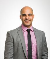
Dr Ramez Ailabouni
Orthopaedic Surgeon
-

Mr Gordon Beadel
Orthopaedic Surgeon
-

Mr Gordon Burgess
Orthopaedic Surgeon
-

Mr James Burn
Orthopaedic Surgeon
-

Ram Chandru (Ramamurthy Chandru)
Orthopaedic Surgeon
-

Dr Tim Chuang
Orthopaedic Surgeon
-

Mr Allen Cockfield
Orthopaedic Surgeon
-

Dr Richard Cowley
Orthopaedic Surgeon
-

Mr Simon Crampton
Orthopaedic Surgeon
-

Mr Mark Cvitanich
Orthopaedic Surgeon
-

Mr Jeremy Evison
Orthopaedic Surgeon
-

Mr Narlaka Jayasekera
Orthopaedic Surgeon
-

Mr Josh Kempthorne
Orthopaedic Surgeon
-

Mr David Kieser
Orthopaedic Surgeon
-

Mr Heath Lash
Orthopaedic Surgeon
-

Dr Alex Lee
Orthopaedic Surgeon
-

Mr Hamish Love
Orthopaedic Surgeon
-

Mr Alexander Malone
Orthopaedic Surgeon
-

Mr Rhett Mason
Orthopaedic Surgeon
-
Mr John McKie
Orthopaedic Surgeon
-

Mr Khalid Mohammed
Orthopaedic Surgeon
-

Mr Ian Penny
Orthopaedic Surgeon
-

Mr Andrew Powell
Orthopaedic Surgeon
-

Mr John Rietveld
Orthopaedic Surgeon
-
Mr Thomas Sharpe
Orthopaedic Surgeon
-

Dr Paul Sharplin
Orthopaedic Surgeon
-

Mr Jonathan Sharr
Orthopaedic Surgeon
-

Dr Bradley Stone
Orthopaedic Surgeon
-

Dr Fiona Timms
Orthopaedic Surgeon
-

Mr Bruce Twaddle
Orthopaedic Surgeon
-

Mr David Whitehead
Orthopaedic Surgeon
Procedures / Treatments
Two or three small incisions (cuts) are made in the ankle and a small telescopic instrument with a tiny camera attached (arthroscope) is inserted. This allows the surgeon to look inside the joint, identify problems and, in some cases, operate. Tiny instruments can be passed through the arthroscope to remove bony spurs, damaged cartilage or inflamed tissue.
Two or three small incisions (cuts) are made in the ankle and a small telescopic instrument with a tiny camera attached (arthroscope) is inserted. This allows the surgeon to look inside the joint, identify problems and, in some cases, operate. Tiny instruments can be passed through the arthroscope to remove bony spurs, damaged cartilage or inflamed tissue.
Two or three small incisions (cuts) are made in the ankle and a small telescopic instrument with a tiny camera attached (arthroscope) is inserted. This allows the surgeon to look inside the joint, identify problems and, in some cases, operate. Tiny instruments can be passed through the arthroscope to remove bony spurs, damaged cartilage or inflamed tissue.
An incision (cut) is made in the front of, and several smaller cuts on the outside of, the ankle. The damaged ankle joint is replaced with a metal and plastic implant.
An incision (cut) is made in the front of, and several smaller cuts on the outside of, the ankle. The damaged ankle joint is replaced with a metal and plastic implant.
An incision (cut) is made in the front of, and several smaller cuts on the outside of, the ankle. The damaged ankle joint is replaced with a metal and plastic implant.
Carpal Tunnel Syndrome is caused by a pinched nerve in the wrist that causes tingling, numbness and pain in your hand. Surgery to relieve carpal tunnel syndrome involves making an incision (cut) from the middle of the palm of your hand to your wrist. Tissue that is pressing on the nerve is then cut to release the pressure.
Carpal Tunnel Syndrome is caused by a pinched nerve in the wrist that causes tingling, numbness and pain in your hand. Surgery to relieve carpal tunnel syndrome involves making an incision (cut) from the middle of the palm of your hand to your wrist. Tissue that is pressing on the nerve is then cut to release the pressure.
Carpal Tunnel Syndrome is caused by a pinched nerve in the wrist that causes tingling, numbness and pain in your hand.
Surgery to relieve carpal tunnel syndrome involves making an incision (cut) from the middle of the palm of your hand to your wrist. Tissue that is pressing on the nerve is then cut to release the pressure.
Discectomy is an operation to remove part or all of a damaged spinal disc that is pressing on nerves, helping to relieve pain and improve movement. Microdiscectomy:a microscope is used by the surgeon to guide tiny instruments to remove the disc or disc fragments.
Discectomy is an operation to remove part or all of a damaged spinal disc that is pressing on nerves, helping to relieve pain and improve movement. Microdiscectomy:a microscope is used by the surgeon to guide tiny instruments to remove the disc or disc fragments.
Discectomy is an operation to remove part or all of a damaged spinal disc that is pressing on nerves, helping to relieve pain and improve movement.
Microdiscectomy:a microscope is used by the surgeon to guide tiny instruments to remove the disc or disc fragments.
Foot and ankle surgeries are operations that help fix problems in your feet and ankles, like broken bones, arthritis, or injuries to the ligaments and tendons. Surgery might be: Arthroscopic: less invasive surgery. The surgeon makes small cuts and uses a tiny camera to see inside. They can then fix or take out the damaged parts. Open: for more complicated problems, the surgeon makes a larger cut to get a better look and fix the damaged parts directly. Joint Replacement (ankle): when the ankle is badly damaged, the surgeon might remove the damaged areas and replace them with parts made of special materials.
Foot and ankle surgeries are operations that help fix problems in your feet and ankles, like broken bones, arthritis, or injuries to the ligaments and tendons. Surgery might be: Arthroscopic: less invasive surgery. The surgeon makes small cuts and uses a tiny camera to see inside. They can then fix or take out the damaged parts. Open: for more complicated problems, the surgeon makes a larger cut to get a better look and fix the damaged parts directly. Joint Replacement (ankle): when the ankle is badly damaged, the surgeon might remove the damaged areas and replace them with parts made of special materials.
Foot and ankle surgeries are operations that help fix problems in your feet and ankles, like broken bones, arthritis, or injuries to the ligaments and tendons. Surgery might be:
- Arthroscopic: less invasive surgery. The surgeon makes small cuts and uses a tiny camera to see inside. They can then fix or take out the damaged parts.
- Open: for more complicated problems, the surgeon makes a larger cut to get a better look and fix the damaged parts directly.
- Joint Replacement (ankle): when the ankle is badly damaged, the surgeon might remove the damaged areas and replace them with parts made of special materials.
Small incisions (cuts) are made in the hip area and a small telescopic instrument with a tiny camera attached (arthroscope) is inserted. This allows the surgeon to look inside the joint, identify problems and, in some cases, operate. Tiny instruments can be passed through the arthroscope to remove loose, damaged or inflamed tissue.
Small incisions (cuts) are made in the hip area and a small telescopic instrument with a tiny camera attached (arthroscope) is inserted. This allows the surgeon to look inside the joint, identify problems and, in some cases, operate. Tiny instruments can be passed through the arthroscope to remove loose, damaged or inflamed tissue.
Small incisions (cuts) are made in the hip area and a small telescopic instrument with a tiny camera attached (arthroscope) is inserted. This allows the surgeon to look inside the joint, identify problems and, in some cases, operate. Tiny instruments can be passed through the arthroscope to remove loose, damaged or inflamed tissue.
An incision (cut) is made on the side of the thigh to allow the surgeon access to the hip joint. The diseased and damaged parts of the hip joint are removed and replaced with smooth, artificial metal ‘ball’ and plastic ‘socket’ parts.
An incision (cut) is made on the side of the thigh to allow the surgeon access to the hip joint. The diseased and damaged parts of the hip joint are removed and replaced with smooth, artificial metal ‘ball’ and plastic ‘socket’ parts.
An incision (cut) is made on the side of the thigh to allow the surgeon access to the hip joint. The diseased and damaged parts of the hip joint are removed and replaced with smooth, artificial metal ‘ball’ and plastic ‘socket’ parts.
Several small incisions (cuts) are made on the knee through which is inserted a small telescopic instrument with a tiny camera attached (arthroscope). This allows the surgeon to look inside the joint, identify problems and, in some cases, make repairs to damaged tissue.
Several small incisions (cuts) are made on the knee through which is inserted a small telescopic instrument with a tiny camera attached (arthroscope). This allows the surgeon to look inside the joint, identify problems and, in some cases, make repairs to damaged tissue.
Several small incisions (cuts) are made on the knee through which is inserted a small telescopic instrument with a tiny camera attached (arthroscope). This allows the surgeon to look inside the joint, identify problems and, in some cases, make repairs to damaged tissue.
An incision (cut) is made on the front of the knee to allow the surgeon access to the knee joint. The damaged and painful areas of the thigh bone (femur) and lower leg bone (tibia), including the knee joint, are removed and replaced with metal and plastic parts.
An incision (cut) is made on the front of the knee to allow the surgeon access to the knee joint. The damaged and painful areas of the thigh bone (femur) and lower leg bone (tibia), including the knee joint, are removed and replaced with metal and plastic parts.
An incision (cut) is made on the front of the knee to allow the surgeon access to the knee joint. The damaged and painful areas of the thigh bone (femur) and lower leg bone (tibia), including the knee joint, are removed and replaced with metal and plastic parts.
Knee surgery is an operation that helps fix problems in your knee, like an injury, arthritis, a torn ligament, or damaged cartilage. Surgery might be: Arthroscopic: less invasive surgery. The surgeon makes small cuts and uses a tiny camera to see inside the knee. They can then fix or take out the damaged parts. Open: for more complicated problems, the surgeon makes a larger cut to get a better look and fix the damaged parts directly. Joint Replacement: when the knee is badly damaged, the surgeon might remove the damaged areas and replace them with parts made of special materials.
Knee surgery is an operation that helps fix problems in your knee, like an injury, arthritis, a torn ligament, or damaged cartilage. Surgery might be: Arthroscopic: less invasive surgery. The surgeon makes small cuts and uses a tiny camera to see inside the knee. They can then fix or take out the damaged parts. Open: for more complicated problems, the surgeon makes a larger cut to get a better look and fix the damaged parts directly. Joint Replacement: when the knee is badly damaged, the surgeon might remove the damaged areas and replace them with parts made of special materials.
Knee surgery is an operation that helps fix problems in your knee, like an injury, arthritis, a torn ligament, or damaged cartilage. Surgery might be:
- Arthroscopic: less invasive surgery. The surgeon makes small cuts and uses a tiny camera to see inside the knee. They can then fix or take out the damaged parts.
- Open: for more complicated problems, the surgeon makes a larger cut to get a better look and fix the damaged parts directly.
- Joint Replacement: when the knee is badly damaged, the surgeon might remove the damaged areas and replace them with parts made of special materials.
Several small incisions (cuts) are made in the shoulder through which is inserted a small telescopic instrument with a tiny camera attached (arthroscope). The surgeon is then able to remove any bony spurs or inflamed tissue and mend torn tendons of the rotator cuff group.
Several small incisions (cuts) are made in the shoulder through which is inserted a small telescopic instrument with a tiny camera attached (arthroscope). The surgeon is then able to remove any bony spurs or inflamed tissue and mend torn tendons of the rotator cuff group.
Several small incisions (cuts) are made in the shoulder through which is inserted a small telescopic instrument with a tiny camera attached (arthroscope). The surgeon is then able to remove any bony spurs or inflamed tissue and mend torn tendons of the rotator cuff group.
This surgery involves making several small incisions (cuts) on the shoulder through which is inserted a small telescopic instrument with a tiny camera attached (arthroscope). This allows the surgeon to look inside the shoulder, identify problems and, in some cases, make repairs to damaged tissue.
This surgery involves making several small incisions (cuts) on the shoulder through which is inserted a small telescopic instrument with a tiny camera attached (arthroscope). This allows the surgeon to look inside the shoulder, identify problems and, in some cases, make repairs to damaged tissue.
This surgery involves making several small incisions (cuts) on the shoulder through which is inserted a small telescopic instrument with a tiny camera attached (arthroscope). This allows the surgeon to look inside the shoulder, identify problems and, in some cases, make repairs to damaged tissue.
Shoulder surgery is an operation that helps fix problems in your shoulder, like torn muscles or tendons or wear and tear from arthritis. Surgery might be: Arthroscopic: less invasive surgery. The surgeon makes small cuts and uses a tiny camera to see inside the shoulder. They can then fix or take out the damaged parts. Open: for more complicated problems, the surgeon makes a larger cut to get a better look and fix the damaged parts directly. Joint Replacement: when the shoulder is badly damaged, the surgeon might remove the damaged areas and replace them with parts made of special materials.
Shoulder surgery is an operation that helps fix problems in your shoulder, like torn muscles or tendons or wear and tear from arthritis. Surgery might be: Arthroscopic: less invasive surgery. The surgeon makes small cuts and uses a tiny camera to see inside the shoulder. They can then fix or take out the damaged parts. Open: for more complicated problems, the surgeon makes a larger cut to get a better look and fix the damaged parts directly. Joint Replacement: when the shoulder is badly damaged, the surgeon might remove the damaged areas and replace them with parts made of special materials.
Shoulder surgery is an operation that helps fix problems in your shoulder, like torn muscles or tendons or wear and tear from arthritis. Surgery might be:
- Arthroscopic: less invasive surgery. The surgeon makes small cuts and uses a tiny camera to see inside the shoulder. They can then fix or take out the damaged parts.
- Open: for more complicated problems, the surgeon makes a larger cut to get a better look and fix the damaged parts directly.
- Joint Replacement: when the shoulder is badly damaged, the surgeon might remove the damaged areas and replace them with parts made of special materials.
An incision (cut) is made over the relevant part of the spine. Two or more vertebrae (the small bones that make up the spinal column) are fused together with bone grafts and/or metal rods to form a single bone.
An incision (cut) is made over the relevant part of the spine. Two or more vertebrae (the small bones that make up the spinal column) are fused together with bone grafts and/or metal rods to form a single bone.
An incision (cut) is made over the relevant part of the spine. Two or more vertebrae (the small bones that make up the spinal column) are fused together with bone grafts and/or metal rods to form a single bone.
An incision (cut) is made over the damaged tendon. The damaged ends of the tendon are sewn together and, if necessary, reattached to surrounding tissue.
An incision (cut) is made over the damaged tendon. The damaged ends of the tendon are sewn together and, if necessary, reattached to surrounding tissue.
An incision (cut) is made over the damaged tendon. The damaged ends of the tendon are sewn together and, if necessary, reattached to surrounding tissue.
Visiting Hours
Weekdays 8:00 to 20:00
Weekends 8:00 to 20:00
Public Transport
The Christchurch City Council provides good public transport information. See here
Parking
Over 60 parking spaces are provided for patients and visitors.
Pharmacy
Contact Details
Southern Cross Christchurch Hospital
Canterbury
-
Phone
(03) 968 3100
-
Fax
(03) 968 3101
Email
Website
131 Bealey Avenue
Christchurch Central
Christchurch
Canterbury 8013
Street Address
131 Bealey Avenue
Christchurch Central
Christchurch
Canterbury 8013
Postal Address
P.O. Box 21-096,
Edgeware,
Christchurch, 8143
Was this page helpful?
This page was last updated at 1:17PM on September 16, 2025. This information is reviewed and edited by Southern Cross Christchurch Hospital - Orthopaedic Surgery.
